
Recommended
More Related Content
What's hot
Similar to Anatomy lungs
Similar to Anatomy lungs (20)
More from surajitkundu
Recently uploaded
Recently uploaded (20)
Anatomy lungs
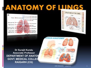
- 1. ANATOMY OF LUNGS Dr Surajit Kundu Associate Professor DEPARTMENT OF ANATOMY GOVT. MEDICAL COLLEGE RAIGARH (CG) 4/15/2021 1 GMC RAIGARH
- 2. A surgeon decides to undergo segmental resection of right Lungs for a patient suffering with Bronchogenic Carcinoma. Use your Anatomical Knowledge to Answer the following: 1. What is Segmental Resection? 2. Cite the Clinical Correlation of knowledge of Broncho pulmonary Segment. 3. Discuss the Anatomy of Broncho pulmonary Segment. 4/15/2021 2 GMC RAIGARH
- 3. 1. Gross Anatomy of Lungs 2. Surfaces and Borders of Lungs 3. Hilum and Root of Lungs 4. Fissures and Lobes of Lungs 5. Bronchopulmonary segments 6. Blood supply of lungs 7. Lymphatics of Lungs 8. Nerve supply of Lungs OBJECTIVES 4/15/2021 3 GMC RAIGARH
- 4. GROSS ANATOMY OF LUNGS CAN BE DISCUSSED AS: 1. INTRODUCTION 2. PRESENTING PARTS (SURFACES & BORDERS) 3. MEDIASTINAL SURFACE OF LUNGS AND ITS IMPRESSIONS 4. FISSURES AND LOBES 5. HILUM AND ROOT OF LUNGS 6. PULMONARY LIGAMENT & ITS CLINICAL SIGNIFICANCE 7. BRONCHIAL TREE 8. BRONCHOPULMONARY SEGMENTS AND 4/15/2021 GMC RAIGARH 4
- 5. GROSS ANATOMY OF LUNGS - INTRODUCTION Lungs are a pair of respiratory organs situated in a thoracic cavity. Right and left lung are separated by the mediastinum. Texture -- Spongy Color – Young – brown Adults -- mottled black due to deposition of carbon particles Weight- Right lung - 600 gms Left lung - 550 gms 4/15/2021 5 GMC RAIGARH
- 6. THORACIC CAVITY 4/15/2021 6 GMC RAIGARH
- 7. - Conical Apex Base 3 Borders 2 Surfaces - anterior - posterior - Inferior - costal - medial - anterior (mediastinal) - posterior (vertebral) 4/15/2021 7 GMC RAIGARH PRESENTING PARTS
- 8. APEX Blunt Lies above the level of anterior end of 1st Rib. Reaches 1-2 cm above medial 1/3rd of clavicle. Coverings – cervical pleura. suprapleural membane Grooved by- Subclavian artery Subclavian vein 4/15/2021 8 GMC RAIGARH What is Supra pleural membrane ??????
- 10. BASE Semilunar and concave. Rests on dome of Diaphragm. Right sided dome is higher than left. 4/15/2021 10 GMC RAIGARH
- 11. APEX & BASE OF LUNGS - RELATIONS 4/15/2021 GMC RAIGARH 11
- 12. BORDERS ANTERIOR BORDER – 1. Corresponds to the anterior (Costomediastinal) line of pleural reflection. 2. It is deeply notched in the left lung posterior to 5th costal cartilage by the pericardium and extends vertically downwards to form Lingula. This is called cardiac notch(percussion in this area gives a dull note as compared to dull note obtained over lung). 4/15/2021 12 GMC RAIGARH
- 13. INFERIOR BORDER Thin and sharp It seperates the base of lung from the costal surface and extends into phrenicocostal sinus. 4/15/2021 13 GMC RAIGARH
- 14. POSTERIOR BORDER Thick and ill defined Fits into deep paravertebral gutter. Extends from C7 to T10. 4/15/2021 14 GMC RAIGARH
- 15. SURFACES OF THE LUNG 1. CostalSurface 2. MedialSurface -It is in contact with costal pleura and overlying thoracic wall. - Posterior / Vertebral Part - Anterior / Mediastinal Part 4/15/2021 15 GMC RAIGARH
- 16. Relations of Posterior Part 1. Vertebral Part 2. Intervertebral Discs 3. Posterior Intercostal Vessels 4. Splanchic Nerves 4/15/2021 16 GMC RAIGARH
- 17. 4/15/2021 GMC RAIGARH 17 MEDIASTINAL SURFACE OF LUNGS AND IMPRESSIONS
- 18. RELATIONS OF ANTERIOR PART 1. Right atrium 2. Small part of RV 3. SVC 4. Right brachiocephalic vein(lower part) 5. Azygos vein 6. Esophagus 7. IVC 8. Trachea 9. Right vagus nerve 10. Right phrenic nerve RIGHT SIDE LEFT SIDE 1. Left ventricle 2. Pulmonary trunk 3. Arch of Aorta 4. Descending thoracic aorta 5. Left Subclavian Artery 6. Thoracic duct 7. Left Brachiocephalic Vein 8. Left vagus nerve 9. Left phrenic nerve 10. Left recurrent laryngeal nerve 4/15/2021 18 GMC RAIGARH
- 22. PULMONARY LIGAMENT AND ITS ANATOMIC SIGNIFICANCE HILUM OF LUNGSVs ROOT OF LUNGS
- 23. HILUM It is a large depressed area that lies near the centre of the medial surface. Various structures enter and leave the lung via its root. 4/15/2021 23 GMC RAIGARH
- 24. ROOT OF THE LUNG The root is enclosed in a short tubular sheet of pleura that joins the pulmonary and mediastinal parts of pleura . It extends inferiorly as a narrow fold - The pulmonary ligament. It lies opposite of the bodies of 5th, 6th and 7th thoracic vertebra 4/15/2021 24 GMC RAIGARH
- 26. STRUCTURES OF THE ROOT Principal Bronchus on the left side. Eparterial and Hyparterial on the right side. One pulmonary artery . Two pulmonary veins - Superior Inferior Bronchial arteries One on right side Two on left side 4/15/2021 26 GMC RAIGARH
- 27. Bronchial veins Anterior and posterior pulmonary plexus of nerves. Lymphatics Bronchopulmonary Lymphnodes Areolar tissue. 4/15/2021 27 GMC RAIGARH
- 28. ARRANGEMENT OF STRUCTURES IN THE ROOT BEFORE BACKWARDS 1. Superior pulmonary vein. 2. Pulmonary artery. 3. Bronchus. 4/15/2021 28 GMC RAIGARH
- 29. ARRANGEMENT OF STRUCTURES IN ABOVE DOWNWARDS A. Right Side 1. Eparterial Bronchus. 2. Pulmonary Artery. 3. Hyparterial Bronchus. 4.Inferior Pulmonary Vein. . THE ROOT 4/15/2021 29 GMC RAIGARH
- 30. ARRANGEMENT OF STRUCTURES IN THE ROOT ABOVE DOWNWARDS B. Left Side 1. Pulmonary artery. 2. Bronchus. 3. Inferior pulmonary vein 4/15/2021 30 GMC RAIGARH
- 32. FISSURES AND LOBES OF LUNGS 4/15/2021 32 GMC RAIGARH
- 33. OBLIQUE FISSURE It begins posteriorly at the level of 5th thoracic vertebra. Passes antero-inferiorly in a spiral course to meet the inferior margin close to 6th costochondral junction. 4/15/2021 33 GMC RAIGARH
- 34. HORIZONTAL FISSURE It extends from anterior margin at the level of 4th costal cartilage. Runs horizontally backwards to meet the oblique fissure in the mid-axillary line. Pulmonary pleura extends into the fissures of the lungs so that the lobes can move on each other during respiration. 4/15/2021 34 GMC RAIGARH
- 37. BRONCHOPULMONARY SEGMENTS ANATOMIC SIGNIFICANCE AETIOLOGIC SIGNIFICANCE (CLASSIFICATION / NOMENCLATURE) RADIOLOGIC SIGNIFICANCE CLINICO-SURGICL SIGNIFICANCE 4/15/2021 GMC RAIGARH 37
- 38. BRONCHO PULMONARY SEGMENTS DEFINITION These are well defined structural, functional, Anatomic and surgical areas of the lung tissue each of which is aerated by a segmental / tertiary bronchus.
- 41. RESPIRATORY PASSAGE (UPPER & LOWER RESPIRATORY TRACT Vs CONDUCTING PART & RESPIRTORY PART OF RESPIRATORY TRACT) 4/15/2021 GMC RAIGARH 41
- 42. Trachea Right and Left Principal Bronchus Lobar Bronchi(Secondary)[2L,3R] Segmental Bronchi(Tertiary)[8L,10R] Terminal Bronchioles(25000 in no.) Respiratory Bronchioles Alveolar ducts ACINUS Alveolar sacs Alveoli 4/15/2021 42 GMC RAIGARH
- 45. The ultimate pulmonary unit from respiratory brochiole to alveoli is called Acinus. There are about 28 orders of division of tracheo-bronchial tree. Total no. of alveoli has been estimated to be between 200 - 600 million, with a total surface area of 40 - 80 meter square. 4/15/2021 45 GMC RAIGARH
- 48. BRONCHOPULMONARY SEGMENTS 3. More in line with trachea at an angle of 15 degrees Right main bronchus Left main bronchus 1. Shorter 1. Longer 2. Wider. 2. Narrower. 3. More oblique than the right at an angle of 45 degrees with the trachea 4/15/2021 48 GMC RAIGARH
- 49. WHAT IS CARINA? 4/15/2021 GMC RAIGARH 49
- 54. BRONCHOPULMONARY SEGMENTS CLASSIFICATION Right Main Bronchus Right upper lobe Bronchus Right Middle lobe Bronchus Right Lower Lobe Bronchus Segmental Bronchi Apical Anterior Posterior Segmental Bronchi Medial Lateral Segmental Bronchi Apical Anterior Posterior Medial and Lateral 4/15/2021 54 GMC RAIGARH
- 55. BRONCHOPULMONARY SEGMENTS CLASSIFICATION Left Main Bronchus Left upper lobe Bronchus Upper Branch Lower Branch Anterior Apico-posterior Superior Lingular Inferior Lingular Left lower lobe bronchus Segmental Bronchi Apical Anterior Posterior Lateral 4/15/2021 55 GMC RAIGARH
- 58. These segments are pyramidal in shape with apex towards the root of lung. Each segment is an independent respiratory unit. Each segment has its own separate artery(branches of pulmonary artery). Pulmonary Veins run in inter-segmental planes between adjoining segments. Thus a bronchopulmonary segment is not a bronchovascular segment as it does not have its own vein. 4/15/2021 58 GMC RAIGARH
- 60. CLINICAL SIGNIFICANCE Segmental resection with minimal destruction to the surrounding lung tissue. To visualize the interior of a bronchi through a bronchoscope when diseases process is limited in a segment. 4/15/2021 60 GMC RAIGARH
- 65. SEGMENTS AND DISEASE DISEASE / FOREIGN BODY BRONCHO PULMONARY SEGMENT 1. FOREIGN BODY ASPIRATION APICAL SEGMENTS OF BOTH LOWER LOBES OR POSTERIOR SEGMENTS OF RIGHT UPPER LOBE 2. TUBERCULOSIS APICAL & POSTERIOR SEGMENTS OF UPPER LOBES 3. LUNG ABSCESS POSTERIOR SEGMENT OF UPPER LOBES OR SUPERIOR / POSTERIOR BASAL SEGMENTS OF LOWER LOBES 4. LEFT LOWER LOBE 4/15/2021 GMC RAIGARH 65
- 67. A surgeon decides to undergo segmental resection of right Lungs for a patient suffering with Bronchogenic Carcinoma. Use your Anatomical Knowledge to Answer the following: 1. What is Segmental Resection? 2. Cite the Clinical Correlation of knowledge of Broncho pulmonary Segment. 3. Discuss the Anatomy of Broncho pulmonary Segment. 4/15/2021 67 GMC RAIGARH
- 68. UNIVERSITY QUESTIONS 1. DISCUSS THE LUNGS AS UNDER: (a) PRESENTING PARTS (b) MEDIASTINAL SURFACE. ADD A NOTE ON BRONCHOPULMONARY SEGMENTS 2. SHORT NOTES: (a) ROOT & HILUM OF LUNG (b)MEDIASTINAL SURFACE OF LUNGS & ITS IMPRESSIONS (c) PULMONARY LIGAMENT (d)BRONCHOPULMONARY SEGMENTS & THEIR CLINICAL RELEVENCE 4/15/2021 GMC RAIGARH 68
- 69. THANK YOU 4/15/2021 69 GMC RAIGARH