
Recommended
More Related Content
Similar to anatomy and physiology of Respiratory System.pdf
Similar to anatomy and physiology of Respiratory System.pdf (20)
Recently uploaded
Recently uploaded (20)
anatomy and physiology of Respiratory System.pdf
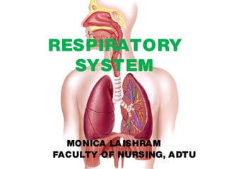
- 1. RESPIRATORY SYSTEM MONICA LAISHRAM FACULTY OF NURSING, ADTU
- 2. Select this paragraph to edit Select this paragraph to edit
- 5. Select this paragraph to edit
- 6. NOSE The right halves and left halves of • the nose is called dorsum. The lower end of the dorsum is • round and is known as tip of nose. The two nostrils are separated by • a soft median, columella. This is continous with nasal septum. Each nostril is bounded laterally • by ala.
- 7. Select this paragraph to edit Select this paragraph to edit
- 8. Function of Nose It is a respiratory passage • It is also the organ of smell. • The secretion of numerous serous • glands make the air moist, while secretion of mucous glands trap dust and other particles. Thus the nose acts as an air conditioner where the inspired air is warmed, moistened and cleaned before it is passed onto the delicate lungs.
- 9. Paranasal sinuses Paranasal sinuses are air filled • spaces present within some bones around the nasal cavities. The sinuses are frontal, maxillary, • sphenoidal and ethmoidal All of them opened to nasal cavity. •
- 10. Select this paragraph to edit
- 11. Function of paranasal sinus Make the skull lighter • Warm up and humidify the inhaled • air These also add resonance to the • voice Infection of the sinus is known as • sinusitis.
- 12. Types: Acute sinusitis and chronic • sinusitis Acute: two or more symptoms • Chronic : Symptoms for 12 weeks • or more Common cold, allergies, nasal • polyps, asthma, nasal septal deviation. Management: Decongestants and • saline nasal washes, antibiotic , steam inhalation
- 13. Select this paragraph to edit Select this paragraph to edit
- 14. Select this paragraph to edit
- 15. Rhinitis: Coryza is the • inflammation of the mucous membrane inside the nose. Common symptoms are stuffy nose, runny nose, sneezing, post nasal drip. Caused by viruses, bacteria, irritants or allergens Can be managed with intranasal • corticosteroids and intranasal anti histamines.
- 16. Select this paragraph to edit Select this paragraph to edit
- 17. Select this paragraph to edit
- 18. PHARYNX The pharynx is a wide muscular • tube, situated behind the nose, the mouth and the larynx. Clinically it is a part of the upper • respiratory passages are common. Parts of pharynx: • The nasal part: Nasopharynx • The oral part: Oropharynx • The laryngeal part: Laryngopharynx • Inflammation of pharynx is known as • pharyngitis.
- 19. Select this paragraph to edit
- 20. The upper part of the pharynx • transmits only air, the lower part only foods but the middle part is a common passage for both air and food. The nasopharynx part • pharynx is connected to the middle ear via the pharyngotympanic tube.
- 21. The eustachian tube ( • pharyngotympanic tube) connects the middle ear cavity with the nasopharynx
- 22. The eustachian tube connects the • middle ear and clears mucus from the middle ear into nasopharynx. Opening and closing of the tube is • important. Normal opening equalizes the • atmospheric pressure in the middle ear: closing of the tube protects the middle from unwanted pressure fluctuations and loud sounds
- 23. Select this paragraph to edit The tube opens when you yawn or • swallow due to contraction of tensor veli palatini muscles.
- 24. Select this paragraph to edit Boundaries: • Superiorly: Base of the skull • Inferiorly: The pharynx is continuous • with esophagus at the level of the 6th cervical vertebra. Posteriorly: The pharynx glides • freely on the pre vertebral fascia which separates it from the cervical vertebral bodies Anteriorly: It communicates with the • nasal cavity, the oral cavity and the larynx.
- 25. Select this paragraph to edit Nasopharynx: It is the upper most part of pharynx. The Eustachian tube is at the lateral wall. This tube equalizes the pressure on the two sides of the tympanic membrane. Air passes from nasopharynx into • the larynx. Air and fluids/food cross each other in to the oropharynx If one shouts or laugh while • eating or drinking , the fluids may enter the larynx.
- 26. Select this paragraph to edit This produces a protective • bout of cough as food/fluid is forbidden inside the larynx/ treachea
- 27. Oropharynx Lies behind oral cavity C2, C3 • vertebrae. It extends between soft palate above to the upper border of epiglottis below. Oropharynx communicates • anteriorly with oral cavity; above with nasopharynx and below with larynopharynx. It gives passage both to air and • food/fluid
- 28. Select this paragraph to edit
- 29. LARYNGOPHARYNX It lies behind larynx opposite • to the C5 and C6 vertebrae. It extends between epiglottis • and cricoid cartilage and anteriorly it is in the inlet of larynx. It gives passage only to • food/fluids.
- 30. WALDEYER’S LYMPHATIC RING It is a ringed arrangement of • lymphoid organs in the pharynx. It surrounds the naso and • oropharynx with tonsilar tissue located above and below the soft palate.
- 31. STRUCTURE 1 pharyngeal tonsil • 2 tubal tonsils on each side • 2 palatine tonsils (tonsil) • Lingual tonsil •
- 32. Select this paragraph to edit
- 33. Select this paragraph to edit
- 34. Select this paragraph to edit
- 35. CLINICAL SIGNIFICANCE Inflammation of tonsil is known as • tonsillitis. Removal of tonsil is known as • tonsillectomy.
- 36. FUNCTIONS OF PHARYNX Gives passage for air, foods and • fluids Warms/ cools and humidified • inspired air Helps in speech as it causes • resonance in voice Helps in hearing • Protects the lymphoid tissue • forming waldeyer ring
- 37. LARYNX
- 38. LARYNX The larynx lies in the anterior • midline of the neck, extending from the root of the tongue to the trachea. The length of the larynx is 44 • mm in male and 36 mm in females.
- 39. Select this paragraph to edit
- 40. Select this paragraph to edit At puberty, the male larynx • grows rapidly and becomes larger ( adam’s apple) which makes voice louder and low pitched. In adult male, it lies in front of • the third to sixth cervical vertebrae. In children and adult female, it • lies at a little higher level ( C1 to C4)
- 41. Select this paragraph to edit The larynx is made up of • skeletal framework of cartilages. The cartilages are connected by joints, ligaments and membranes.
- 42. Select this paragraph to edit
- 43. CARTILAGES OF LARYNX The larynx • contains nine cartilages, of which three are unpaired and three are paired.
- 44. Select this paragraph to edit Paired cartilages: • Arytenoid cartilage • Corniculate • Cuneiform • Unpaired cartilage: • Thyroid cartilage • Cricoid cartilage • Epiglotic •
- 45. CAVITY OF LARYNX Within the cavity of larynx, there are two folds of mucous membrane on each side. The upper fold is the vestibular fold and the lower fold is the vocal fold The space between the right and left vestibular folds is the rima vestibuli The space between the vocal folds is the rima glottidis.
- 46. FUNCTION OF LARYNX Acts as sphincter for lower • respiratory passage Produces voice/ sound •
- 47. TRACHEA
- 48. Select this paragraph to edit The trachea is a non • collapsible , wide tube forming the beginning of the lower respiratory passage due to C shaped cartilaginous ring The posterior wall of trachea • is deficient of cartilage. This is made of muscles and fibrous tissue for expansion of the esophagus during passage of food.
- 49. Select this paragraph to edit
- 50. Select this paragraph to edit The trachea is about 10-15 cm • long. Its upper half lies in the neck and its lower half in the superior mediastinum. The diameter is about 2 cm in • male and 1.5 cm in female.
- 51. Select this paragraph to edit The trachea is supplied by • branches from the inferior thryroid arteries. Its vein drain into the left brachiocephalic vein. Lymphatic drain into the pre • tracheal and para tracheal nodes.
- 52. HISTOLOGY OF TRACHEA The trachea is lined by pseudo • stratified ciliated columnar epithelium. The cells are of varying height, giving a false appearance of more than a layer of cells. Deep to the epithelium are mucus • and serous glands. The main bulk is formed by the C • shaped hyaline cartilages to keep it permanently patent.
- 53. Select this paragraph to edit FUNCTION OF C SHAPED CARTILAGE: C shaped cartilage keep the airway patent. The smooth muscles joining 2 ends of c helps the esophagus to dilate during the passage of bolus. These provide flexibility to trachea Ciliary escalator helps to remove the mucus swallowed into laryngopharynx or expectorated.
- 54. COUGH REFLEX If irritated, the nerve endings in • larynx, trachea, bronchi pass impulses by 10th nerve to respiratory center in brain stem. There is deep inspiration, closure • of vocal cords, contraction of abdominal and thoracic respiratory muscles and the increased pressure in lungs leads to abduction of vocal cords to expel the irritant through mouth.
- 55. LUNGS Lungs are two voluminous • cone shaped organs occupying most of the thoracic cavity leaving a small space for the heart. Each lung cavity is enclosed • within pleural cavity which contain serous fluid which helps in expansion and contraction of lungs.
- 56. Select this paragraph to edit Pleura: It is a closed • serous sac which encloses the lungs. It has two layers, parietal pleura and visceral pleura. The pleural fluid prevent • friction during breathing.
- 57. PARTS OF LUNGS
- 58. Select this paragraph to edit Apex: It is rounded and rise • into the root of neck about 2.5 cm above the level of the middle third of clavicle. Base: It is the concave part • related with diahpragm. Costal surface: It is the • surface related with ribs, intercostal muscles. Mediastinal surface: It is • related to the hilum.
- 59. Right lung vs left lung It has 2 • fissures and 3 lobes Larger and • heavier, weighs about 700 gms Shorter and • broader Anterior • border is straight It has 1 fisure • and 2 lobes Smaller and • lighter , weighs about 600 gms Longer and • narrower Anterior • border is interrupted by cardiac notch
- 60. PORTION OF LUNGS Lungs are made up of two • portions, a conducting portion and respiratory portion CONDUCTING PORTION: The • primary bronchus enters the lung at the hilum.
- 61. Select this paragraph to edit In the right lung it divides into • three secondary bronchi for each three lobes i.e upper, middle and lower separated by obligue and horizontal fissures. The three secondary bronchi are • divided into 10 segmental bronchi. Left lung contains two lobes that • is upper lobe and lower lobe separated by oblique fissures. Therefore there are only two secondary bronchi. There are 10 segmental bronchi in upper and lower lobe
- 62. Select this paragraph to edit
- 63. Select this paragraph to edit Each segmental bronchus divides • repeatedly till its diameter becomes 1.0 mm. At this level it is called terminal • bronchiole, with no cartilage. RESPIRATORY PORTION: terminal • bronchiole divides further and forms respiratory bronchiole, alveolar sac and finally the alveoli. Here the wall becomes thinner. Alveoli are lined by two types of •
- 64. Select this paragraph to edit There are as many as 700 million • alveoli in each lungs, where they facilitate gaseous of oxygen and carbon dioxide exchange. Type 1 pneumocytes • Types 2 pneumocytes which • secretes fluid called surfactant which prevent the alveoli from collapsing by decreasing surface tension
- 65. PULMONARY SURFACTANT Because of the huge force of • surface tension in the lungs, the airway may collapse after expiration. It can further can make re- • inflation during inspiration much more difficult. So the type II pneumocytes • secrete a substance called SURFACTANT to reduce the force of surface tension due to water
- 66. Select this paragraph to edit The fetus in the womb receives • oxygen from the mother. The lungs of the fetus are not fully functional, the lungs don’t produce surfactant until 35 weeks of fetal development. This is the reason that premature • newborn infants are at an increased risk of respiratory distress syndrome due to airway collapse
- 67. Select this paragraph to edit It could be fetal and result in • dead. This can be treated through • pulmonary surfactant replacement therapy and mechanical ventilation. Collapse of the lungs is known as • atelactasis.(complete or partial collapse of a lung or section of (lobe) Anaesthesia, blockage of air • passage (bronchus or
- 68. MUSCLES OF RESPIRATION For quiet inspiration, • daiphragm, external intercostal muscles. Deep inspiration – erector • spinae, scalene muscles, pectoral muscles.
- 69. Select this paragraph to edit
- 70. PULMONARY VENTILATION It is the movement of air in • and out of alveoli It is the process of flow of air • into the lungs during inspiration and out of the lungs during expiration Air flows because of pressure • differences between the atmosphere and the gases
- 71. Select this paragraph to edit The primary function of • pulmonary ventilation is to make oxygen available to the blood, which is transported by the cardiovascular system throughout the body to all the cells
- 72. Select this paragraph to edit Air like other gases, flows from a • region with higher pressure to a region with lower pressure. Involves 3 pressures • Atmospheric pressure: pressure • of air outside the body Intraalveolar (intrapleural • )pressure: pressure within the alveoli. Intrapleural pressure: pressure • within the pleural cavity
- 73. Select this paragraph to edit During inspiration, the diaphragm • contracts and the thoracic cavity increases its volume. This decreases the intraalveolar pressure so that air flows into the lungs. During expiration, the relaxation • of the diaphragm and elastic recoils of the tissues decreases the thoracic volume and increases the intraalveolar pressure. This pushes the air out of the lungs.
- 74. Select this paragraph to edit
- 75. Select this paragraph to edit
- 76. PHYSIOLOGY OF RESPIRATION The respiratory cycle consists of • inspiration, expiration and diffusion of gases. In normal quite breathing, there are about 15 complete respiratory cycles per minutes, i.e respiratory rate of normal adult is 12-16 breathes/minute.
- 77. Respiratory movements The anteroposterior diameter is • increased The transverse diameter is • increased The vertical diameter is increased •
- 78. Select this paragraph to edit Before inspiration, intrapulmonary • pressure equals atmospheric pressure at about 760 mmHg
- 79. PULMONARY CIRCULATION Pulmonary circulation moves • the blood between the heart and the lungs. It transport deoxygenated blood to the lungs to absorb oxygen and release CO2. The oxygenated blood the flows back to the heart.
- 80. Select this paragraph to edit Pulmonary artery • divides into two, each branch carrying deoxygenated blood to the lungs. Within the lung tissues, the pulmonary artery divides and redivides into dense capillary network around
- 81. Select this paragraph to edit The pulmonary circulation • begins at the pulmonary valve, making the vascular exit from the right side of the heart and extends to the orifices of the pulmonary veins in the wall of left atrium, which marks the entrance into the left side of the heart.
- 82. Select this paragraph to edit The pulmonary circulation is • supplied with both sympathetic and para symphathetic innervation The exchange of gases between • air in the alveoli and the blood in the capillaries takes place Capillaries join up to form • pulmonary veins in each lung. They leave the hilum and carry oxygenated blood to heart.
- 83. REGULATION OF RESPIRATION Physiologically, the respiration is • controlled by respiratory center – group of neurons present bilaterally in medulla oblongata and pons. Respiratory control is an involuntary • process. The respiratory center: The • respiratory center is formed by group of nerve cells( medulla oblongata and pons) which controls the rate and depth of respiration.
- 84. Select this paragraph to edit The active inspiratory center • nerves to the inspiratory muscles, the phrenic nerve to the diaphragm and intercostal nerves to the external intercostal muscles. These muscles contract and lead to expansion of the thorax and the air is drawn into the lungs.
- 85. Select this paragraph to edit When the inspiratory center • neurons are not active, the stimulus to the muscles stops and the muscles relax. At this point expiration occurs passively. This rhythmic pattern continues till any other stimulus affects the neurons of inspiratory center.
- 86. Select this paragraph to edit Chemical control: Irritant receptors: • These are present between the epithelial cells. These receptors get stimulated by irritant gases or dust particles. Activation of receptors causes coughing and increased mucus secretion Chemoreceptors: In the body, few • receptors are present which respond to changes in PH, pO2 and pCO2. The sensitivity of chemoreceptors to raised arterial CO2 concentration is most important factor in maintaining homeostasis of blood gases.
- 87. Select this paragraph to edit The chemoreceptors are located in the medulla oblongata. They monitor the level of CO2 and O2. The types of chemoreceptors are peripheral chemoreceptors and central chemoreceptors: The peripheral chemoreceptors are present in the aortic bodies( located in the arch of aorta) and carotid bodies( located at common carotid artery).
- 88. Select this paragraph to edit They are sensitive to change in • H+, PCO2 and PO2 in blood. Nerve impulses from the peripheral chemoreceptors are conveyed by the glossopharyngeal and vagus nerves to the medulla. It stimulates the respiratory center. The result is immediate increase in the depth and rate of respiration.
- 89. Select this paragraph to edit The central chemoreceptors: • They are present on the surface of medulla oblongata. They respond to changes in PCO2 and H+ in CSF. These respond by stimulating the respiratory center. They increases the ventilation of lungs and reduce arterial PCO2 . This control normal blood gas levels.
- 90. OTHER FACTORS THAT INFLUENCE RESPIRATION Limbic system: Emotional anxiety or • anticipation of activity, both may stimulate the limbic system. This ends the input to the inspiratory center. It increases the rate and depth of ventilation. Blood pressure: The carotid and aortic sinuses • contain baroreceptors. It detects the change in blood pressure. It also affects respiration.
- 91. Select this paragraph to edit For example, increase in blood • pressure decreases the respiration. Decrease in blood pressure increases respiration. Temperature: Increase in body • temperature will increase the rate of respiration. A decrease in body temperature decreases the respiratory rate. Pain: sudden, severe pain causes • apnea. Prolonged pain results in increase of respiratory rate.
- 92. Select this paragraph to edit Irritation of airway: • Mechanical irritation of the pharynx or larynx stoppage of breathing for a while. It leads to coughing or sneezing Stretching of the anal • sphincter muscle: This increases the respiratory rate.
- 93. LUNG VOLUMES AND CAPACITIES Lung volume is the static volume • of air breathed by an individual i.e volume of air present in lung under specific position of the thorax. Lung volumes depends on age, weight and gender and body position. When two or more volumes combine this is called capacity.
- 94. Select this paragraph to edit Dead space: It is constituted • by air, which doesn’t participate in diffusion,i.e air present in nose, trachea and bronchial tree. Normally it is 150 ml. Tidal volume: Volume of gas • inspired or expired in each breath during normal quiet respiration. It is 400-500 ml.
- 95. Select this paragraph to edit Alveolar ventilation: It is the • volume of air that moves into and out of alveoli per minutes. Alveolar ventilation: Tidal volume- anatomical dead space)*respiratory rate 500-150*15=5.25L/min Inspiratory reserve volume: It is the maximum volume of gas, which a person can inhale from end inspiratory position. Its 2,400- 2,600ml.
- 96. Select this paragraph to edit Expiratory reserve volume: • Maximum volume of gas that can be exhaled after normal expiration. It is 1200-1500ml
- 97. Select this paragraph to edit Vital capacity: It is the • maximum amount of gas that can be exhaled after maximum inhalation, i.e it is IRV+TV+ERV, 4,200- 4,500 ML. Residual volume: It is the volume of gas still present in lungs after maximal expiration. It is 1,200-1,500 ml
- 98. LUNG CAPACITIES Inspiratory capacity: Amount of • gas inspired into the lungs after normal tidal expiration. IC=TV+IRV Functional residual capacity • (FRC): Amount of gas remains in the lungs after normal expiration. FRC=ERV+RV (2500ml) Vital capacity (VC): Amount of gas • expired from the lungs after a maximum inspiration.
- 99. Select this paragraph to edit Total lung capacity (TLC max): • Amount of gas inspired to expand the lungs to its maximum extends. ( TLC= TV+IRV+ERV+RV) 6000ML Minute volume : It is the tidal • volume*respiratory rate. It is 500*12=6000 ml/min Total lung volume: • IRV+TV+ERV+RV. It is 5,500- 6,000ml.
- 100. Select this paragraph to edit Select this paragraph to edit
- 101. MECHANISM OF RESPIRATION Select this paragraph to edit
- 102. PULMONARY FUNCTION TEST PFTs are a group of tests that • measures how well your lungs work that is how well the lungs take in and exhale air, and how efficiently they transfer oxygen into the blood. They are useful in assessing the • functional and pathological conditions. It is based on the measurement of lung volumes of air breathed in and out in normal breathing and forced breathing. It
- 103. PURPOSES OF PFTs Detect disease and serves as a • diagnostic tool, and has investigation role Evaluates severity, extent and • monitor the course of disease Evaluates treatment • Measures effects and results of • treatment exposures.
- 104. Select this paragraph to edit Helps to diagnose asthma, • chronic bronchitis, respiratory infections, lung fibrosis, bronchiectasis, allergy, emphysema, cystic fibrosis, asbestosis which is the inflammation of lungs, liver, lymph nodes, eyes, skin Used to evaluate physiological • aspect of breathing from respiratory muscle function to the diffusion of gas at the alveolar
- 105. Select this paragraph to edit Helps physiotherapy to distinguish • between obstructive and restrictive lung problem and to select appropriate treatment Measures the effect of the given • treatment
- 106. PROCEDURE Sit up straight • Get a good seal around the mouth • piece Rapidly inhale maximally • Without any delay blow out as • hard as fast as possible Continue the exhale until the • patient cannot blow no more
- 107. Select this paragraph to edit Expiration should continue at • least 6 sec in adult and 3 sec children under 10 years Repeat at least three technically • acceptable times ( without cough, air leak and false start)
- 108. SPIROMETRY In order to do spirometry, an • instrument that is spirometer is used. Spirometer measures the air capacity of the lungs during controlled ventilatory maneuvers. Spirometer is used to measure the • air flow, ventilatory regulation, ventilatory mechanics and lung volume during a forced expiratory maneuver from full inspiration.
- 109. LUNG EXAMINATION AND LUNG SOUNDS Lung examination includes vital • signs and cardiac examination. There are four elements- observation, palpation, percussion, ascultation. Lung sounds: • Bronchial breath sounds: Normal • sounds when listemning over large airways like trachea is bronchial breath sounds
- 110. Select this paragraph to edit It can be present when a person is in the • setting of dense com