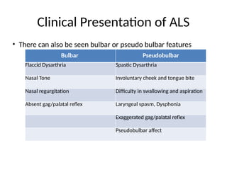
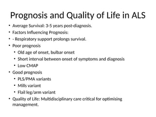
Amyotrophic Lateral Sclerosis
(ALS)
Presented by: Dr. Omkar Kulkarni
Guide: Dr. Pravin Soni Sir
Dr. Ram Mundhe sir
Date: 04/10/2024
Lou Gehrig aka The Iron Horse
American baseball Player
Stephen Hawking
English theoretical physicist and
cosmologist
Introduction
• ALS is the most common progressive motor
neuron disease.
• The most devastating of the neurodegenerative
disorders.
• First Described: Jean-Martin Charcot in 1869.
• It is also called as Lou Gehrig's disease or
Charcot’s Disease.
Types of ALS
• Sporadic ALS/SALS - 90%
• Familial ALS/FALS - 10%
• It has a variant which presents in younger
age group - Juvenile ALS
• It has slower progression than SALS
• Stephen Hawking was affected with Juvenile
ALS
Pathology of ALS
• The pathologic hallmark of motor neuron
degenerative disorders is death of lower motor
neurons(consisting of anterior horn cells in the
spinal cord and their brainstem homologues
innervating bulbar muscles) and upper, or
corticospinal, motor neurons (originating in
layer five of the motor cortex and descending
via the pyramidal tract
Pathology of ALS
• At the onset ALS may have loss of either UMN or LMN but
ultimately causes progressive loss of both UMN and LMN.
• In other MNDs only particular subset of neurons is involved.
• In bulbar palsy and spinal muscular atrophy (SMA; also
called progressive muscular atrophy), the lower motor
neurons of brainstem and spinal cord, respectively, are most
severely involved.
• While pseudobulbar palsy, primary lateral sclerosis (PLS),
and hereditary spastic paraplegia (HSP) affect only upper
motor neurons innervating the brainstem and spinal cord.
Pathology of ALS
• the affected motor neurons undergo shrinkage, often
with accumulation of the pigmented lipid (lipofuscin)
• Focal enlargements are frequent in proximal motor
axons; ultrastructurally, these “spheroids” are composed
of accumulations of neurofilaments and other proteins.
• the affected neurons demonstrate ubiquitin-positive
aggregates, typically associated with the protein TDP43
• Bunina bodies(specific), skein like inclusion, rounded
inclusions can be seen in Betz cells
Pathology of ALS
Bunina Bodies
Skein like inclusions
IHC staining for TDP43
Pathology of ALS
• The death of the peripheral motor neurons in the brainstem
and spinal cord leads to denervation and atrophy of the
corresponding muscle fibers.
• As denervation progresses, muscle atrophy occurs -
Amyotrophy
• The loss of cortical motor neurons results in thinning of the
corticospinal tracts that travel via the internal capsule and
pyramidal tracts in the brainstem to the lateral and anterior
white matter columns of the spinal cord.
• The loss of fibers in the lateral columns and resulting fibrillary
gliosis impart a particular firmness - lateral sclerosis
Epidemiology of ALS
• Incidence of 1–3 per 100,000 and a prevalence of 3–
5 per 100,000.
• Male to female ratio is 1.4:1
• In cases of bulbar onset ALS Female slightly more
than males
• Juvenile ALS may start at around 12 hrs of age
• Average survival after diagnosis of SALS- 3 YEARS
• 1/5th will survive 5 years
• In that 1/10th will survive to 10 yrs
ALS: Genetics
• Familial ALS (fALS): 5-10% of ALS cases.
• Key Mutations:
• C9ORF72 (Most common among FALS- 30%):
• Causes toxic RNA foci and dipeptide repeat proteins.
• Young onset with rapid progression.
• SOD1 (Superoxide dismutase 1)2nd most common:
• Gain of function mutation
• Causes oxidative stress and motor neuron death.
• A/w ALS1; Late onset(30yrs)
• TDP43 - a/w ALS 10, 12
• FUS - a/w ALS 6
• Mechanisms: Protein misfolding, RNA metabolism disruption, axonal
transport dysfunction.
ALS juvenile : Genetics
• ALS 2,4,5,16
• Age of onset- 25 yrs
• ALS2 - ALSin gene, onset 12 yrs
• ALS4 - AD mutation of Senataxin gene
• ALS5 - SPASTICIN Gene
• ALS16 - SIGMARI Gene
• ALS 12 - OPTN gene
• ALS17 - CHMP2B gene
Etiology of ALS: Sporadic Causes
• Sporadic ALS (sALS): 90-95% of ALS cases.
• Risk Factors:
• - Toxins: Heavy metals (lead), pesticides, BMAA.
• - Physical trauma: Increased risk in athletes.
• - Military service: Veterans show higher ALS
incidence.
• Multifactorial Hypothesis: Interaction of genetic
predisposition and environmental exposures.
Pathophysiology of ALS: Motor Neuron
Degeneration
• Motor Neurons Affected:
• - Upper Motor Neurons (UMNs): Located in the
motor cortex. Leads to spasticity, hyperreflexia.
• - Lower Motor Neurons (LMNs): Located in the
spinal cord and brainstem. Leads to weakness,
atrophy.
Pathophysiology of ALS: Cellular
Mechanisms
• Mechanisms of Neuronal Death:
• - Glutamate Excitotoxicity: Excess glutamate
causes calcium influx and neuronal death.
• - Oxidative Stress: Mutant SOD1 increases free
radicals.
• - Protein Aggregation: Misfolded proteins (TDP-43,
SOD1) form inclusions in neurons.
• - Neuroinflammation: Activated microglia release
pro-inflammatory cytokines, exacerbating damage.
Clinical Presentation of ALS
• LMN + UMN symptoms-
• Wasting, fasciculation, hyperreflexia, hypertonia.
• Wasting >>> weakness
• Starts asymmetrically and distally.
• Spread to contiguous areas.
• Loss of thenar muscles > hypothenar- split hand
• Excessive forced yawning
• Head drop
• ALS cachexia
• Atypical for ALS - Extrapyramidal involvement, autonomic and
extraoccular muscles involvement.
Clinical Presentation of ALS
Severe muscle atrophy
Split hand with thenar atrophy
Tongue atrophy
Clinical Presentation of ALS
• There can also be seen bulbar or pseudo bulbar features
Bulbar Pseudobulbar
Flaccid Dysarthria Spastic Dysarthria
Nasal Tone Involuntary cheek and tongue bite
Nasal regurgitation Difficulty in swallowing and aspiration
Absent gag/palatal reflex Laryngeal spasm, Dysphonia
Exaggerated gag/palatal reflex
Pseudobulbar affect
Clinical Presentation of ALS
• Initial Symptoms:
• - Limb-Onset ALS (~70%): Asymmetric weakness in
one limb, fasciculations, cramps.
• - Bulbar-Onset ALS (~25%): Slurred speech
(dysarthria), swallowing difficulties (dysphagia).
• Progression: Atrophy and weakness spread,
respiratory muscles eventually affected.
• Cognitive Impairment: Present in ~50% of cases,
linked to frontotemporal degeneration.
Variants of ALS
• LMN onset
• UMN onset
• Bulbar onset
• Respiratory onset
• Spinal
Variants of ALS
• Based on area involved
• Pseudo neurotic / flail leg with foot drop
• Flail arm with wrist drop
• Monomelic - only one limb involved
• Man in barrel - b/l upper limbs are wasted
and cannot be moved
Investigations and Diagnosis for ALS
• VDRL, HIV, TFT, PTH, Serum Ca2+ should be done
• Total CK is increased
• MRI - principally to exclude other pathologies, such as
cerebral lesions, skull base lesions, cervical spondylotic
myelopathy, other myelopathy, conus lesions, and
thoracolumbar/sacral radiculopathy
• MRI in ALS show hyperintensity on T2-weighted imaging
along the corticospinal tracts.
• The loss of signal on GRE and SWI of the precentral gyrus,
known as the "motor band sign" is seen in ~50%.
Investigations and Diagnosis for ALS
Hyper intensities in posterior limb of IC
Motor Band Sign
Investigations and Diagnosis for ALS
• EMG Studies
• Spontaneous/insertional activities-
fibrillations, fasciculations, positive sharps
• MUAP- giant MUAPs, polyphasic/serrated
pattern
• Recruitment- reduced
• Interference pattern - less dense(Picket-
fence pattern)
Investigations and Diagnosis for ALS
B. Picket-fence pattern of ALS
Diagnostic criteria for ALS
Therapeutic Strategies for ALS
• No treatment arrests the underlying pathologic process in
ALS.
• The drug Riluzole (100 mg/d) was approved for ALS
because it produces a modest lengthening of survival.
• Exact mechanism is unknown but riluzole may reduce
excitotoxicity by diminishing glutamate release.
• second drug, Edaravone, has also been approved by the
U.S. Food and Drug Administration.
• It is administered in a dose of 16mg via recurring monthly
10-day series of daily intravenous infusions.
Emerging Therapies for ALS
• Gene Therapy:
• - Antisense Oligonucleotides (ASOs): Target SOD1,
reduce toxic proteins.
• - CRISPR/Cas9: Gene editing approaches for
correcting pathogenic mutations.
• Stem Cell Therapy: Mesenchymal stem cells (MSCs)
for neuroprotection and immunomodulation.
• Neuroprotective Drugs: Trofinetide targeting
neuroinflammation and mitochondrial dysfunction.
Prognosis and Quality of Life in ALS
• Average Survival: 3-5 years post-diagnosis.
• Factors Influencing Prognosis:
• - Respiratory support prolongs survival.
• Poor prognosis
• Old age of onset, bulbar onset
• Short interval between onset of symptoms and diagnosis
• Low CMAP
• Good prognosis
• PLS/PMA variants
• Mills variant
• Flail leg/arm variant
• Quality of Life: Multidisciplinary care critical for optimising
management.

ALS_Detailed_Academic_Presentation .pptx

  • 1.
    Amyotrophic Lateral Sclerosis (ALS) Presentedby: Dr. Omkar Kulkarni Guide: Dr. Pravin Soni Sir Dr. Ram Mundhe sir Date: 04/10/2024
  • 2.
    Lou Gehrig akaThe Iron Horse American baseball Player Stephen Hawking English theoretical physicist and cosmologist
  • 3.
    Introduction • ALS isthe most common progressive motor neuron disease. • The most devastating of the neurodegenerative disorders. • First Described: Jean-Martin Charcot in 1869. • It is also called as Lou Gehrig's disease or Charcot’s Disease.
  • 4.
    Types of ALS •Sporadic ALS/SALS - 90% • Familial ALS/FALS - 10% • It has a variant which presents in younger age group - Juvenile ALS • It has slower progression than SALS • Stephen Hawking was affected with Juvenile ALS
  • 5.
    Pathology of ALS •The pathologic hallmark of motor neuron degenerative disorders is death of lower motor neurons(consisting of anterior horn cells in the spinal cord and their brainstem homologues innervating bulbar muscles) and upper, or corticospinal, motor neurons (originating in layer five of the motor cortex and descending via the pyramidal tract
  • 6.
    Pathology of ALS •At the onset ALS may have loss of either UMN or LMN but ultimately causes progressive loss of both UMN and LMN. • In other MNDs only particular subset of neurons is involved. • In bulbar palsy and spinal muscular atrophy (SMA; also called progressive muscular atrophy), the lower motor neurons of brainstem and spinal cord, respectively, are most severely involved. • While pseudobulbar palsy, primary lateral sclerosis (PLS), and hereditary spastic paraplegia (HSP) affect only upper motor neurons innervating the brainstem and spinal cord.
  • 7.
    Pathology of ALS •the affected motor neurons undergo shrinkage, often with accumulation of the pigmented lipid (lipofuscin) • Focal enlargements are frequent in proximal motor axons; ultrastructurally, these “spheroids” are composed of accumulations of neurofilaments and other proteins. • the affected neurons demonstrate ubiquitin-positive aggregates, typically associated with the protein TDP43 • Bunina bodies(specific), skein like inclusion, rounded inclusions can be seen in Betz cells
  • 8.
    Pathology of ALS BuninaBodies Skein like inclusions IHC staining for TDP43
  • 9.
    Pathology of ALS •The death of the peripheral motor neurons in the brainstem and spinal cord leads to denervation and atrophy of the corresponding muscle fibers. • As denervation progresses, muscle atrophy occurs - Amyotrophy • The loss of cortical motor neurons results in thinning of the corticospinal tracts that travel via the internal capsule and pyramidal tracts in the brainstem to the lateral and anterior white matter columns of the spinal cord. • The loss of fibers in the lateral columns and resulting fibrillary gliosis impart a particular firmness - lateral sclerosis
  • 10.
    Epidemiology of ALS •Incidence of 1–3 per 100,000 and a prevalence of 3– 5 per 100,000. • Male to female ratio is 1.4:1 • In cases of bulbar onset ALS Female slightly more than males • Juvenile ALS may start at around 12 hrs of age • Average survival after diagnosis of SALS- 3 YEARS • 1/5th will survive 5 years • In that 1/10th will survive to 10 yrs
  • 11.
    ALS: Genetics • FamilialALS (fALS): 5-10% of ALS cases. • Key Mutations: • C9ORF72 (Most common among FALS- 30%): • Causes toxic RNA foci and dipeptide repeat proteins. • Young onset with rapid progression. • SOD1 (Superoxide dismutase 1)2nd most common: • Gain of function mutation • Causes oxidative stress and motor neuron death. • A/w ALS1; Late onset(30yrs) • TDP43 - a/w ALS 10, 12 • FUS - a/w ALS 6 • Mechanisms: Protein misfolding, RNA metabolism disruption, axonal transport dysfunction.
  • 12.
    ALS juvenile :Genetics • ALS 2,4,5,16 • Age of onset- 25 yrs • ALS2 - ALSin gene, onset 12 yrs • ALS4 - AD mutation of Senataxin gene • ALS5 - SPASTICIN Gene • ALS16 - SIGMARI Gene • ALS 12 - OPTN gene • ALS17 - CHMP2B gene
  • 13.
    Etiology of ALS:Sporadic Causes • Sporadic ALS (sALS): 90-95% of ALS cases. • Risk Factors: • - Toxins: Heavy metals (lead), pesticides, BMAA. • - Physical trauma: Increased risk in athletes. • - Military service: Veterans show higher ALS incidence. • Multifactorial Hypothesis: Interaction of genetic predisposition and environmental exposures.
  • 14.
    Pathophysiology of ALS:Motor Neuron Degeneration • Motor Neurons Affected: • - Upper Motor Neurons (UMNs): Located in the motor cortex. Leads to spasticity, hyperreflexia. • - Lower Motor Neurons (LMNs): Located in the spinal cord and brainstem. Leads to weakness, atrophy.
  • 15.
    Pathophysiology of ALS:Cellular Mechanisms • Mechanisms of Neuronal Death: • - Glutamate Excitotoxicity: Excess glutamate causes calcium influx and neuronal death. • - Oxidative Stress: Mutant SOD1 increases free radicals. • - Protein Aggregation: Misfolded proteins (TDP-43, SOD1) form inclusions in neurons. • - Neuroinflammation: Activated microglia release pro-inflammatory cytokines, exacerbating damage.
  • 16.
    Clinical Presentation ofALS • LMN + UMN symptoms- • Wasting, fasciculation, hyperreflexia, hypertonia. • Wasting >>> weakness • Starts asymmetrically and distally. • Spread to contiguous areas. • Loss of thenar muscles > hypothenar- split hand • Excessive forced yawning • Head drop • ALS cachexia • Atypical for ALS - Extrapyramidal involvement, autonomic and extraoccular muscles involvement.
  • 17.
    Clinical Presentation ofALS Severe muscle atrophy Split hand with thenar atrophy Tongue atrophy
  • 18.
    Clinical Presentation ofALS • There can also be seen bulbar or pseudo bulbar features Bulbar Pseudobulbar Flaccid Dysarthria Spastic Dysarthria Nasal Tone Involuntary cheek and tongue bite Nasal regurgitation Difficulty in swallowing and aspiration Absent gag/palatal reflex Laryngeal spasm, Dysphonia Exaggerated gag/palatal reflex Pseudobulbar affect
  • 19.
    Clinical Presentation ofALS • Initial Symptoms: • - Limb-Onset ALS (~70%): Asymmetric weakness in one limb, fasciculations, cramps. • - Bulbar-Onset ALS (~25%): Slurred speech (dysarthria), swallowing difficulties (dysphagia). • Progression: Atrophy and weakness spread, respiratory muscles eventually affected. • Cognitive Impairment: Present in ~50% of cases, linked to frontotemporal degeneration.
  • 20.
    Variants of ALS •LMN onset • UMN onset • Bulbar onset • Respiratory onset • Spinal
  • 21.
    Variants of ALS •Based on area involved • Pseudo neurotic / flail leg with foot drop • Flail arm with wrist drop • Monomelic - only one limb involved • Man in barrel - b/l upper limbs are wasted and cannot be moved
  • 22.
    Investigations and Diagnosisfor ALS • VDRL, HIV, TFT, PTH, Serum Ca2+ should be done • Total CK is increased • MRI - principally to exclude other pathologies, such as cerebral lesions, skull base lesions, cervical spondylotic myelopathy, other myelopathy, conus lesions, and thoracolumbar/sacral radiculopathy • MRI in ALS show hyperintensity on T2-weighted imaging along the corticospinal tracts. • The loss of signal on GRE and SWI of the precentral gyrus, known as the "motor band sign" is seen in ~50%.
  • 23.
    Investigations and Diagnosisfor ALS Hyper intensities in posterior limb of IC Motor Band Sign
  • 24.
    Investigations and Diagnosisfor ALS • EMG Studies • Spontaneous/insertional activities- fibrillations, fasciculations, positive sharps • MUAP- giant MUAPs, polyphasic/serrated pattern • Recruitment- reduced • Interference pattern - less dense(Picket- fence pattern)
  • 25.
    Investigations and Diagnosisfor ALS B. Picket-fence pattern of ALS
  • 26.
  • 27.
    Therapeutic Strategies forALS • No treatment arrests the underlying pathologic process in ALS. • The drug Riluzole (100 mg/d) was approved for ALS because it produces a modest lengthening of survival. • Exact mechanism is unknown but riluzole may reduce excitotoxicity by diminishing glutamate release. • second drug, Edaravone, has also been approved by the U.S. Food and Drug Administration. • It is administered in a dose of 16mg via recurring monthly 10-day series of daily intravenous infusions.
  • 28.
    Emerging Therapies forALS • Gene Therapy: • - Antisense Oligonucleotides (ASOs): Target SOD1, reduce toxic proteins. • - CRISPR/Cas9: Gene editing approaches for correcting pathogenic mutations. • Stem Cell Therapy: Mesenchymal stem cells (MSCs) for neuroprotection and immunomodulation. • Neuroprotective Drugs: Trofinetide targeting neuroinflammation and mitochondrial dysfunction.
  • 29.
    Prognosis and Qualityof Life in ALS • Average Survival: 3-5 years post-diagnosis. • Factors Influencing Prognosis: • - Respiratory support prolongs survival. • Poor prognosis • Old age of onset, bulbar onset • Short interval between onset of symptoms and diagnosis • Low CMAP • Good prognosis • PLS/PMA variants • Mills variant • Flail leg/arm variant • Quality of Life: Multidisciplinary care critical for optimising management.