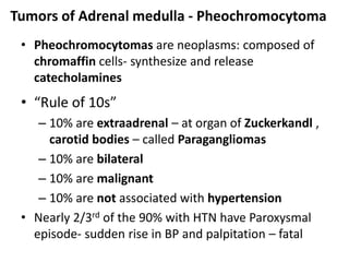
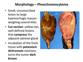
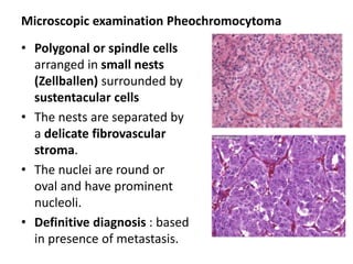
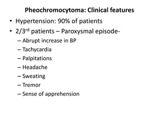
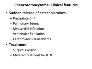
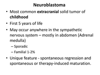
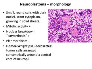
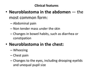
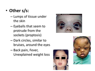
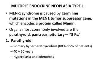
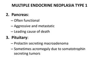
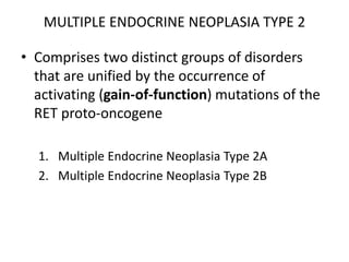
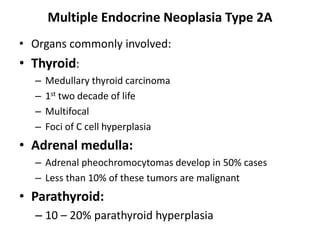
Adrenal Medulla
Dr. Reetu Baral MD (Pathology)
Department of Pathology
Nobel medical college
Adrenal Medulla
• Adrenal medulla cells derived from:
– Neural crest (chromaffin cells)
– Their supporting (sustentacular) cells
• The chromaffin cells are so named because of their brown-
black color after exposure to potassium dichromate
• They synthesize and secrete catecholamines in response to
signals from preganglionic nerve fibers in the sympathetic
nervous system
• The most important diseases of the adrenal medulla are
neoplasms:
– Neuroblastomas
– Pheochromocytomas
Tumors of Adrenal medulla - Pheochromocytoma
• Pheochromocytomas are neoplasms: composed of
chromaffin cells- synthesize and release
catecholamines
• “Rule of 10s”
– 10% are extraadrenal – at organ of Zuckerkandl ,
carotid bodies – called Paragangliomas
– 10% are bilateral
– 10% are malignant
– 10% are not associated with hypertension
• Nearly 2/3rd of the 90% with HTN have Paroxysmal
episode- sudden rise in BP and palpitation – fatal
Morphology – Pheochromocytoma
• Small, circumscribed
lesion to large
haemorrhagic masses
weighing several kilos
• Cut section: yellow-tan,
well-defined lesions
that compress the
adjacent adrenal gland
• Incubation of the fresh
tissue with potassium
dichromate solutions
turns the tumor dark
brown
Microscopic examination Pheochromocytoma
• Polygonal or spindle cells
arranged in small nests
(Zellballen) surrounded by
sustentacular cells
• The nests are separated by
a delicate fibrovascular
stroma.
• The nuclei are round or
oval and have prominent
nucleoli.
• Definitive diagnosis : based
in presence of metastasis.
Pheochromocytoma: Clinical features
• Hypertension: 90% of patients
• 2/3rd patients – Paroxysmal episode-
– Abrupt increase in BP
– Tachycardia
– Palpitations
– Headache
– Sweating
– Tremor
– Sense of apprehension
Pheochromocytoma: Clinical features
• Sudden release of catecholamines-
– Precipitate CHF
– Pulmonary Edema
– Myocardial infarction
– Ventricular fibrillation
– Cerebrovascular accidents
• Treatment
– Surgical excision
– Medical treatment for HTN
Neuroblastoma
• Most common extracranial solid tumor of
childhood
• First 5 years of life
• May occur anywhere in the sympathetic
nervous system – mostly in abdomen (Adrenal
medulla)
– Sporadic
– Familial 1-2%
• Unique feature - spontaneous regression and
spontaneous or therapy-induced maturation.
Neuroblastoma – morphology
• Small, round cells with dark
nuclei, scant cytoplasm,
growing in solid sheets.
• Mitotic activity +
• Nuclear breakdown
“karyorrhexis” +
• Pleomorphism +
• Homer-Wright pseudorosettes:
tumor cells arranged
concentrically around a central
core of neuropil
Clinical features
• Neuroblastoma in the abdomen — the
most common form:
– Abdominal pain
– Non tender mass under the skin
– Changes in bowel habits, such as diarrhea or
constipation
• Neuroblastoma in the chest:
– Wheezing
– Chest pain
– Changes to the eyes, including drooping eyelids
and unequal pupil size
• Other s/s:
– Lumps of tissue under
the skin
– Eyeballs that seem to
protrude from the
sockets (proptosis)
– Dark circles, similar to
bruises, around the eyes
– Back pain, Fever,
Unexplained weight loss
Multiple Endocrine Neoplasia (MEN) syndromes
• The MEN syndromes are a group of inherited
diseases caused by proliferative lesions (hyperplasias,
adenomas, and carcinomas) of multiple endocrine
organs.
• Distinctive features of MEN:
– Younger age
– They arise in multiple endocrine organs: synchronously or
metachronously
– Multifocal – even in one organ
– Aggressive and recur
– Preceded by an asymptomatic stage of endocrine
hyperplasia involving the cell of origin of the tumor (C-cell
hyperplasia followed by medullary thyroid carcinoma)
MULTIPLE ENDOCRINE NEOPLASIA TYPE 1
• MEN-1 syndrome is caused by germ line
mutations in the MEN1 tumor suppressor gene,
which encodes a protein called Menin.
• Organs most commonly involved are the
parathyroid, pancreas, pituitary— “3 Ps.”
1. Parathyroid:
– Primary hyperparathyroidism (80%–95% of patients)
– 40 – 50 years
– Hyperplasia and adenomas
MULTIPLE ENDOCRINE NEOPLASIA TYPE 1
2. Pancreas:
– Often functional
– Aggressive and metastatic
– Leading cause of death
3. Pituitary:
– Prolactin secreting macroadenoma
– Sometimes acromegaly due to somatotrophin
secreting tumors
MULTIPLE ENDOCRINE NEOPLASIA TYPE 2
• Comprises two distinct groups of disorders
that are unified by the occurrence of
activating (gain-of-function) mutations of the
RET proto-oncogene
1. Multiple Endocrine Neoplasia Type 2A
2. Multiple Endocrine Neoplasia Type 2B
Multiple Endocrine Neoplasia Type 2A
• Organs commonly involved:
• Thyroid:
– Medullary thyroid carcinoma
– 1st two decade of life
– Multifocal
– Foci of C cell hyperplasia
• Adrenal medulla:
– Adrenal pheochromocytomas develop in 50% cases
– Less than 10% of these tumors are malignant
• Parathyroid:
– 10 – 20% parathyroid hyperplasia
Multiple Endocrine Neoplasia Type 2B
• A single amino acid change in RET, distinct
from the mutations that are seen in MEN-2A,
seems to be responsible for virtually all cases
of MEN-2B.
• Patients develop medullary thyroid
carcinomas, which are usually multifocal and
more aggressive than in MEN-2A, and
pheochromocytomas.
Multiple Endocrine Neoplasia Type 2B
• Primary hyperparathyroidism does not develop In
patients with MEN-2B
• Extraendocrine manifestations are characteristic in
patients with MEN-2B
• These include ganglioneuromas of mucosal sites
(gastrointestinal tract, lips, tongue)
• Marfanoid habitus, in which overly long bones of the
axial skeleton give an appearance resembling that in
Marfan syndrome
Next class
• Diabetes mellitus

Adrenal Medulla.ppt

  • 1.
    Adrenal Medulla Dr. ReetuBaral MD (Pathology) Department of Pathology Nobel medical college
  • 2.
    Adrenal Medulla • Adrenalmedulla cells derived from: – Neural crest (chromaffin cells) – Their supporting (sustentacular) cells • The chromaffin cells are so named because of their brown- black color after exposure to potassium dichromate • They synthesize and secrete catecholamines in response to signals from preganglionic nerve fibers in the sympathetic nervous system • The most important diseases of the adrenal medulla are neoplasms: – Neuroblastomas – Pheochromocytomas
  • 3.
    Tumors of Adrenalmedulla - Pheochromocytoma • Pheochromocytomas are neoplasms: composed of chromaffin cells- synthesize and release catecholamines • “Rule of 10s” – 10% are extraadrenal – at organ of Zuckerkandl , carotid bodies – called Paragangliomas – 10% are bilateral – 10% are malignant – 10% are not associated with hypertension • Nearly 2/3rd of the 90% with HTN have Paroxysmal episode- sudden rise in BP and palpitation – fatal
  • 4.
    Morphology – Pheochromocytoma •Small, circumscribed lesion to large haemorrhagic masses weighing several kilos • Cut section: yellow-tan, well-defined lesions that compress the adjacent adrenal gland • Incubation of the fresh tissue with potassium dichromate solutions turns the tumor dark brown
  • 5.
    Microscopic examination Pheochromocytoma •Polygonal or spindle cells arranged in small nests (Zellballen) surrounded by sustentacular cells • The nests are separated by a delicate fibrovascular stroma. • The nuclei are round or oval and have prominent nucleoli. • Definitive diagnosis : based in presence of metastasis.
  • 6.
    Pheochromocytoma: Clinical features •Hypertension: 90% of patients • 2/3rd patients – Paroxysmal episode- – Abrupt increase in BP – Tachycardia – Palpitations – Headache – Sweating – Tremor – Sense of apprehension
  • 7.
    Pheochromocytoma: Clinical features •Sudden release of catecholamines- – Precipitate CHF – Pulmonary Edema – Myocardial infarction – Ventricular fibrillation – Cerebrovascular accidents • Treatment – Surgical excision – Medical treatment for HTN
  • 8.
    Neuroblastoma • Most commonextracranial solid tumor of childhood • First 5 years of life • May occur anywhere in the sympathetic nervous system – mostly in abdomen (Adrenal medulla) – Sporadic – Familial 1-2% • Unique feature - spontaneous regression and spontaneous or therapy-induced maturation.
  • 9.
    Neuroblastoma – morphology •Small, round cells with dark nuclei, scant cytoplasm, growing in solid sheets. • Mitotic activity + • Nuclear breakdown “karyorrhexis” + • Pleomorphism + • Homer-Wright pseudorosettes: tumor cells arranged concentrically around a central core of neuropil
  • 10.
    Clinical features • Neuroblastomain the abdomen — the most common form: – Abdominal pain – Non tender mass under the skin – Changes in bowel habits, such as diarrhea or constipation • Neuroblastoma in the chest: – Wheezing – Chest pain – Changes to the eyes, including drooping eyelids and unequal pupil size
  • 11.
    • Other s/s: –Lumps of tissue under the skin – Eyeballs that seem to protrude from the sockets (proptosis) – Dark circles, similar to bruises, around the eyes – Back pain, Fever, Unexplained weight loss
  • 12.
    Multiple Endocrine Neoplasia(MEN) syndromes • The MEN syndromes are a group of inherited diseases caused by proliferative lesions (hyperplasias, adenomas, and carcinomas) of multiple endocrine organs. • Distinctive features of MEN: – Younger age – They arise in multiple endocrine organs: synchronously or metachronously – Multifocal – even in one organ – Aggressive and recur – Preceded by an asymptomatic stage of endocrine hyperplasia involving the cell of origin of the tumor (C-cell hyperplasia followed by medullary thyroid carcinoma)
  • 13.
    MULTIPLE ENDOCRINE NEOPLASIATYPE 1 • MEN-1 syndrome is caused by germ line mutations in the MEN1 tumor suppressor gene, which encodes a protein called Menin. • Organs most commonly involved are the parathyroid, pancreas, pituitary— “3 Ps.” 1. Parathyroid: – Primary hyperparathyroidism (80%–95% of patients) – 40 – 50 years – Hyperplasia and adenomas
  • 14.
    MULTIPLE ENDOCRINE NEOPLASIATYPE 1 2. Pancreas: – Often functional – Aggressive and metastatic – Leading cause of death 3. Pituitary: – Prolactin secreting macroadenoma – Sometimes acromegaly due to somatotrophin secreting tumors
  • 15.
    MULTIPLE ENDOCRINE NEOPLASIATYPE 2 • Comprises two distinct groups of disorders that are unified by the occurrence of activating (gain-of-function) mutations of the RET proto-oncogene 1. Multiple Endocrine Neoplasia Type 2A 2. Multiple Endocrine Neoplasia Type 2B
  • 16.
    Multiple Endocrine NeoplasiaType 2A • Organs commonly involved: • Thyroid: – Medullary thyroid carcinoma – 1st two decade of life – Multifocal – Foci of C cell hyperplasia • Adrenal medulla: – Adrenal pheochromocytomas develop in 50% cases – Less than 10% of these tumors are malignant • Parathyroid: – 10 – 20% parathyroid hyperplasia
  • 17.
    Multiple Endocrine NeoplasiaType 2B • A single amino acid change in RET, distinct from the mutations that are seen in MEN-2A, seems to be responsible for virtually all cases of MEN-2B. • Patients develop medullary thyroid carcinomas, which are usually multifocal and more aggressive than in MEN-2A, and pheochromocytomas.
  • 18.
    Multiple Endocrine NeoplasiaType 2B • Primary hyperparathyroidism does not develop In patients with MEN-2B • Extraendocrine manifestations are characteristic in patients with MEN-2B • These include ganglioneuromas of mucosal sites (gastrointestinal tract, lips, tongue) • Marfanoid habitus, in which overly long bones of the axial skeleton give an appearance resembling that in Marfan syndrome
  • 19.