INTRODUCTION
• Multiple endocrineneoplasia are groups of rare, genetically distinct
familial diseases characterized by adenomatous transformation or
malignant tumor formation in several endocrine glands.
• It is characterized by the simultaneous occurrence of tumors involving a
number of endocrine glands
• These tumours usually originate from 2 or more endocrine or neural
tissues which produce peptide hormones
• These diseases are inherited in an autosomal dominant manner with a
high degree of penetrance, variable expressivity and significant
pleitropism.
3.
• It usuallyarises from the expression of
oncogenic mutations.
• Affected individuals may pass the mutation to
their offspring in the germ cell, but for the
disease to become evident, a somatic
mutation must also occur such as deletion or
loss of normal homologous chromosomes
4.
CLASSIFICATION
• Multiple endocrineneoplasia are of 2 types. The clinical
manifestations usually occur within the 1st
decade of life although it
may also occur as late as the 7th
decade of life
• Clinical features depend on the type of endocrine tumour present.
Not all the features are present at the same time
• MEN are subdivided into:
MEN 1 (Wermer’s syndrome)
MEN 2:
MEN 2a (Sipple’s syndrome)
MEN 2b
5.
MEN1 (WERMER’S SYNDROME)
•MEN1 is a rare hereditary endocrine cancer syndrome characterized primarily by tumours of the :
- parathyroid glands
- pancreatic glands/ endocrine gastroenteropancreatic (GEP) tract
- anterior pituitary glands
• Other endocrine and non endocrine neoplasms including adrenocortical and thyroid tumors, visceral and
cutaneous lipomas, meningiomas, facial angiofibromas and collagenomas, and gastric, thymic and
bronchial carcinoids also occur
• MEN1 should be suspected in:
1. patients with an endocrinopathy of 2 or 3 characteristic affected organs
2. patients with an endocrinopathy in one of these organs + a first degree relative
affected by MEN1 sydrome
. MEN1 patients usually have a family history of MEN1. Inheritance of MEN1 is autosomal dominant
. Any affected parent has a 50% chance to transmit the disease to his/her offspring
6.
MEN1 CONTD
• TheMEN1 gene encodes a 610 amino acid protein called MENIN
• The defect in MEN1 is in the menin protein located in the long arm of chromosome 11 (11q13)
• Menin is as a tumor supressor gene, its downregulation due to mutation leads to tumorogenesis
• Menin normally represses a transcription factor (JunD). Lack of JunD suppression leads to
decreased apoptosis and hence oncogenesis
• MEN1 gene mutation can be identified in 70-95% of MEN1 patients
• Many endocrine tumors in MEN1 are benign and cause symptoms by overproduction of
hormones or by local mass effect.
• MEN 1 tumours are also associated with an elevated risk for malignancy
7.
EPIDERMIOLOGY OF MEN1
•MEN1 syndrome occurs in approximately 1 in 30,000
individuals
• There is an equal sex distribution of MEN1
• MEN1 has no ethnic or racial predilection
• Patients with MEN1 are usually diagnosed between 8-81
years of age, but diagnosis before the age of 10 is rare
8.
PATHOPHYSIOLOGY OF MEN1
• MEN1 gene encodes a 610 amino acid protein callen menin
• MEN1 follows Knudson’s two hit model for tumor suppressor gene carcinogenesis
• People with MEN 1 gene carry 1 mutant gene and 1 wild type gene (ie are heterozygous)
• The 1st
hit is a heterozygous MEN1 germline mutation, inherited from 1 parent (familial cases) or developed in an
early embryonic stage (sporadic cases) and hence present in all cells at birth. This accounts for the mutant gene
• The 2nd
hit is a MEN1 somatic mutation , usually a large deletion, that occurs in the predisposed endocrine cells as
a loss of the remaining wild type allelle and gives cells the survival advantage needed for tumor development.
• Menin acts as a tumor supressor gene. The wild type menin binds to and represses JunD activated transcription.
• This two fold genetic defect in MEN1 affects menin function in repressing JunD which hence leads to decreaded
apoptosis and hence oncogenesis. Disruption of menin-JunD seems to be a component of the mechanism of
tumorogenesis in MEN1 syndrome
9.
CLINICAL FEATURES OFMEN1
• Patients most often present with multiple
tumors of the parathyroids, anterior pituitary
adenomas and tumors of the neuroendocrine
cells in the GEP tract which constitute the
typical clinical features of this syndrome
• Other endocrine and non-endocrine lesions can
also occur in varying combinations in patients
10.
PARATHYROID GLANDS
• Primaryhyperparathyroidism is the most common endocrinopathy
• It affects nearly 100% of patients by the age of 50
• It is the 1st endocrine MEN1 manifestation in 90% of patients and may be recognized as
early as 8 yrs in rare cases.
• MEN1 hyperparathyroidism can be differentited from sporadic hyperparathyroidism by
its earlier age of onset (typically between 20-25yrs of age vs 50yrs)
• Also MEN1 hyperparathyroidism is usually characterized by multiglandular hyperplasia
and usually all parathyroids are affected while sporadic hyperparathyroidism are
usually single adenomas.
• Parathyroid carcinoma is more common in sporadic than in MEN1 hyperparathyroidism
11.
• The clinicalfeatures of both sporadic and MEN1 hyperparathyroidism are similar.
• There is usually long period of asymptomatic hypercalcaemia and a low morbidity
• Hypercalcaemia may also increase gastrin secretion from a gastrinoma, precipitating or
exacerbating Zollinger-Ellison syndrome.
• Most common clinical manifestations of hypercalcaemia include-
CNS- altered mental status, lethargy, depression, decreased alertness, confusion
GIT- anorexia, constipation, nausea, vomiting, occasionally peptic ulcers
KIDNEYS- altered diuresis, impaired concentrating ability, dehydration, hypercalcuria with
increased risk of renal stones
SKELETON- increased bone resorption with higher fracture risk especially in women who
manifest before 35 years of age
CVS- hypertension, shortened QT interval
12.
GASTROENTEROPANCREATIC (GEP) TRACT
NEUROENDOCRINETUMORS
• These occur in like 30-80% of MEN1 patients and are the 2nd
most frequent clinical
manifestation of the disorder
• Unlike sporadic GEP tumors, they are characterized by multiple nodular lesions that
develop usually a decade earlier than their sporadic counterparts.
• The multiple adenomas scattered throughout the whole pancreas may be very numerous
(up to 100 in some cases) and range in size from microadenomas slightly larger than
unaffected islets of langerhans to macroadenomas larger than 0.5 cm.
• 2/3 of these tumors produce excessive amounts of hormones ( gastrin, insulin,
somatostatin, glucagos, neurotensin or vasoactive intestinal peptide(VIP) ) and are
associated with distinct clinical syndromes.
• The most common functional pancreatic tumours are Gastrinomas (54%) and
Insulinomas (15%)
13.
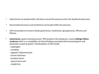
• Gastrinomas arelocated within soft tissue around the pancreas and in the duodenal submucosa .
• Non functional tumours and insulinomas are located within the pancreas
• GEP neuroendocrine tumors include gastrinomas, insulinomas, glucagonomas, VIPomas and
PPomas
• Gastrinomas- gastrin secreting tumors. 90% located in the duodenum. Causes Zollinger-Ellison
syndrome which is a constellation of clinical findings associated with increased gastric acid
production caused by gastrin. Manifestations of ZES include-
- esophagitis
- vomitting
- epigastric abdominal pain
- chronic diarrhoea
- duodenal ulcers
- jejunal ulcers and
- weight loss
14.
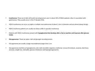
• Insulinomas- Theseare b-islet cell insulin secreting tumors seen in about 10% of MEN1 patients, often in association with
gastrinomas. They usually occur in the 3rd
decade of life.
• MEN1 insulinomas can occur as single or multiple macroadenomas of about 1-4cm in diameter and are almost always benign
• MEN1 insulinoma patients are usually not obese unlike in sporadic insulinomas
• Patients with MEN1 insulinomas present with hypoglycemia that develops after a fast or exertion and improves after glucose
intake.
• Glucagonomas- These are alpha- islet cell glucagon secreting tumors
• Glucagonomas are usually a single macroadenoma larger than 3 cm.
• Glucagonomas manifest as hyperglycemia, skin rash( necrolytic migratory erythema), venous thrombosis, anaemia, diarrhoea,
anorexia, weight loss, stomatitis, glucose intolerance and hyperglucagonaemia
15.
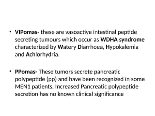
• VIPomas- theseare vasoactive intestinal peptide
secreting tumours which occur as WDHA syndrome
characterized by Watery Diarrhoea, Hypokalemia
and Achlorhydria.
• PPomas- These tumors secrete pancreatic
polypeptide (pp) and have been recognized in some
MEN1 patients. Increased Pancreatic polypeptide
secretion has no known clinical significance
16.
ANTERIOR PITUITARY TUMORS
•Anterior pituitary adenomas have been reported to occur in 15-90% of MEN1 patients
• They are the 1st
manifestation on MEN1 in 25% of sporadic and 10% of familial cases
• MEN1 anterior pituitary adenomas are usually single
• They are invasive only in 10-15% of cases and malignant degeneration is very rare
• Symptoms depend on both the secreted pituitary hormone and/or the compressive effects due to size of
the tumor
• Pituitary macroadenomas may :
- compress the optic chiasm causing bitemporal hemianopia and other
visual field defects, blurred vision and headaches
- compress the adjacent normal pituitary tissue causing hypopituitarism
. 60% of MEN1-associated pituitary tumours secrete prolactin (prolactinomas), 25% secrete Growth
Hormone and 3% secrete adenocorticotropic hormone (ACTH)
17.
• Prolactinomas –these prolactin secreting umors (with or without simultaneous GH over-secretion) are the most common
pituitary tumors in MEN1
• Symptoms of prolactinomas include galactorrhoea, amenorrhoea and infertility in women and Hypogonadism, sexual dysfunction,
reduction in libido, impotence and rarely gynaecomastia in men
• GH secreting tumors- these are the 2nd
most frequent MEN1 anterior pituitary tumors after prolactinomas.
• the increased secretion of growth hormone is responsible for the development of gigantism in children and acromegaly in adults
• Other MEN1 associated endocrine tumors include:
- Adreno-cortical tumors
- Pheochromocytoma
- Thyroid tumours
MEN1 associated non-endocrine tumours include
- Carcinoid tumors
- Collagenomas and facial angiofibromas
- Lipomas
- Leiomyomas
- CNS tumors- meningiomas and ependymomas
18.
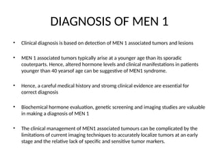
DIAGNOSIS OF MEN1
• Clinical diagnosis is based on detection of MEN 1 associated tumors and lesions
• MEN 1 associated tumors typically arise at a younger age than its sporadic
couterparts. Hence, altered hormone levels and clinical manifestations in patients
younger than 40 yearsof age can be suggestive of MEN1 syndrome.
• Hence, a careful medical history and stromg clinical evidence are essential for
correct diagnosis
• Biochemical hormone evaluation, genetic screening and imaging studies are valuable
in making a diagnosis of MEN 1
• The clinical management of MEN1 associated tumours can be complicated by the
limitations of current imaging techniques to accurately localize tumors at an early
stage and the relative lack of specific and sensitive tumor markers.
19.
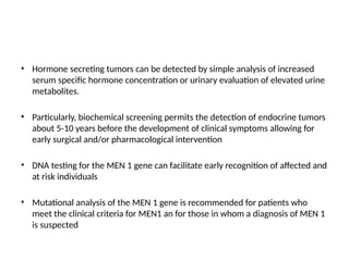
• Hormone secretingtumors can be detected by simple analysis of increased
serum specific hormone concentration or urinary evaluation of elevated urine
metabolites.
• Particularly, biochemical screening permits the detection of endocrine tumors
about 5-10 years before the development of clinical symptoms allowing for
early surgical and/or pharmacological intervention
• DNA testing for the MEN 1 gene can facilitate early recognition of affected and
at risk individuals
• Mutational analysis of the MEN 1 gene is recommended for patients who
meet the clinical criteria for MEN1 an for those in whom a diagnosis of MEN 1
is suspected
20.
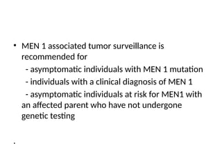
• MEN 1associated tumor surveillance is
recommended for
- asymptomatic individuals with MEN 1 mutation
- individuals with a clinical diagnosis of MEN 1
- asymptomatic individuals at risk for MEN1 with
an affected parent who have not undergone
genetic testing
.
21.
MANAGEMENT
• MEN 1treatment options are generally limited to surgical intervention
• Surgery is mostly effective when performed early before malignant
transformation or metastasis
• Chemotherapy and radiation play a limited role in treatment
• Parathyroid tumors (primary hyperparathyroidism)- subtotal
parathyroidectomy (surgical ablation of 3 parathyroid glands and part of
the 4th
gland) or total parathyroidectomy (removal of all 4 parathyroid
glands and thymic tissue) are indicated
• subtotal parathroidectomy avoids permanent hypoparathyroidism
22.
• GEP neuroendocrinetumors- the treatment for non metastatic gastrinoma is
surgical resection
• Unfortunately about 50% of MEN 1 gastrinomas have already metastasized
before diagnosis
• Surgical resection include duodenectomy, subtotal pancreatectomy and
pancreatoduodectomy (whipple procedure)
• Treatment for multiple and/or disseminated gastrinomas is therapy with human
somatostatin analogue octreotide and PPI omeprazole (60mg bd with titration
upwards if necessary)
• Chemotherapy with 5-fluorouracil and streptazocin can also be used
23.
• Insulinomas- surgicalresection is usually the indicated approach for insulinomas
• The best surgical approach is intraoperative localization of nodules greater than
0.5cm in diameter by palpation or intraoperative ultrasound followed either by
enucleation of these nodules or by pancreatic resection if multiple large deep
tumors are present
• Chemotherapy with streptazocine or octreotide is used for metastatic disease
• Anterior pituitary tumors- pituitary adenomas are classified as micro(< 10mm)
or macro (>10mm).
• Trans- sphenoidal resection, endoscopic resection and/or radioablation are the
treatment of choice for macroadenomas
24.
• Dopamine agonists( cabergoline, bromocryptine, pergolide and
quinagolide) are the preferred treatment for prolactin secreting
microadenomas
• Somatostatin analogues are the medical therapy of choice for
treatment of growth hormone secreting microadenomas
• For non- secreting microadenomas, surgery is the treatment of
choice
• Perioperatively, treatment with potent dopamine agonists or
somatostatin analogues shrinks 5-15% of tumors
25.
MULTIPLE ENDOCRINE NEOPLASIATYPE 2
(MEN2)
• MEN2 is an inherited disorder caused by mutation of the RET proto-oncogene located on
chromosome 10. It is inherited in an autosomal dominant fashion
• The RET gene encodes for a transmenbrane glycoprotein receptor
• MEN2 is classified into 2 subtypes which include :
-MEN 2A(Sipple’s syndrome)
-MEN 2B
• All subtypes of MEN 2 have a high risk of development of medullary carcinoma of the thyroid
gland (MTC) and pheochromocytoma
• Medullary carcinoma of the thyroid gland typically occurs in early adulthood in MEN 2A and
in early childhood in MEN 2B
• MEN 2A has an increased risk for parathyroid adenomas or hyperplasia
26.
PATHOPHYSIOLOGY OF MEN2
• Mutations in RET, a transmembrane proto-oncogene localized in the long arm of
chromosome 10 (10q11) is responsible for MEN 2
• The RET protein is critical during embryonic developoment of the enteric nervous
system and kidneys
• RET consists of 3 domains, a cysteine rich extracellular receptor domain, a
hydrophobic transmembrane domain and an intracellular tyrosine kinase catalytic
domain
• For MEN 2A, the transmembrane glycoprotein receptor is in the extracellular
domain, while in MEN 2B its in the intracellular domain
• Mutation and upregulation of the RET proto-oncogene hence interfers with normal
development and promotes oncogenesis.
27.
MEN 2A (SIPPLE’SSYNDROME)
• This subtype constitutes approximately 70-80% of MEN2 cases
• Tumours arise from the thyroid C cells, the parathyroid glands and the adrenal medulla
• Tumors associated with this type include
- medullary carcinoma of the thyroid(95% of
individuals)
- pheochromocytoma, usually bilateral (50% of
individuals)
- hyperparathyroidism from parathyroid adenoma or
hyperplasia (20-30% of cases)
. MEN 2A is diagnosed clinically by the occurrence of 2 or more specific endocrine tumors in a single
individual or in close relatives.
. A small number of families with MEN 2A have pruritic cutaneous lichen amyloidosis (PCLA) , which is
usually located over the upper portion of the back and may appear before the onset of Medullary
Carcinoma of the thyroid.
28.
MEDULLARY CARCINOMA OFTHE THYROID
(MTC)
• This is generally the first manifestation of MEN 2A. It produces calcitonin
• Patients usually present with neck mass or neck pain before 35 years of age.
• Up to 70% of cases already have cervical lymph node metatasis before 35yrs of
age
• Diarrhoea is the most frequent systemic syptom in these patients. It occurs in
patients with a plasma calcitonin concentration of >10ng/dl and implies a poor
prognosis.
• All individuals with an MTC- predisposing pathogenic variant who have not had a
prophylactic thyroidectomy done demonstrate biochemical evidence of MTC by
35yrs of age
29.
PHEOCHROMOCYTOMA
• This isa neuroendocrine tumor of the medulla of the adrenal glands originating in the
chromaffin cells which secretes high amounts of catecholamines mostly norepinephrine plus
epinephrine to a lesser extent.
• This usually presents after MTC or concomitantly
• However they are the first sign in 13-27% of patients with MEN 2A.
• Pheochromocytoma in patients with MEN 2A are usually diagnosed at an earlier age, have
subtler symptoms and are more likely to be bilateral than in sporadic pheochromocytoma.
• Malignant transformation occurs in about 4% of cases
• Patients usually present with clinical features indicative of sympathetic nervous system
hyperactivity which include elevated blood pressure, elevated heart rate, flank pains, skin
sensations, palpitations, anxiety, diaphoresis, headaches, weakness, pallor and weight loss.
30.
HYPERPARATHYROIDISM
• Hyperparathyroidism inMEN 2A, is typically mild and may range
from a single adenoma to marked hyperplasia
• The average age of onset is usually 38yrs of age
• Most individuals with hyperparathyroidism have no symptoms
however hypercalcuria and renal calculi may occur.
• Hyperparathyroidism in MEN 2A usually presents many years
after the diagnosis of Medullary carcinoma of the thyroid.
31.
MEN 2B
• TheMEN 2B subtype accounts for approximately 5% of cases of MEN 2.
• The tumours associated with this type include:
- Medullary carcinoma of the thyroid (MTC)
- Multiple mucosal neuromas (on the lips, tongue,
conjuctiva, eyelids and gut)
- Pheochromocytoma
• It is characterized by the early development of an aggressive form of MTC in all affected
individuals
• Individuals with MEN 2B, who do not undergo prophylactic thyroidectomy before 1yr of age
are likely to develop metastatic MTC at an early age.
• Prior to intervention with early prophylactic thyroidectomy, the average age of death in
individuals with MEN 2B, was 21 yrs of age
32.
• Pheochromocytoma occursin 50% of individuals with MEN 2B
• About half of cases with pheochromocytoma are multiple and bilateral.
• Clinically significant parathyroid disease is absent in MEN 2B
• Individuals with MEN 2B may be identified in infancy and early childhood by the presence of
mucosal neuromas on the anterior dorsal surface of the tongue, palate or pharynx with a
distinct facial appearance.
• The lips of such individuals become prominent over time and submucosal nodules may be
present in the vermilion border of the lips
• Neuromas of the eyelids may cause thickening and eversion of the upper eyelid margins
• Prominent thickened corneal nerves may be seen by slit lamp examination
33.
• About 40%of affected individuals have diffuse
ganglioneuromatosis of the gastrointestinal tract. Associated
symptoms include abdominal distention, megacolon,
constipation or diarrhoea.
• About 75% of individuals have a marfanoid habitus, often
with kyphoscoliosis or lordosis, joint laxity and decreased
subcutaneous fat.
• Proximal muscle wasting and weakness can also be seen in
such cases.
34.
PREVENTION OF MEN2
• Prophylactic thyroidectomy is the primary preventive measure for individuals with an
identified germline RET pathogenic variant
• For all individuals with RET pathogenic variant who have not had a thyroidectomy, annual
biochemical screening is recommended with immediate thyroidectomy if results are
abnormal
• Annual serum calcitonin screening should begin at 6months of age for children with MEN
2B and 3-5years of age for children with MEN 2A
• Before any surgery, the presence of any functioning pheochromocytoma should be
excluded by appropriate biochemical screening in any individual with MEN 2A or MEN 2B.
• If pheochromocytoma is present, adrenalectomy should be performed before
thyroidectomy to avoid intraoperative catecholamine crisis
35.
TREATMENT OF MEN2
• Medullary thyroid carcinoma- standard treattment for MTC is surgical
removal of the thyroid and lymph node dissection
• Radiation therapy which includes external beam radiation therapy (EBRT) or
intensity modulated radiation therapy (IMRT) is indicated for incomplete
tumor resection or extrathyroidal extension with positive margins
• Two kinase inhibitors vandetanib and cabozantinib have been shown to
improve progression-free survival and in some cases cause disease
regression in unresectable or advanced metastatic MTC
• All individuals who have undergone thyroidectomy need thyroid hormone
replacement therapy
36.
• Pheochromocytomas- surgicalremoval by adrenalectomy which
may be performed using video assisted laparoscopy
• Unilateral aderenalectomy in unilateral tumors and cortical sparing
adrenal surgery with close monitoring of the remnant tissue in
bilateral pheochromocytoma
• Bilateral adrenalectomy is no longer adviced because of the risk of
adrenal insufficiency and addisonian crisis
• Hyperparathyroidism from parathyroid adenoma or hyperplasia -
subtotal or total parathyroidectomy is indicated


![谷歌留痕技术 [ 𝙩𝙤𝙥 𝟮𝟯𝟯. 𝙘 𝙤𝙢 ]](https://cdn.slidesharecdn.com/ss_thumbnails/top233-260130174328-3833018c-thumbnail.jpg?width=640&height=640&fit=bounds)


