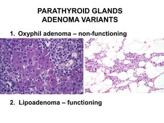
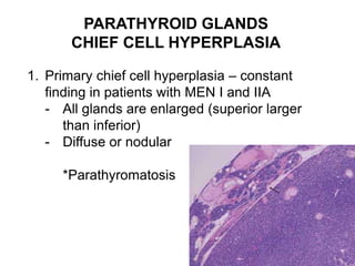
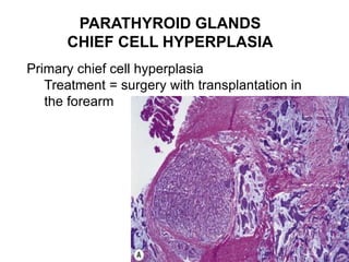
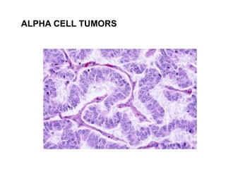
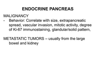
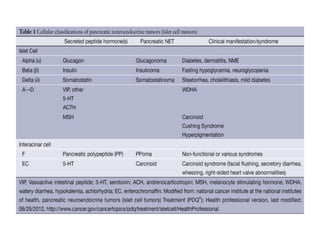
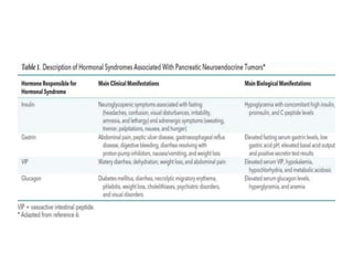
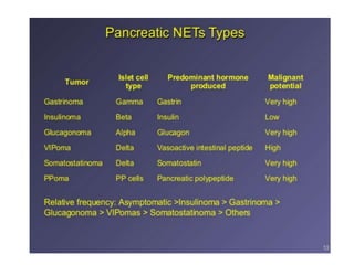
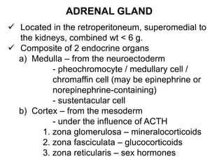
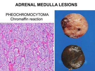
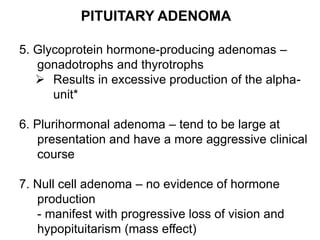
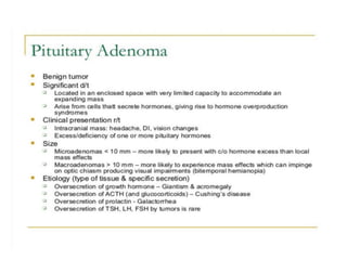
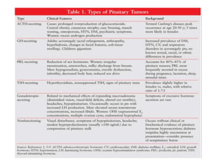
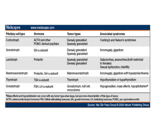
ENDOCRINE SYSTEM
November 29, 2017
Aida Isabel G. Mendoza MD DPSP
Course Objectives:
• Review the different
functions of selected
organs in endocrine
system
• Identify and describe
the histologic features
of different neoplastic
lesions of selected
organs in endocrine
system
• Correlate the lesions
clinically
Outline
• Parathyroid Gland
• Endocrine
Pancreas
• Adrenal Gland
• Pituitary Gland
PARATHYROID GLANDS
1. Chief cell – with variable amount of glycogen
particles and secretory droplets
- Secretes parathormone (PTH) and PTH-
related protein
- (+) anti PTH E11* strong in normal
2. Oxyphil cell – with a deeply granular and
eosinophilic cytoplasm
- many mitochondria but few secretory granules
- Appear as nodules
3. Water-clear cells
4. Transitional oxyphil and transitional water- clear
cells – more common in hyperfunctioning glands
PARATHYROID GLANDS
NORMAL HISTOLOGY
Before puberty: Gland is composed of chief
cells (no fat)
At puberty: oxyphil cells and fat (tiny droplets)
appear (until age 40)
Adult parathyroid – 40% fat but variable,
parenchyma is always constant
Some parathyroids (50%) contain follicles and
cysts with colloid-like material (thyroid vs
parathyroid*)
+ glycogen-parathyroid
+ CaOx (bifringent in polarized light)-thyroid
PARATHYROID GLANDS
PARATHYROID GLANDS
NORMAL PHYSIOLOGY
- Mediate their endocrine function through the
production of PTH
Physiologic actions of PTH
1. Increased renal excretion of phosphate
2. Increased renal tubular absorption of
calcium
3. Increased intestinal absorption of calcium
4. Direct effects in bone – increased
osteoclastic activity
PARATHYROID GLAND ADENOMA
o M<F, ave. age: 4TH decade
o Majority are Solitary (75% involve one of the
inferior glands)
o Other glands have a normal or atrophic
appearance
o Usually oval with slight lobulation, and
surrounded by a thin capsule
o With a rim of compressed non-neoplastic
tissue
o PTH staining is weaker in the adenoma than
in the residual normal gland
PARATHYROID GLAND ADENOMA
PARATHYROID GLANDS
ADENOMA VARIANTS
1. Oxyphil adenoma – non-functioning
2. Lipoadenoma – functioning
PARATHYROID GLANDS
CHIEF CELL HYPERPLASIA
1. Primary chief cell hyperplasia – constant
finding in patients with MEN I and IIA
- All glands are enlarged (superior larger
than inferior)
- Diffuse or nodular
*Parathyromatosis
PARATHYROID GLANDS
CHIEF CELL HYPERPLASIA
Primary chief cell hyperplasia
Treatment = surgery with transplantation in
the forearm
PARATHYROID GLANDS
CHIEF CELL HYPERPLASIA
2. Secondary chief cell hyperplasia – secondary
to impairment of renal function or chronic
malabsorption
- higher number of oxyphil cells compared to
primary form
PARATHYROID GLANDS
WATER-CLEAR CELL HYPERPLASIA
 No familial incidence and not associated
with MEN
 Extreme enlargement of all parathyroid
tissue (>100g)
 Strangely, this disorder disappeared in
recent times (unknown reason)
 Superior larger than inferior
 Have a typical brown color
 With pseudopods
PARATHYROID GLANDS
WATER-CLEAR CELL HYPERPLASIA
PARATHYROID GLANDS
CARCINOMA
 Presents with hyperparathyroidism
 Clinical features: very high serum calcium or
PTH, palpable cervical mass, vocal cord
paralysis, recurrence of hyperparathyroidism a
short time after surgery
 Gross: hard, surrounded by a dense fibrous
reaction, and adherent to adjacent structures
PARATHYROID GLANDS
CARCINOMA
HYPERPARATHYROIDISM – persistent
production of PTH
1. PRIMARY HYPERPARATHYROIDISM
 there is no evidence of previous
parathyroid stimulation by chronic renal or
intestinal disease
 caused by adenoma (>80%), chief cell
hyperplasia, carcinoma, or water-clear
cell hyperplasia
 Hypercalcemia, hypercalciuria,
hypophosphatemia
HYPERPARATHYROIDISM
PRIMARY HYPERPARATHYROIDISM
a) Osseous manifestations – always
generalized
- osteitis fibrosa cystica or
Recklinghausen’s disease or brown
tumor*
- G: with alternation of solid and cystic
areas
- M: combination of osteoblastic and
osteoclastic activity
PRIMARY HYPERPARATHYROIDISM
a) Osseous manifestations
- expansile, multilocular masses
- osteitis fibrosa cystica or
Recklinghausen’s disease or
brown tumor
Jaw-preferred location
HYPERPARATHYROIDISM
PRIMARY HYPERPARATHYROIDISM
b) Renal manifestations – renal stones,
nephrocalcinosis, polyuria, polydipsia and
impairment of renal function
- still progress after the removal of the
parathyroid lesion
c) Other manifestations – hypertension,
peptic ulcer, pancreatitis, mental
disturbances
*Parathyroid crisis - fatal
HYPERPARATHYROIDISM
2. SECONDARY HYPERPARATHYROIDISM
- occurs as a consequence of renal disease
or intestinal malabsorption
- with Vitamin D resistance*
- with mild bone changes
- cutaneous calciphylaxis*
- chief cell hyperplasia-parathyroid
manifestation
HYPERPARATHYROIDISM
3. TERTIARY HYPERPARATHYROIDISM
- secondary to chronic renal disease or
intestinal malabsorption, in which one or more
of the stimulated parathyroid glands seem to
become autonomous
- nonsuppressible, autonomous, or refractory
hyperparathyroidism, reversible
- microscopic…chief cell hyperplasia but more
nodular
THERAPY OF HYPERPARATHYROIDISM
Primary chief cell hyperplasia – subtotal
parathyroidectomy*
Primary and secondary chief cell hyperplasia –
total parathyroidectomy with
autotransplantation into the forearm muscle
Adenoma – local excision of the tumor*
Carcinoma – excision of the tumor and
surrounding soft tissues and thyroid
lobectomy
PANCREAS
Endocrine tumors of the pancreas – islet cell
tumors, pancreatic endocrine neoplasm
 most common location: body and tail
 G: more cellular, more pink
 Do not have a well-defined capsule
 M: small cuboidal cells with centrally
located nuclei and finely granular
cytoplasm
 May be non-functioning or functioning (esp
malignant)
ENDOCRINE PANCREAS
IMMUNOHISTOCHEMISTRY
(+) epithelial markers
(+) panendocrine markers
Special stain for islet cell – Silver technique
PATTERNS OF GROWTH
I A solid
II B gyriform – beta or alpha cells
III C glandular – G or VIP cell
IV D nondescript
ENDOCRINE PANCREAS
1. BETA CELL TUMORS – insulinomas (if functional)
- most common
- majority are benign
- usually solitary (90%), with gyriform pattern
Whipple triad-characteristic of the tumor
a) Mental confusion, weakness, fatigue,
convulsions
b) FBS <50mg%
c) Relief of symptoms by the administration of
glucose
Treatment: surgical (subtotal pancreatectomy)
ENDOCRINE PANCREAS
2. ALPHA CELL TUMORS
A) Glucagonomas – solitary and large
- high incidence of malignancy
- non descript pattern
- IHC: few cells positive for glucagon
B) Not associated with the glucagonoma
syndrome - multiple and small
- gyriform pattern
- nearly always benign
- strongly immunoreactive for glucagon
ENDOCRINE PANCREAS
ALPHA CELL TUMORS
A) Glucagonomas – solitary and large
- high incidence of malignancy
ALPHA CELL TUMORS
ENDOCRINE PANCREAS
ALPHA CELL TUMORS
Components of Glucagonoma Sndrome
a) abnormal glucose tolerance test
b) normocytic normochromic anemia
c) skin rash – necrolytic migratory erythema
- heals with hyperpigmentation
d) sore red tongue, angular stomatitis, DVT,
overwhelming infections
ENDOCRINE PANCREAS
3. G-CELL TUMORS – gastrinomas
- produce the Zollinger-Ellison syndrome
(gastric hyperacidity with gastric, duodenal
or jejunal ulcers)
- pancreas > duodenum > antrum
a) Sporadic – solitary, clinically malignant,
pancreas (assoc with ZES)
b) Familial – component of MEN I
- multicentric, less likely to be clinically
malignant, duodenum
Treatment: total excision of tumor and/or
removal of target organ
ENDOCRINE PANCREAS
4. VIP-PRODUCING TUMORS – associated with
diarrhea or cholera-like syndrome
5. DELTA CELL TUMORS – clinically silent because
somatostatin is an inhibitory hormone
S/Sx: Somatostatinoma syndrome (diabetes,
cholecystolithiasis, steatorrhea, indigestion,
hypochloridia)
6. CARCINOID TUMORS – arise from
Kulchitsky’s (serotonin-producing) cells
ENDOCRINE PANCREAS
MALIGNANCY
- Behavior: Correlate with size, extrapancreatic
spread, vascular invasion, mitotic activity, degree
of Ki-67 immunostaining, glandular/solid pattern,
METASTATIC TUMORS – usually from the large
bowel and kidney
MULTIPLE ENDOCRINE NEOPLASIA
 Autosomal dominant
 Hyperplastic or neoplastic proliferation of >1
endocrine gland
MEN type I (Werner’s syndrome) 3Ps
• Mutation of MEN 1 gene (chrom. 11)
• Pituitary adenomas, pancreatic tumor (G-cell
tumor, 50%), Chief cell hyperplasia of the
parathyroids
• S/Sx; acromegaly or hypopituitarism, Zollinger-
Ellison syndrome, hyperparathyroidism
MULTIPLE ENDOCRINE NEOPLASIA
MEN II
 Mutation of RET proto-oncogene
 Medullary thyroid carcinoma
a) MEN IIA
b) MEN IIB
c) Familial medullary thyroid carcinoma
MULTIPLE ENDOCRINE NEOPLASIA II
a) MEN IIA (Sipple syndrome)
 medullary thyroid carcinoma (multiple &or bil)
 pheochromocytoma of adrenal (bil)
 C-cell hyperplasia
 Chief cell hyperplasia of parathyroid
b) MEN IIB (Gorlin’s syndrome)
 medullary carcinoma of the thyroid
 pheochromocytoma of adrenal
 Mucosal neuromas (most constant feature)
 Skeletal abnormalities (marfanoid habitus)
c) Familial medullary thyroid carcinoma
 In at least 4 family members
 Combined with pancreatic endocrine tumors,
paraganglioma, etc
ADRENAL GLAND
 Located in the retroperitoneum, superomedial to
the kidneys, combined wt < 6 g.
 Composite of 2 endocrine organs
a) Medulla – from the neuroectoderm
- pheochromocyte / medullary cell /
chromaffin cell (may be epinephrine or
norepinephrine-containing)
- sustentacular cell
b) Cortex – from the mesoderm
- under the influence of ACTH
1. zona glomerulosa – mineralocorticoids
2. zona fasciculata – glucocorticoids
3. zona reticularis – sex hormones
LESIONS OF ADRENAL CORTEX
 HETEROTOPIA – MC site is the retroperitoneal fat
 CONGENITAL HYPERPLASIA – autosomal
recessive, inborn error of metabolism
- Responsible for adrenogenital syndrome
developing within the first year of life
- Defect in an enzyme required to synthesize
cortisol from cholesterol
- 95% absence of 21-hydroxylase (virilization)
- Deficiency of 11β-hydroxylase (virilization and
hypertension)
- Pathology: Diffuse cortical hyperplasia esp. the
zona reticularis
- Treatment: Replacement with cortisol and surgical
correction of the external sex organ
LESIONS OF ADRENAL CORTEX
 ACQUIRED HYPERPLASIA – bilateral
- Diffuse or a nodular enlargement of the adrenal
gland (>6g)
 Diffuse: Due to ACTH hyperproduction by the
pituitary gland or ACTH-producing neoplasm in
the lung, ACTH dependent
 Nodular : ACTH independent or adrenal
dependent
- Pathology: increased thickness of the zona
fasciculata and reticularis
LESIONS OF ADRENAL CORTEX
ACQUIRED HYPERPLASIA – bilateral
- Diffuse or a nodular enlargement of the adrenal
gland (>6g)
ADRENOCORTICAL TUMORS
• Adenomas and carcinomas
• Usually in adults (50 years old ave), M=F
• Asymptomatic or with signs related to hormonal
dysfunction
• Highly necrotic carcinomas may result in fever
• Palpable=malignant
• Gross: solitary
• Adenomas-small
• Carcinoma-> 10 g
• Diagnosis: Computed tomography
• Treatment: Surgical excision
ADRENOCORTICAL TUMORS
1. Adenomas - <5cm, encapsulated, yellow
homogenous
- IHC: (+) LMWK
- treated by excision
2. Carcinoma - >10g, variegated, with necrosis and
hemorrhage
- mitotic rate >5 per 50 HPF
- absence of clear cells
- with capsular invasion
- spreads to the retroperitoneum and
infiltrates the kidney
- overexpression of p53
- IHC: strongly (+) for vimentin
ADRENOCORTICAL TUMORS
 Adenoma
 Carcinoma
ADRENAL CORTEX
FUNCTIONAL MANIFESTATIONS
 Non-functioning lesions – usually carcinomas
- due to deletion in some of the enzymes
for cortisol synthesis
 Primary aldosteronism – Conn’s syndrome
- results in loss of potassium, retention of
sodium, hypertension, suppressed renin
levels, muscle weakness
- 70% is due to adenoma
- spironolactone bodies are seen in patients
treated with spironolactone
Primary aldosteronism - spironolactone bodies
ADRENAL CORTEX
FUNCTIONAL MANIFESTATIONS
 Cushing syndrome
- due to hyperproduction of cortisol
- only 20% has primary adrenal cause (80%F)
- may be diffuse/nodular hyperplasia (bilat),
adenoma or carcinoma (both unilat)
- majority of the cases of diffuse cortical
hyperplasia are the result of pituitary
hyperfunction (Cushing disease)
- Other ACTH –producing neoplasms: small cell
ca of the lung, endocrine tumors of the
foregut, pheochromocytoma, certain ovarian
tumors
ADRENAL CORTEX
FUNCTIONAL MANIFESTATIONS
 Adrenogenital syndrome
- excess androgens bring about changes towards
adult musculinity in male or female children and
toward masculinity in female adults.
- excess of dehydroisoandrosterone
CAH- MC cause of childhood virilization
- hyperplasia > carcinoma > adenoma
If with neoplasm presentcarcinoma
Malignant  70% virilization in adult woman
 100% adult male with fem
ADRENAL MEDULLA
Paraganglion system – numerous collections of
neuroepithelial cells scattered throughout the body,
having numerous cytoplasmic neurosecretory
granules containing catecholamines
- appear as chief cells arranged in well-defined nests
(“Zelballen”) encircled by a thin layer of S-100
protein-positive sustentacular cells
a) adrenal medulla
b) parasympathetic system
and sympathetic system
ADRENAL MEDULLA
Paraganglia
ADRENAL MEDULLA TUMORS
2 Categories
1. Embryonal neural (sympathetic) – children
 Neuroblastoma
 Ganglioneuroblastoma
 Ganglioneuroma
2. Adult neuroendocrine tumors and tumor-like
conditions
 Pheochromocytoma
 Adrenal medullary hyperplasia
ADRENAL MEDULLA TUMORS
NEUROBLASTOMA
• 80% are detected in children <4 years
• Usually present as an abdominal mass first
noted by the parents
• G: large, soft and relatively well
circumscribed with areas of calcification,
hemorrhage and necrosis
• Homer Wrights rosettes are seen in ¼ to 1/3
of cases (tumor cells arranged around a
central area filled with a fibrillary material*
without a central lumen)
ADRENAL MEDULLA TUMORS
NEUROBLASTOMA
• IHC: neuron-specific enolase,
neurofilaments
• Genetic events: loss of a region on chrom.
1 and amplification of N-myc
• Metastasize to liver (Pepper syndrome),
skeletal system (Hutchinson syndrome)
• Poor prognosis: <2 y/o, high Ki-67 scores,
presence of CD44
• Treatment: surgical excision,
chemotherapy and bone marrow
transplantation
ADRENAL MEDULLA TUMORS
NEUROBLASTOMA
ADRENAL MEDULLA TUMORS
NEUROBLASTOMA
ADRENAL MEDULLA TUMORS
GANGLIONEUROBLASTOMA
• Malignant ganglioneuroma
• young children
• most are located in the retroperitoneum
rather than the adrenal gland
• Better prognosis than neuroblastoma
ADRENAL MEDULLA TUMORS
GANGLIONEUROMA
• Benign
• Seen in older age group
• Most common neoplasm of the sympathetic
nervous system in adults
• be multiple and/or associated with other
independent types of neural/neuroendocrine
neoplasms,
• MC location is the posterior mediastinum
and retroperitoneum
• Catecholamine synthesis is a constant
feature
ADRENAL MEDULLA TUMORS
GANGLIONEUROMA
ADRENAL MEDULLA LESIONS
ADRENAL MEDULLARY HYPERPLASIA
Usually occur as a component of MEN 2b
PHEOCHROMOCYTOMA
• Paraganglia of the adrenal medulla
• 10% tumor (extra-adrenal, children, bilateral,
malignant)
• Triad of sweating attacks, tachycardia and
headaches
• Secrete epinephrine and norepinephrine
(paragangliomas secrete norepinephrine)
• Confirmation of Dx: measuring urinary
catecholamines or their catabolites
ADRENAL MEDULLA LESIONS
PHEOCHROMOCYTOMA
• Takes on a dark brown appearance when
immersed in dichromate solution *
(chromaffin reaction)
• Triad:3P
• G: encapsulated, soft
• M: tumor cells are arranged in well-defined
nests (“Zellballen”) bound by a delicate
fibrovascular stroma
- with sustentacular cells at the
periphery, (+) for S-100
ADRENAL MEDULLA LESIONS
PHEOCHROMOCYTOMA
Chromaffin reaction
ADRENAL MEDULLA LESIONS
PHEOCHROMOCYTOMA
• Malignant tumors metastasize to the skeletal
system, particularly the ribs and spine
• Treatment: surgical excision
PITUITARY ADENOMA
• Arise from the adenohypophysis
• Usually benign
• 70% are functioning
• Some present with insidious symptoms of
hypopituitarism
• Suprasellar extension produce neurologic signs
• Pituitary apoplexy (abrupt onset of headache,
ocular deficits and altered consciousness)
• Dx: MRI
PITUITARY ADENOMA
1. PRL adenoma – MC
 Usually present with the galactorrhea-
amenorrhea syndrome (Chiari-Frommel
syndrome or Forbes-Albright syndrome)
 Dx: serum PRL >200 ng/mL
 M: chromophobic or slightly acidophilic
 Treatment: dopamine agonists
(bromocriptine)
PITUITARY ADENOMA
2. GH cell adenoma
 May result in gigantism or acromegaly
3. Mixed GH-cell-PRL-cell adenoma
 Mammosomatotroph adenoma
 Associated with acromegaly and slight
hyperprolactinemia
PITUITARY ADENOMA
GH cell adenoma
- 2nd MC type
- May result in gigantism
or acromegaly
- Densely vs sparsely
granulated GH
adenomas
PITUITARY ADENOMA
4. ACTH cell adenoma
 Corticotroph adenoma
 Patients have Cushing’s disease, Nelson
syndrome, Pituitary apoplexy, or non-
functioning
 Usually basophilic
 PAS-positive due to proopiomelanocortin
 With perinuclear microfilaments – an important
diagnostic feature (Crooke’s hyalinization)
PITUITARY ADENOMA
ACTH cell adenoma
Crooke’s hyalinization
PITUITARY ADENOMA
5. Glycoprotein hormone-producing adenomas –
gonadotrophs and thyrotrophs
 Results in excessive production of the alpha-
unit*
6. Plurihormonal adenoma – tend to be large at
presentation and have a more aggressive clinical
course
7. Null cell adenoma – no evidence of hormone
production
- manifest with progressive loss of vision and
hypopituitarism (mass effect)
PITUITARY ADENOMA
TREATMENT
 Surgical excision (transsphenoidal)
 radiation
 Medical – bromocriptine, ocreotide
PITUITARY GLAND
OTHER LESIONS
LYMPHOCYTIC HYPOPHYSITIS
o Primary or secondary
o rare autoimmune endocrine disease
o With polymorphic lymphoplasmacytic infiltration
associated with destruction of the anterior
pituitary cells
o Tx: Corticosteroid
PITUITARY GLAND
OTHER LESIONS
RATHKE’S CLEFT CYST
o Cyst of the hypophyseal cleft
o Become clinically apparent with
compression of the hypothalamus and
optic chiasm
o M: cysts are lined by
predominantly columnar
ciliated cells
o Tx: surgical excision and
o drainage
o MRI findings of an anteriorly
o displaced pituitary stalk, a
o ‘posterior ledge sign’
PITUITARY GLAND
OTHER LESIONS
RATHKE’S CLEFT CYST
PITUITARY GLAND
SELLAR/SUPRASELLAR LESIONS
CRANIOPHARYNGIOMA
o Constitutes 3% of brain tumors
o Sx: hydrocephalus, pressure on the optic
chiasm and optic tracts
1. Adamantinomatous form – with peripheral
palisaded nuclei, ghost cells, focal calcification,
wet keratin
- children
2. Papillary craniopharyngioma – adults
- with papillary, nonkeratinizing stratified
squamous epithelium
PITUITARY GLAND OTHER LESIONS
CRANIOPHARYNGIOMA
PITUITARY GLAND
OTHER LESIONS
PITUICYTOMA
o Arise from the pituicyte, a specialized glial cell
found in posterior pituitary
o Sx: headache, visual field defect
Copy_of_endocrine.pptx

Copy_of_endocrine.pptx

  • 1.
    ENDOCRINE SYSTEM November 29,2017 Aida Isabel G. Mendoza MD DPSP
  • 2.
    Course Objectives: • Reviewthe different functions of selected organs in endocrine system • Identify and describe the histologic features of different neoplastic lesions of selected organs in endocrine system • Correlate the lesions clinically
  • 3.
    Outline • Parathyroid Gland •Endocrine Pancreas • Adrenal Gland • Pituitary Gland
  • 4.
    PARATHYROID GLANDS 1. Chiefcell – with variable amount of glycogen particles and secretory droplets - Secretes parathormone (PTH) and PTH- related protein - (+) anti PTH E11* strong in normal 2. Oxyphil cell – with a deeply granular and eosinophilic cytoplasm - many mitochondria but few secretory granules - Appear as nodules 3. Water-clear cells 4. Transitional oxyphil and transitional water- clear cells – more common in hyperfunctioning glands
  • 5.
    PARATHYROID GLANDS NORMAL HISTOLOGY Beforepuberty: Gland is composed of chief cells (no fat) At puberty: oxyphil cells and fat (tiny droplets) appear (until age 40) Adult parathyroid – 40% fat but variable, parenchyma is always constant Some parathyroids (50%) contain follicles and cysts with colloid-like material (thyroid vs parathyroid*) + glycogen-parathyroid + CaOx (bifringent in polarized light)-thyroid
  • 6.
  • 7.
    PARATHYROID GLANDS NORMAL PHYSIOLOGY -Mediate their endocrine function through the production of PTH Physiologic actions of PTH 1. Increased renal excretion of phosphate 2. Increased renal tubular absorption of calcium 3. Increased intestinal absorption of calcium 4. Direct effects in bone – increased osteoclastic activity
  • 8.
    PARATHYROID GLAND ADENOMA oM<F, ave. age: 4TH decade o Majority are Solitary (75% involve one of the inferior glands) o Other glands have a normal or atrophic appearance o Usually oval with slight lobulation, and surrounded by a thin capsule o With a rim of compressed non-neoplastic tissue o PTH staining is weaker in the adenoma than in the residual normal gland
  • 9.
  • 10.
  • 11.
    PARATHYROID GLANDS ADENOMA VARIANTS 1.Oxyphil adenoma – non-functioning 2. Lipoadenoma – functioning
  • 12.
    PARATHYROID GLANDS CHIEF CELLHYPERPLASIA 1. Primary chief cell hyperplasia – constant finding in patients with MEN I and IIA - All glands are enlarged (superior larger than inferior) - Diffuse or nodular *Parathyromatosis
  • 13.
    PARATHYROID GLANDS CHIEF CELLHYPERPLASIA Primary chief cell hyperplasia Treatment = surgery with transplantation in the forearm
  • 14.
    PARATHYROID GLANDS CHIEF CELLHYPERPLASIA 2. Secondary chief cell hyperplasia – secondary to impairment of renal function or chronic malabsorption - higher number of oxyphil cells compared to primary form
  • 15.
    PARATHYROID GLANDS WATER-CLEAR CELLHYPERPLASIA  No familial incidence and not associated with MEN  Extreme enlargement of all parathyroid tissue (>100g)  Strangely, this disorder disappeared in recent times (unknown reason)  Superior larger than inferior  Have a typical brown color  With pseudopods
  • 16.
  • 17.
    PARATHYROID GLANDS CARCINOMA  Presentswith hyperparathyroidism  Clinical features: very high serum calcium or PTH, palpable cervical mass, vocal cord paralysis, recurrence of hyperparathyroidism a short time after surgery  Gross: hard, surrounded by a dense fibrous reaction, and adherent to adjacent structures
  • 18.
  • 19.
    HYPERPARATHYROIDISM – persistent productionof PTH 1. PRIMARY HYPERPARATHYROIDISM  there is no evidence of previous parathyroid stimulation by chronic renal or intestinal disease  caused by adenoma (>80%), chief cell hyperplasia, carcinoma, or water-clear cell hyperplasia  Hypercalcemia, hypercalciuria, hypophosphatemia
  • 20.
    HYPERPARATHYROIDISM PRIMARY HYPERPARATHYROIDISM a) Osseousmanifestations – always generalized - osteitis fibrosa cystica or Recklinghausen’s disease or brown tumor* - G: with alternation of solid and cystic areas - M: combination of osteoblastic and osteoclastic activity
  • 21.
    PRIMARY HYPERPARATHYROIDISM a) Osseousmanifestations - expansile, multilocular masses - osteitis fibrosa cystica or Recklinghausen’s disease or brown tumor Jaw-preferred location
  • 22.
    HYPERPARATHYROIDISM PRIMARY HYPERPARATHYROIDISM b) Renalmanifestations – renal stones, nephrocalcinosis, polyuria, polydipsia and impairment of renal function - still progress after the removal of the parathyroid lesion c) Other manifestations – hypertension, peptic ulcer, pancreatitis, mental disturbances *Parathyroid crisis - fatal
  • 23.
    HYPERPARATHYROIDISM 2. SECONDARY HYPERPARATHYROIDISM -occurs as a consequence of renal disease or intestinal malabsorption - with Vitamin D resistance* - with mild bone changes - cutaneous calciphylaxis* - chief cell hyperplasia-parathyroid manifestation
  • 24.
    HYPERPARATHYROIDISM 3. TERTIARY HYPERPARATHYROIDISM -secondary to chronic renal disease or intestinal malabsorption, in which one or more of the stimulated parathyroid glands seem to become autonomous - nonsuppressible, autonomous, or refractory hyperparathyroidism, reversible - microscopic…chief cell hyperplasia but more nodular
  • 25.
    THERAPY OF HYPERPARATHYROIDISM Primarychief cell hyperplasia – subtotal parathyroidectomy* Primary and secondary chief cell hyperplasia – total parathyroidectomy with autotransplantation into the forearm muscle Adenoma – local excision of the tumor* Carcinoma – excision of the tumor and surrounding soft tissues and thyroid lobectomy
  • 26.
    PANCREAS Endocrine tumors ofthe pancreas – islet cell tumors, pancreatic endocrine neoplasm  most common location: body and tail  G: more cellular, more pink  Do not have a well-defined capsule  M: small cuboidal cells with centrally located nuclei and finely granular cytoplasm  May be non-functioning or functioning (esp malignant)
  • 27.
    ENDOCRINE PANCREAS IMMUNOHISTOCHEMISTRY (+) epithelialmarkers (+) panendocrine markers Special stain for islet cell – Silver technique PATTERNS OF GROWTH I A solid II B gyriform – beta or alpha cells III C glandular – G or VIP cell IV D nondescript
  • 28.
    ENDOCRINE PANCREAS 1. BETACELL TUMORS – insulinomas (if functional) - most common - majority are benign - usually solitary (90%), with gyriform pattern Whipple triad-characteristic of the tumor a) Mental confusion, weakness, fatigue, convulsions b) FBS <50mg% c) Relief of symptoms by the administration of glucose Treatment: surgical (subtotal pancreatectomy)
  • 29.
    ENDOCRINE PANCREAS 2. ALPHACELL TUMORS A) Glucagonomas – solitary and large - high incidence of malignancy - non descript pattern - IHC: few cells positive for glucagon B) Not associated with the glucagonoma syndrome - multiple and small - gyriform pattern - nearly always benign - strongly immunoreactive for glucagon
  • 30.
    ENDOCRINE PANCREAS ALPHA CELLTUMORS A) Glucagonomas – solitary and large - high incidence of malignancy
  • 31.
  • 32.
    ENDOCRINE PANCREAS ALPHA CELLTUMORS Components of Glucagonoma Sndrome a) abnormal glucose tolerance test b) normocytic normochromic anemia c) skin rash – necrolytic migratory erythema - heals with hyperpigmentation d) sore red tongue, angular stomatitis, DVT, overwhelming infections
  • 33.
    ENDOCRINE PANCREAS 3. G-CELLTUMORS – gastrinomas - produce the Zollinger-Ellison syndrome (gastric hyperacidity with gastric, duodenal or jejunal ulcers) - pancreas > duodenum > antrum a) Sporadic – solitary, clinically malignant, pancreas (assoc with ZES) b) Familial – component of MEN I - multicentric, less likely to be clinically malignant, duodenum Treatment: total excision of tumor and/or removal of target organ
  • 34.
    ENDOCRINE PANCREAS 4. VIP-PRODUCINGTUMORS – associated with diarrhea or cholera-like syndrome 5. DELTA CELL TUMORS – clinically silent because somatostatin is an inhibitory hormone S/Sx: Somatostatinoma syndrome (diabetes, cholecystolithiasis, steatorrhea, indigestion, hypochloridia) 6. CARCINOID TUMORS – arise from Kulchitsky’s (serotonin-producing) cells
  • 35.
    ENDOCRINE PANCREAS MALIGNANCY - Behavior:Correlate with size, extrapancreatic spread, vascular invasion, mitotic activity, degree of Ki-67 immunostaining, glandular/solid pattern, METASTATIC TUMORS – usually from the large bowel and kidney
  • 39.
    MULTIPLE ENDOCRINE NEOPLASIA Autosomal dominant  Hyperplastic or neoplastic proliferation of >1 endocrine gland MEN type I (Werner’s syndrome) 3Ps • Mutation of MEN 1 gene (chrom. 11) • Pituitary adenomas, pancreatic tumor (G-cell tumor, 50%), Chief cell hyperplasia of the parathyroids • S/Sx; acromegaly or hypopituitarism, Zollinger- Ellison syndrome, hyperparathyroidism
  • 40.
    MULTIPLE ENDOCRINE NEOPLASIA MENII  Mutation of RET proto-oncogene  Medullary thyroid carcinoma a) MEN IIA b) MEN IIB c) Familial medullary thyroid carcinoma
  • 41.
    MULTIPLE ENDOCRINE NEOPLASIAII a) MEN IIA (Sipple syndrome)  medullary thyroid carcinoma (multiple &or bil)  pheochromocytoma of adrenal (bil)  C-cell hyperplasia  Chief cell hyperplasia of parathyroid b) MEN IIB (Gorlin’s syndrome)  medullary carcinoma of the thyroid  pheochromocytoma of adrenal  Mucosal neuromas (most constant feature)  Skeletal abnormalities (marfanoid habitus) c) Familial medullary thyroid carcinoma  In at least 4 family members  Combined with pancreatic endocrine tumors, paraganglioma, etc
  • 43.
    ADRENAL GLAND  Locatedin the retroperitoneum, superomedial to the kidneys, combined wt < 6 g.  Composite of 2 endocrine organs a) Medulla – from the neuroectoderm - pheochromocyte / medullary cell / chromaffin cell (may be epinephrine or norepinephrine-containing) - sustentacular cell b) Cortex – from the mesoderm - under the influence of ACTH 1. zona glomerulosa – mineralocorticoids 2. zona fasciculata – glucocorticoids 3. zona reticularis – sex hormones
  • 45.
    LESIONS OF ADRENALCORTEX  HETEROTOPIA – MC site is the retroperitoneal fat  CONGENITAL HYPERPLASIA – autosomal recessive, inborn error of metabolism - Responsible for adrenogenital syndrome developing within the first year of life - Defect in an enzyme required to synthesize cortisol from cholesterol - 95% absence of 21-hydroxylase (virilization) - Deficiency of 11β-hydroxylase (virilization and hypertension) - Pathology: Diffuse cortical hyperplasia esp. the zona reticularis - Treatment: Replacement with cortisol and surgical correction of the external sex organ
  • 46.
    LESIONS OF ADRENALCORTEX  ACQUIRED HYPERPLASIA – bilateral - Diffuse or a nodular enlargement of the adrenal gland (>6g)  Diffuse: Due to ACTH hyperproduction by the pituitary gland or ACTH-producing neoplasm in the lung, ACTH dependent  Nodular : ACTH independent or adrenal dependent - Pathology: increased thickness of the zona fasciculata and reticularis
  • 47.
    LESIONS OF ADRENALCORTEX ACQUIRED HYPERPLASIA – bilateral - Diffuse or a nodular enlargement of the adrenal gland (>6g)
  • 48.
    ADRENOCORTICAL TUMORS • Adenomasand carcinomas • Usually in adults (50 years old ave), M=F • Asymptomatic or with signs related to hormonal dysfunction • Highly necrotic carcinomas may result in fever • Palpable=malignant • Gross: solitary • Adenomas-small • Carcinoma-> 10 g • Diagnosis: Computed tomography • Treatment: Surgical excision
  • 49.
    ADRENOCORTICAL TUMORS 1. Adenomas- <5cm, encapsulated, yellow homogenous - IHC: (+) LMWK - treated by excision 2. Carcinoma - >10g, variegated, with necrosis and hemorrhage - mitotic rate >5 per 50 HPF - absence of clear cells - with capsular invasion - spreads to the retroperitoneum and infiltrates the kidney - overexpression of p53 - IHC: strongly (+) for vimentin
  • 50.
  • 51.
    ADRENAL CORTEX FUNCTIONAL MANIFESTATIONS Non-functioning lesions – usually carcinomas - due to deletion in some of the enzymes for cortisol synthesis  Primary aldosteronism – Conn’s syndrome - results in loss of potassium, retention of sodium, hypertension, suppressed renin levels, muscle weakness - 70% is due to adenoma - spironolactone bodies are seen in patients treated with spironolactone
  • 52.
    Primary aldosteronism -spironolactone bodies
  • 53.
    ADRENAL CORTEX FUNCTIONAL MANIFESTATIONS Cushing syndrome - due to hyperproduction of cortisol - only 20% has primary adrenal cause (80%F) - may be diffuse/nodular hyperplasia (bilat), adenoma or carcinoma (both unilat) - majority of the cases of diffuse cortical hyperplasia are the result of pituitary hyperfunction (Cushing disease) - Other ACTH –producing neoplasms: small cell ca of the lung, endocrine tumors of the foregut, pheochromocytoma, certain ovarian tumors
  • 54.
    ADRENAL CORTEX FUNCTIONAL MANIFESTATIONS Adrenogenital syndrome - excess androgens bring about changes towards adult musculinity in male or female children and toward masculinity in female adults. - excess of dehydroisoandrosterone CAH- MC cause of childhood virilization - hyperplasia > carcinoma > adenoma If with neoplasm presentcarcinoma Malignant  70% virilization in adult woman  100% adult male with fem
  • 55.
    ADRENAL MEDULLA Paraganglion system– numerous collections of neuroepithelial cells scattered throughout the body, having numerous cytoplasmic neurosecretory granules containing catecholamines - appear as chief cells arranged in well-defined nests (“Zelballen”) encircled by a thin layer of S-100 protein-positive sustentacular cells a) adrenal medulla b) parasympathetic system and sympathetic system
  • 56.
  • 57.
    ADRENAL MEDULLA TUMORS 2Categories 1. Embryonal neural (sympathetic) – children  Neuroblastoma  Ganglioneuroblastoma  Ganglioneuroma 2. Adult neuroendocrine tumors and tumor-like conditions  Pheochromocytoma  Adrenal medullary hyperplasia
  • 58.
    ADRENAL MEDULLA TUMORS NEUROBLASTOMA •80% are detected in children <4 years • Usually present as an abdominal mass first noted by the parents • G: large, soft and relatively well circumscribed with areas of calcification, hemorrhage and necrosis • Homer Wrights rosettes are seen in ¼ to 1/3 of cases (tumor cells arranged around a central area filled with a fibrillary material* without a central lumen)
  • 59.
    ADRENAL MEDULLA TUMORS NEUROBLASTOMA •IHC: neuron-specific enolase, neurofilaments • Genetic events: loss of a region on chrom. 1 and amplification of N-myc • Metastasize to liver (Pepper syndrome), skeletal system (Hutchinson syndrome) • Poor prognosis: <2 y/o, high Ki-67 scores, presence of CD44 • Treatment: surgical excision, chemotherapy and bone marrow transplantation
  • 60.
  • 61.
  • 62.
    ADRENAL MEDULLA TUMORS GANGLIONEUROBLASTOMA •Malignant ganglioneuroma • young children • most are located in the retroperitoneum rather than the adrenal gland • Better prognosis than neuroblastoma
  • 63.
    ADRENAL MEDULLA TUMORS GANGLIONEUROMA •Benign • Seen in older age group • Most common neoplasm of the sympathetic nervous system in adults • be multiple and/or associated with other independent types of neural/neuroendocrine neoplasms, • MC location is the posterior mediastinum and retroperitoneum • Catecholamine synthesis is a constant feature
  • 64.
  • 65.
    ADRENAL MEDULLA LESIONS ADRENALMEDULLARY HYPERPLASIA Usually occur as a component of MEN 2b PHEOCHROMOCYTOMA • Paraganglia of the adrenal medulla • 10% tumor (extra-adrenal, children, bilateral, malignant) • Triad of sweating attacks, tachycardia and headaches • Secrete epinephrine and norepinephrine (paragangliomas secrete norepinephrine) • Confirmation of Dx: measuring urinary catecholamines or their catabolites
  • 66.
    ADRENAL MEDULLA LESIONS PHEOCHROMOCYTOMA •Takes on a dark brown appearance when immersed in dichromate solution * (chromaffin reaction) • Triad:3P • G: encapsulated, soft • M: tumor cells are arranged in well-defined nests (“Zellballen”) bound by a delicate fibrovascular stroma - with sustentacular cells at the periphery, (+) for S-100
  • 67.
  • 68.
    ADRENAL MEDULLA LESIONS PHEOCHROMOCYTOMA •Malignant tumors metastasize to the skeletal system, particularly the ribs and spine • Treatment: surgical excision
  • 69.
    PITUITARY ADENOMA • Arisefrom the adenohypophysis • Usually benign • 70% are functioning • Some present with insidious symptoms of hypopituitarism • Suprasellar extension produce neurologic signs • Pituitary apoplexy (abrupt onset of headache, ocular deficits and altered consciousness) • Dx: MRI
  • 70.
    PITUITARY ADENOMA 1. PRLadenoma – MC  Usually present with the galactorrhea- amenorrhea syndrome (Chiari-Frommel syndrome or Forbes-Albright syndrome)  Dx: serum PRL >200 ng/mL  M: chromophobic or slightly acidophilic  Treatment: dopamine agonists (bromocriptine)
  • 71.
    PITUITARY ADENOMA 2. GHcell adenoma  May result in gigantism or acromegaly 3. Mixed GH-cell-PRL-cell adenoma  Mammosomatotroph adenoma  Associated with acromegaly and slight hyperprolactinemia
  • 72.
    PITUITARY ADENOMA GH celladenoma - 2nd MC type - May result in gigantism or acromegaly - Densely vs sparsely granulated GH adenomas
  • 73.
    PITUITARY ADENOMA 4. ACTHcell adenoma  Corticotroph adenoma  Patients have Cushing’s disease, Nelson syndrome, Pituitary apoplexy, or non- functioning  Usually basophilic  PAS-positive due to proopiomelanocortin  With perinuclear microfilaments – an important diagnostic feature (Crooke’s hyalinization)
  • 74.
    PITUITARY ADENOMA ACTH celladenoma Crooke’s hyalinization
  • 75.
    PITUITARY ADENOMA 5. Glycoproteinhormone-producing adenomas – gonadotrophs and thyrotrophs  Results in excessive production of the alpha- unit* 6. Plurihormonal adenoma – tend to be large at presentation and have a more aggressive clinical course 7. Null cell adenoma – no evidence of hormone production - manifest with progressive loss of vision and hypopituitarism (mass effect)
  • 79.
    PITUITARY ADENOMA TREATMENT  Surgicalexcision (transsphenoidal)  radiation  Medical – bromocriptine, ocreotide
  • 80.
    PITUITARY GLAND OTHER LESIONS LYMPHOCYTICHYPOPHYSITIS o Primary or secondary o rare autoimmune endocrine disease o With polymorphic lymphoplasmacytic infiltration associated with destruction of the anterior pituitary cells o Tx: Corticosteroid
  • 81.
    PITUITARY GLAND OTHER LESIONS RATHKE’SCLEFT CYST o Cyst of the hypophyseal cleft o Become clinically apparent with compression of the hypothalamus and optic chiasm o M: cysts are lined by predominantly columnar ciliated cells o Tx: surgical excision and o drainage o MRI findings of an anteriorly o displaced pituitary stalk, a o ‘posterior ledge sign’
  • 82.
  • 83.
    PITUITARY GLAND SELLAR/SUPRASELLAR LESIONS CRANIOPHARYNGIOMA oConstitutes 3% of brain tumors o Sx: hydrocephalus, pressure on the optic chiasm and optic tracts 1. Adamantinomatous form – with peripheral palisaded nuclei, ghost cells, focal calcification, wet keratin - children 2. Papillary craniopharyngioma – adults - with papillary, nonkeratinizing stratified squamous epithelium
  • 84.
    PITUITARY GLAND OTHERLESIONS CRANIOPHARYNGIOMA
  • 85.
    PITUITARY GLAND OTHER LESIONS PITUICYTOMA oArise from the pituicyte, a specialized glial cell found in posterior pituitary o Sx: headache, visual field defect