
Recommended
More Related Content
Similar to 5.GINGIVAL_INFECTIONS.ppt dental patholo
Similar to 5.GINGIVAL_INFECTIONS.ppt dental patholo (20)
Recently uploaded
Recently uploaded (20)
5.GINGIVAL_INFECTIONS.ppt dental patholo
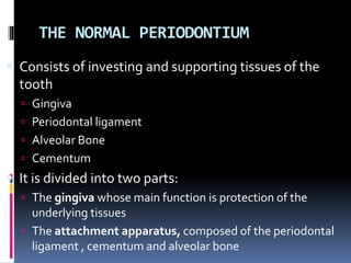
- 1. THE NORMAL PERIODONTIUM Consists of investing and supporting tissues of the tooth Gingiva Periodontal ligament Alveolar Bone Cementum It is divided into two parts: The gingiva whose main function is protection of the underlying tissues The attachment apparatus, composed of the periodontal ligament , cementum and alveolar bone
- 18. Types of gingivitis Depending on course and duration Acute gingivitis: is of sudden onset and short duration and can be painful Subacute: is less severe than acute Recurrent : reappears after treatment Chronic gingivitis: slow in onset, long duration, usually painless and the most common
- 19. Depending on distribution Localized or generalized : a group of teeth or entire mouth Marginal : limited to the marginal gingiva Papillary : limited to the interdental papilla Diffuse : when the inflammation spreads to the attached gingiva
- 23. OTHER GINGIVALINFECTIONS Acute lesions of sudden onset, limited duration and well-defined clinical features. Traumatic lesions, both physical and chemical Viral infections Bacterial infections Fungal infections Gingival abscess Aphthous ulcers Erythema multiforme Drug allergy
- 24. Traumatic lesions Physical injury Mechanical or thermal Chemical damage Asprin burn Silver nitrate Hydrogen peroxide Careless use of caustics by dentist e.g. chlorophenol, tetra acetic acid –Asprin burn
- 25. Traumatic injuries The diagnosis is easy since the patient is usually aware of the accident and suffers from immediate and severe pain. A localized area of inflammation and ulceration may form.
- 26. Healing of traumatic lesions is fairly quick and begins with epithelium covering the ulcer. Does not need active treatment Healing can be complicated by secondary infection. This may be accompanied by lymph gland enlargement and malaise, then an antibiotic may be needed.
- 27. Viral infections Acute herpetic gingivostomatitis Herpangina Hand foot and mouth disease Measles Herpes varicella/zoster virus infections Glandular fever HIV infection and AIDS
- 28. Acute herpetic gingivostomatitis Primary infection by the herpes simplex virus type 1. Usually occurs in children (1-10) but may affect older children or even adults Infection in neonates can produce encephalitis or meningitis In children or adults it produces a febrile illness or subclinical infection Incubation period is 5 days
- 29. CONT. Symptoms appear abruptly with fever and temperatures as high as 39.4 C Lymph gland enlargement, malaise and the mouth and throat may be painful Irritability in children, refusal to eat and increase salivation Small vesicles form on the gingivae, tongue, buccal mucosa and lips Vesicles burst and form round or irregular ulcers with grey membrane and surrounded by a red mucosa There is acute gingivitis with redness swelling and bleeding
- 30. Herpetic gingivostomatitis Symptoms subside in 10-21 days About 30% of patients develop recurrent infections later in life. The recurrence is usually herpes labialis and it is a reactivation of the latent virus in the trigeminal ganglion
- 31. Herpes labialis
- 32. Treatment Symptomatic and supportive In infants, milk of magnesia Benzocaine lozenges are useful in the older child Aspirin or paracetamol Phenergan a sedative in the child In severe cases acyclovir tabs 200mg four times daily or suspension 5ml four times daily Acyclovir cream can be used as a preventive measure
- 33. Hand, foot and mouth disease Acute febrile illness Coxsackie virus A type 16 Sporadic outbreak affecting mainly children Maculopapular and vesicular lesions appear on the skin and oral mucosa Skin lesions affect the hands, arms and feet The oral vesicles break into ulcers Uneventful recovery in 10-14 days
- 35. Measles Severe febrile illness affecting mainly children Presents with fever, malaise, cough, conjunctivitis, photophobia and lacrimation Blotchy macular rash Oral lesions present as Koplik’s spots which are bluish-white specks surrounded by a bright red margin on the buccal mucosa mainly Oral lesions precede skin lesions by a few days. Recovery in 2-3 weeks in healthy, fit well fed children Can be serious in poor and malnourished previously unexposed children Vaccination is available against measles
- 37. Herpes varicella/zoster virus infections Varicella or chickenpox is an acute febrile illness mainly in children Widespread maculopapular or vesicular eruptions on the skin Small vesicles also form on the oral mucosa, tongue and gingival Recovery in 2-3 weeks
- 38. Herpes zoster or shingles is caused by reactivation of latent varicella virus and common in older adults. Affects sensory nerves and produces severe neuralgia Vesicular eruptions on the skin or mucosa innervated by the affected sensory nerve
- 39. Herpes zoster
- 42. BACTERIAL INFECTIONS Acute necrotizing ulcerative gingivitis (NUG) Tuberculosis syphilis
- 45. Syphilis Secondary syphilis occurs 6 weeks after primary infection and produces skin rash and oral eruption. The ulcers are either mucous patches or snail track
- 48. GINGIVALABSCESS Abscess formation may follow damage by toothpick or fish bone if the foreign object is not removed. Gingival abscess associated with physical damage can also arise from wall of a gingival pocket where drainage has been impeded localized shiny, painful, red swelling and associated teeth TTP may drain spontaneously or require the removal of foreign object hot salt water mouthwashes and antibiotics if systemic involvement
- 49. Aphthous ulcers Most common type of recurrent ulcerative condition Possible defect of cell-mediated immune response painful lesions which appear without any reason, last for several days to heal and, after sometime recur Cause unknown but may be auto-immunity to a component of the oral mucosa. Related factors may be stress and hormonal changes In some patients related to the menstrual cycle Relationship between ulceration, iron-deficiency anaemia, folic acid deficiency and Vitamin B12
- 50. Three types minor aphthous ulcers, major aphthous ulcers herpetiform ulcers
- 51. MINOR APHTHOUS ULCERS Clinical features Well circumscribed white-yellow round lesion with red margin Usually less than 5 mm in size Affects non-keratinized oral mucosa Moderate to severe pain Heals without scarring within 7 to 10 days Affected patients report a history of aphthous ulcers usually for “as long as they can remember”
- 52. Management Inquire about type of toothpaste used SLS (sodium lauryl sulphate)-containing toothpastes may aggravate condition) Discontinue any SLS-containing toothpaste for 1month. Discontinue use of any mouthwash Rx topical steroid (e.g., fluocinonide, clobetasol) to be applied tid as soon as prodromal Symptoms occur (may reduce duration of ulcer by half) Always instruct patient to come back if ulcer not healed after 3 weeks
- 53. MAJORAPHTHOUS ULCERS Clinical features Larger, more severe, less common than minor Ulcers 10 to 30 mm in diameter Extremely painful, heals with scarring within weeks or even months Affected patients report a history of aphthous ulcers usually for “as long as they can remember” Major “aphthous-like”ulcers occur among immunosuppressed patients (HIV+; transplant recipients) with no prior history of aphthous ulcers
- 54. MANAGEMENT Topical steroids: Fluocinonide ointment 0.05% mixed 1/1 with orabase B(apply tid) Clobetasol ointment 0.05% mixed 1/1 with orabase B(apply tid) Tablets of hydrocortisone Treatment topical anaesthetics Antibacterial rinses to clear secondary bacterial infection, Tetracycline caps (250mg): dissolve in 1 Tbsp water, then rinse for 1 minute + expectorate qid
- 55. Copyright © 2003, Elsevier Science (USA). All rights reserved.
- 56. 56
- 58. Periodontitis Defined as:- “an inflammatory disease of the supporting tissues of the teeth caused by specific micro- organisms resulting in progressive destruction of the periodontal ligament and alveolar bone with pocket formation, recession, or both”
- 59. Periodontitis Chronic periodontitis Aggressive periodontitis Periodontitis as a manifestation of systemic diseases
- 61. Chronic Periodontitis Prevalent in adults but can occur in children Amount of destruction consistent with local factors Associated with variable microbial pattern Subgingival calculus frequently found Slow to moderate rate of progression with possible periods of rapid progression Possibly modified by: systemic factors such as diabetes and HIV; local factors: enviromental factors such as smoking and emotional stress
- 62. Localized or generalized Slight – 1-2mm of clinical attachment loss Moderate – 3-4mm CAL Severe - ≥ 5mm CAL
- 64. Aggressive periodontitis Otherwise clinically healthy patient Rapid attachment loss and bone destruction A/o microbial deposits inconsistent with disease severity Familial aggregation of diseased individuals
- 65. Localized form Circumpubertal onset of disease Localized to first molars or incisors with proximal attachment loss on at least two permanent teeth one of which is a first molar
- 66. Generalized form Usually affecting persons under 30 years of age (maybe older) Generalized proximal attachment loss affecting at least three teeth other than first molars and incisors Pronounced episodic nature of periodontal destruction Poor serum antibody response to infecting agents
- 67. Periodontitis as a manifestation of systemic diseases Haematological disorders 1. Acquired neutropenia 2. Leukaemias 3. Other Genetic disorders a. Familial and cyclic neutropenia b. Down syndrome c. Leukocyte adhesion deficiency syndrome d. Papillon-Lefevre syndrome e. Chediak-Higashi syndrome f. Histiocytosis syndromes g. Glycogen storage disease h. Infantile genetic agranulocytosis i. Cohen syndrome j. Ehlers-Danlos syndrome (types IV andVIIIAD) k. Hypophosphatasia. etc
- 68. Necrotizing periodontal diseases Necrotizing ulcerative gingivitis Necrotizing ulcerative periodontitis Tissue necrosis is the primary clinical feature.
- 70. Abscesses of the periodontium A periodontal abscess is a localized purulent infection of periodontal tissues and is classified by its tissue of origin Gingival abscess Periodontal abscess Pericoronal abscess
- 72. DrugAllergy Can be provoked by:- Penicillin Diazepam Local anaesthetic Codeine Tetracycline Barbiturates Others
- 73. Drug allergy and contact hypersensitivity Two types:- Those following systemic administration of a drug or chemical Those following direct contact with the oral mucosa
- 74. Manifestation depends on the type of allergic response provoked, ranging from simple drying of the mouth to the most severe response, anaphylactic shock, which is potentially fatal. Hypersensitivity to drugs may involve type 1, II, III and IV mechanisms.
- 75. Symptoms Burning sensation of oral mucosa, swelling, redness of the tongue, lips and gingivae Peeling epithelium leaving very sore ulcerated areas Gingivae, bright red and sensitive. Poor oral hygiene due to the sensitivity in brushing
- 76. Contact hypersensitivity Reactions of the oral mucosa have been reported to:- Chewing gum Toothpaste Mouthwashes Sweets Cosmetics Topical anaesthetic Topical antibiotics Periodontal dressings Flavouring agents e.g. peppermint, menthol cinnamon and eugenol
- 77. Management Implicated substance immediately withdrawn. Antihistamine Injection of hydrocortisone hemisuccinate In anaphylactic shock, intramuscular injection of 0.5ml of 1:1000 adrenaline is necessary Frequent warm saline rinses.
- 79. DEFINITION PERICORONITIS (From the Greek peri, “around”, Latin corona “crown” and itis, “inflammation”) also known as operculitis, is the inflammation of soft tissues surrounding the crown of a partially erupted tooth, including the gingiva (gums) and the dental follicles.
- 80. ANATOMIC RELATIONSHIP The occlusal surface of an involved tooth may be partly covered by a flap of tissue, the operculum, which exists during the eruption of the tooth and may persist afterwards. Varying degrees of eruption, malposition, or impaction may further complicate the soft tissue architecture
- 82. CONT… An accumulation of bacteria and debris beneath the operculum Mechanical trauma (e.g. biting the operculum with the opposing tooth). Often associated with partially erupted and impacted mandibular third molars. Periodontal pain. Pulpitis from dental caries (tooth decay). Acute myofascial pain in the temporomandibular joint disorder.
- 85. CLINICAL FEATURES The symptoms vary based on whether the condition is acute or chronic. ACUTE Sever pain near the back teeth Swelling of gum tissue due to fluid accumulation Pain when swallowing The discharge of pus
- 86. CHRONIC Bad breath A bad taste in the mouth A mild or dull ache lasting for one or two days Swelling of the lymph nodes in the neck Infection Fever Loss of appetite
- 87. COMPLICATIONS The involvement may become localized in the form of a pericoronitis abscess. It may spread posteriorly into the oropharyngeal area and medially to the base of the tongue, making swallowing difficult. Involvement of sub maxillary, cervical, deep cervical and retropharyngeal lymph nodes.
- 88. CONT… The partially erupted or impacted mandibular third molar is the most common site of pericoronitis. The space between the crown of the tooth and overlying gingival flap is an ideal area for the accumulation of food debris and bacterial growth. An influx of the inflammatory fluid and cellular exudates results in an increase in the bulk of the flap.
- 89. CONT… Swelling of the cheek in the region of the angle of the jaw and lymphadenitis. Mandibular movement is limited (Trismus). Toxic systemic complications – fever, leucocytosis and malaise. Foul taste and an inability to close the jaws. Radiating pain to the ear, throat, and floor of the mouth.
- 90. RISK FACTORS Occur in young adults in their mid 20s who are experiencing poorly erupting wisdom teeth. Poor oral hygiene. Excess gum tissue. Fatigue and emotional stress
- 91. TREATMENTS Depending on the severity of the condition, the treatment option may vary. Gently flush the area with warm water or antiseptic to remove debris and exudates Pain management and resolving the pericoronal inflammation and/or infection Antibiotics can be prescribed in severe cases. Minor oral surgery to remove the overlapping gingival tissue Wisdom tooth or teeth removal