Clinical Discussion
Dr. AnjirAnwar
FCPS student
Paediatric Neurology & Development
Bangabandhu Sheikh Mujib Medical University
2.
Case 1
Naima, 8yrsold girl, admitted with
complaints of fever & headache for 1
month, altered level of consciousness for 10
days. She had no H/O seizure, TB contact.
O/E: Naima is drowsy, disoriented, GCS-
8/15, signs of meningeal irritation-present,
Cranial nerve-intact. Muscle bulk & tone-
normal, muscle power-2/5, DTR-diminished
in both upper & lower limbs, planta-
extensor, Signs of cerebellar lesion –present
Investigations: CSF study-cell-300(85%-
lymphocyte),protein-1g/dl, ADA-10.7U/L
Dx: TBM with ADEM
4.
Case 2
• Naznin,11yrs old, admitted with complaints of high grade fever
for 13 days, H/O GTCS for several times for 5days, H/O
unconsciousness
• O/E: She is unconscious, GCS-4/15, having decerebrate
posture, signs of meningeal irritation present, UMN sign in both
upper & lower limbs, plantar-extensor.
6.
Case 3
Zabir, 5years,male
Unable to walk for 9 days
Weakness of both upper limb for 2 days
H/O back pain & leg pain, excessive
sweating, urinary incontinence
Irritability
H/O fever 10days back
O/E: Hypotonia & areflexia of all 4limbs
present, bilateral plantar extensor
Investigations: CSF-Albumino-cytological
dissociation
NCV-AMAN variety of GBS
Dx: GBS with ADEM
8.
Acute Disseminated Encephalomyelitis
•Acute disseminated encephalomyelitis (ADEM) is usually a
monophasic, autoimmune, inflammatory, demyelinating
disease of the CNS defined by a polysymptomatic
presentation and encephalopathy.
• It is often seen 7-14 days following a viral infection or
immunization .
9.
The annual incidenceof ADEM is reported to be 0.4–0.8
per 100,000
More commonly affects children and young adults,
probably related to the high frequency of exanthematous
and other infections and vaccination in this age group.
10.
Patients with ADEMtend to be prepubertal, with 80 % of
childhood cases occurring in those aged 10 years or
younger.
Peak incidence at 5–8 years.
Seasonal predilection for fall-to-winter occurrence.
Often a history of preceding infection or vaccination.
11.
PRECEDING
INFECTIOUS
ILLNESSES:
Viral :
Measles
Mumps
Influenza A or B
Hepatitis A or B
Herpes simplex
Varicella, rubella.
Epstein-Barr
Cytomegalovirus
HIV
Dengue
Others :
M. Tuberculosis
Mycoplasma
pneumoniae
Chlamydia
Legionella.
Campylobacter
S.Pyogenes
Brucella
Borrelia
Salmonella Typhi
VACCINE:
Rabies
Diphtheria
Tetanus
Pertussis
Smallpox
Measles
Japanese B
encephalitis
Polio
Hepatitis B
Influenza
Meningococcal
A&C
12.
Pathophysiology
• The pathophysiologyinvolves transient autoimmune
response directed at myelin or other self-antigens(e.g.,
myelin basic protein, myelin oligodendrocyte protein,
proteolipid protein), possibly by molecular mimicry or by
nonspecific activation of autoreactive T-cell clones
Neurology. 2001 May 22; 56(10):1308-12.
13.
Genetic susceptibility explainswhy encephalomyelitic
complications develop in only a small minority of patients.
Human leucocyte antigen class II genes have the most
significant influence.
Immunopathological events leading to ADEM can be
divided into two major phases-
1.Initial T cell priming and activation
2.Subsequent recruitment and effector phase
14.
Anti–myelin oligodendroglial glycoprotein(MOG) antibodies have
been detected in 18%–35% of children with a first acute episode of
inflammatory demyelination ( “Immunopathophysiology of pediatric
CNS inflammatory demyelinating disease”) .
The transient detection of anti-MOG antibodies is more frequent in
children at onset of ADEM, optic neuritis, and relapsing optic
neuritis, and anti-MOG positivity predicts a non-MS disease course.
15.
• MOG-positive childrenhave clinical and radiologic features typical
for ADEM and elevated lymphocyte counts in CSF, suggesting a
high degree of inflammation. Persistence of anti-MOG antibodies
has been associated with MS, although fewer than 25% of pediatric
patients with MS have detectable antiMOG antibodies.
16.
Pathology
Pathological hallmark :-
Areas of perivenous demyelination and infiltration of lymphocytes
and macrophages.
Other changes - hyperaemia, endothelial swelling, and vessel wall
invasion by inflammatory cells, perivascular oedema, and
haemorrhage.
Present in the small blood vessels of both white and grey matter.
Postinfectious encephalomyelitis typically involves the white matter,
lesions in grey matter can also been seen.
Summary of 2012International Pediatric
Multiple Sclerosis Study Group definitions
• Pediatric acute disseminated encephalomyelitis (ADEM )All
are required:
A first polyfocal, clinical CNS event with presumed
inflammatory demyelinating cause.
An encephalopathy that cannot be explained by fever.
No new clinical or MRI findings 3 months or more after onset.
Brain MRI is abnormal during the acute (3 months) phase with
typically diffuse, poorly demarcated large lesions involving
predominantly the cerebral white matter.
20.
Summary of 2012International Pediatric
Multiple Sclerosis Study Group definitions
• Pediatric clinically isolated syndrome (CIS) (all are required):
A clinical CNS event with presumed inflammatory
demyelinating cause.
Absence of a clinical history of CNS demyelinating disease (if
any, see pediatric MS).
No encephalopathy except as readily explained by fever. Criteria
for MS diagnosis on baseline MRI are not met.
21.
Pediatric MS
• Anyof the following
Two or more CIS separated by more than 30 days involving more than one area of
CNS.
One CIS associated with MRI findings consistent with criteria of dissemination in
space (DIS) and in which a follow-up MRI shows at least one new lesion consistent
with dissemination in time (DIT) criteria.
One ADEM attack followed by 1 CIS 3 or more months after symptom onset that is
associated with new MRI findings consistent with criteria for DIS.
A CIS whose MRI findings are consistent with criteria for DIS and DIT (at least 1
T2 lesion in at least 2 of 4 areas: spinal cord, infratentorial, juxtacortical, and
periventricular [DIS] associated with a simultaneous presence of asymptomatic
gadolinium-enhancing and nonenhancing lesions [DIT] if the patient is $12 years
old).
24.
Radiologically isolated syndrome(RIS)
• RIS refers to individuals with incidental MS-typical MRI findings in whom
clinical history or signs of MS are lacking. The following adult criteria8 are
believed to be appropriate for pediatric RIS but should be validated:
1.MRI showing ovoid, well-circumscribed, and homogenous T2 hyperintensities
fulfilling at least 3 Barkhof criteria (at least 1 gadoliniumenhancing lesion or 9 T2-
hyperintense lesions; at least 1 infratentorial lesion; at least 1 juxtacortical lesion; at
least 3 periventricular lesions).
2. No historical account of remitting symptoms of neurologic dysfunction indicating
MS.
3. MRI findings do not account for symptoms for which the individual was imaged.
4. MRI findings are not better explained by another disease process.
26.
Clinical Features
Fever, headache,vomiting, and meningismus are
often seen at the time of initial presentation.
Encephalopathy is a characteristic feature
Progression of initial neurologic signs to maximum
deficits usually occurs within 4 to 7 days.
The level of consciousness ranges from subtle
lethargy to frank coma.
27.
Clinical Features
Longtract signs
Acute hemiparesis
Cerebellar ataxia
Cranial neuropathies, including optic neuritis, and
Spinal cord dysfunction (transverse myelitis)
Symptoms of optic neuritis include vision loss, pain with eye movement,
and an afferent pupillary defect
Hallucinations, Psychiatric abnormalities,Headache,Language
disturbances
Meningeal signs
Nystagmus
Ophthalmoparesis
28.
Clinical Features
Boweland bladder involvement secondary to spinal cord disease
results in constipation and urinary retention.
Retention of urine, urinary frequency, urgency, or incontinence may
occur during the acute stage and lower urinary tract dysfunction
may persist even after disappearance of other neurological deficits
The arms can be involved if the demyelinating lesion affects the
cervical cord.
Respiratory failure may appear with high cervical lesions that
extend into the brainstem.
29.
ADEM may havevarious atypical presentations.
Behavioral disturbances may occasionally be the sole symptom.
Presence of flaccidity and areflexia in an otherwise typical case
of ADEM betrays additional PNS involvement, which is most
commonly at the level of the spinal roots; this picture is frequently
seen in antirabies vaccination-related ADEM.
• Neurology 2005;1057-65
30.
Combined CNS andPNS demyelination may suggest the
possibility of shared pathological epitopes.
There is evidence to suggest that central (ADEM) and
peripheral (acute and chronic inflammatory demyelinating
polyradiculoneuropathy) demyelinating disorders represent two
ends of a spectrum and overlap of clinical features may occur.
Chronic inflammatory demyelinating polyradiculoneuropathy with
tumefactive central demyelination. Muscle Nerve 2006;33:283-8.
31.
Extrapyramidal syndromes suchas tremor, chorea, dystonia or
rigidity, may appear, though only sporadical.
Post-measles ADEM develops abruptly, usually 3 to 7 days
after the appearance of skin exanthema and after the
amelioration or vanishing of measles rash.
Rarely ADEM may present with features of intracranial space
occupying lesion, with tumefactive demyelinating lesions.
32.
• Certain clinicalpresentations may be specific with certain
infections:
Cerebellar ataxia for varicella infection
Myelitis for mumps,
Myeloradiculopathy for Semple antirabies vaccination,
Explosive onset with seizures and mild pyramidal dysfunction
for rubella.
Extrapyramidal manifestations such as chorea and dystonia are
rare but may be prominent in ADEM following group A
streptococcal infection.
34.
Recurrent ADEM
New eventof ADEM occurs with recurrence of the initial
symptoms and signs 3 or more months after the first ADEM
event without involvement of new clinical areas by history,
examination, or neuroimaging.
Event does not occur while on steroids and occurs at least
1 month after completing therapy.
MRI shows no new lesions; original lesions may have
enlarged.
No better explanation exists.
35.
Multiphasic ADEM
ADEM isfollowed by a new clinical event also meeting criteria
for ADEM, but involving new anatomic areas of the CNS as
confirmed by history, neurologic examination, and
neuroimaging.
Subsequent event must occur
(1) at least 3 months after the onset of the initial ADEM
event and
(2) at least 1 month after completing steroid therapy.
Subsequent event must include a polysymptomatic
presentation, including encephalopathy, with neurologic
symptoms or signs that differ from the initial event (mental
status changes may not differ from the initial event).
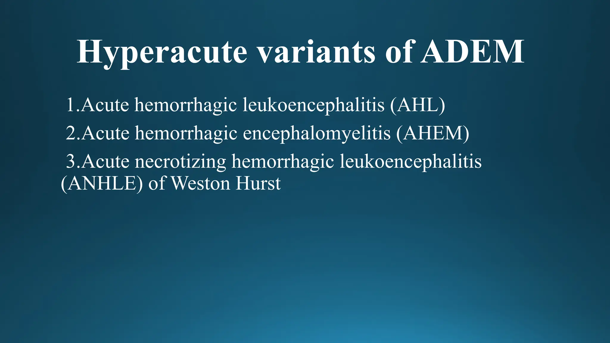
Acute hemorrhagic leukoencephalitis
MRI:T2* hypointensity within areas of T2 hyperintense signal change
Large tumefactive lesions involving the white matter and sparing the
cortex
Associated punctate hemorrhages and extensive mass effect and
surrounding edema.
Possible involvement of ganglia and thalami
Detection of cerebral microhemorrhages by GRE or the more
sensitive (SWI) is an important finding and may allow for
differentiation from ADEM.
39.
Acute hemorrhagic leukoencephalitis
Symptomatologyis similar to ADEM, with meningismus, headache,
seizures, multifocal neurologic signs, asymmetrical neurologic
deficits, and coma.
They typically follow an upper respiratory infection. (similar to
ADEM)
CSF typically shows both white and red blood cells, with increased
protein concentration.
40.
(a) FLAIR sequence
showsasymmetrical
heterogeneous
hyperintense lesions in
bilateral periventricular
regions. (b) GRE
sequence shows
"blooming" (i.e,. dark)
foci suggestive of
hemorrhage
Cerebrospinal fluid analysis
Abnormalitiesin 50% - 80% of patients with ADEM.
These findings may include lymphocytic pleocytosis (with a white
blood cell count of fewer than 100 cells/mL) and a slightly elevated
CSF protein (Fewer than 70 mg/dL).
An elevated level of cerebrospinal fluid (CSF) myelin basic protein
on CSF analysis. This is a sign of demyelination in the CNS.
• Acute Disseminated Encephalomyelitis (ADEM)Arayamparambil C.
Anilkumar; Lisa A. Foris; Prasanna Tadi.Last Update: August 14, 2019.
43.
May contain increasedamounts of gamma globulin and IgG
and raised levels of myelin basic protein.
Glucose content is usually normal.
Rarely oligoclonal band of IgG may be demonstrated-58% of
adult and 29% of pediatric cases.
Production of intrathecal oligoclonal IgG almost ceases as the
patient improves.
44.
MRI of Brain
Multiple,bilateral but asymmetric, poorly
demarcated, areas of increased signal on T2-
weighted and FLAIR sequences which can affect
both white and gray matter
White matter lesions are usually asymmetric and
most frequently situated subcortically, in the
cerebellum, brain stem, and spinal cord.
Thalamic and basal ganglia lesions are seen in
nearly one-third of cases and may be symmetric.
Corpus callosum may be affected when
involvement is extensive.
45.
MRI of Brain
Enhancementafter contrast administration is
variable and occurs in acute lesions due to
disruption of the blood-brain barrier. Lesions
may show complete ring, incomplete ring
('open-ring sign'), nodular, gyral, or spotty
patterns of enhancement.
Tumefactive demyelination appears as a large
white matter lesion with mass effect.
46.
Spinal cord MRI
•Spinal cord MRI may show confluent
intramedullary lesion(s) with variable
enhancement, in addition to abnormal
brain MRI findings specified
previously.
47.
Though clinically amonophasic illness, new lesions may
appear serially over several weeks and hence, may appear to be
of varying ages on MRI.
Occasionally, initial MR images may be normal and lesions
may appear in images repeated later in the course of the disease
or even during the stage of clinical improvement.
Though lesions generally resolve with treatment,
hyperintensities may persist in MRI long after clinical recovery
and is due to astrocytic hyperplasia, gliosis, or cystic changes.
Resolution of MRI abnormalities within six months of the
demyelinating episode favors the diagnosis of ADEM.
48.
• ADVANCED NEUROIMAGINGTECHNIQUES :
Diffusion tensor imaging (DTI) and magnetic transfer imaging
(MTI), may provide a better assessment of the underlying
histopathology than an increase in T2W signal on conventional
MRI.
Magnetization transfer and diffusion tensor MR imaging may
also be helpful in identifying involvement of the so- called
‘‘normal-appearing white matter’’ ,(NAWM and NAGM )
Double inversion recovery improve detection of cortical lesions.
49.
• Postcontrast MRIT1 images showings scattered hypointense rounded and oval lesions,
mostly situated at the junction of deep cortical gray and subcortical white matter. The
immediate periventricular region is spared. Lesions are mostly larger than MS lesions
and did not show contrast enhancement. Some lesions are seen encroaching upon the
cortical grey matter. In general lesions did not have marked mass effect.
50.
• Multifocal cortical/
subcortical lesions are
sparing the periventricular
region. The ADEM
lesions are hypointense
on MRI T1 images and
hyperintense on MRI T2
and FLAIR images.
ADEM lesions, though
large, exert mild mass
effect.
51.
• T1 C+(Gd): punctate,
ring or arc enhancement
(open ring sign) is often
demonstrated along the
leading edge of
inflammation; absence
of enhancement does not
exclude the diagnosis
52.
• DWI: therecan be
peripherally restricted
diffusion; the center of the
lesion, although high on T2
and low on T1, does not have
increased restriction on DWI
(cf. cerebral abscess), nor
does it demonstrate absent
signal on DWI as one would
expect from a cyst; this is due
to increase in extracellular
water in the region of
demyelination.
53.
• Contrast enhancement
whichis characteristic of
acute lesions. Also
notice that many lesions
are situated at the
junction of deep cortical
gray and subcortical
white matter which is
characteristic of ADEM
The lesions inADEM often have poorly defined margins, whereas MS
lesions have well defined “plaque-like” margins.
Periaqueductal, corpus callosum, and periventricular white matter
lesions are characteristic of MS.
By contrast, in ADEM the lesions tend to be in the deeper white matter
with periventricular sparing (only 29–60% of ADEM patients have
periventricular lesions) .
When the spinal cord is involved in ADEM, the lesion is typically
large, swollen, and thoracic.
The spinal cord lesions in MS are typically smaller, more discrete, and
cervical.
Although the white matter is classically involved in both disorders, the
grey matter (both cortical and deep grey/basal ganglia) is frequently
involved in ADEM (in contrast to MS).
57.
• Post-streptococcal ADEMshows particular predisposition to
basal ganglia lesions. A recent MRI study of 116 children with a
first episode of inflammatory demyelination showed that
perpendicular corpus callosum lesions and the sole presence of
well defined lesions were the most specific predictive factors
for relapse (although they had a low sensitivity).
58.
• (A &B) MRI brain (T2
weighted) in MS
showing well
demarcated lesions in
the region of the
periventricular white
matter.
• (C) MRI brain in
ADEM showing
multiple large lesions
with poorly defined
margins & cloudy
appearance.
60.
EEG
• Electroencephalography :
Abnormalitiesare common but are usually non-specific.
Mild generalised slowing, to severe generalised slowing with
infrequent focal slowing and epileptiform discharges.
• Computed tomography:
Generally normal at onset and usually becomes abnormal 5–14
days later.
Typical computed tomographic appearance is that of low
attenuation, multifocal lesions in the subcortical white matter.
61.
Supportive Care
Airway protectionin patients with altered mental status and
mechanical ventilation if required.
Antiseizure medication in patients with seizures
Correction of fluid and electrolyte disturbances
Prophylactic anticoagulation for prevention of deep vein
thrombosis in patients with high risk.
62.
Immunomodulation
• A typicaltreatment regimen consists of IV methylprednisolone at a
dose of 30 mg/kg/d (maximally 1,000 mg/d) for 5 days, followed
by an oral taper over 4–6 weeks with a starting dose of prednisone
of 1–2 mg/kg/d. An increased risk of relapse was observed with
steroid taper of ≤3 weeks.
• Acute disseminated encephalomyelitis, multiphasic disseminated
encephalomyelitis and multiple sclerosis in children. Brain 2000;12:2407–
2422
63.
• With thismodality of treatment, full recovery has been reported
in 50%–80% of patients. Methylprednisolone-treated patients
had significantly better outcome with respect to disability status
when compared with those treated with dexamethasone.
• Any type of vaccination should be avoided during the first 6
months following recovery.
64.
• The putativemechanism of action includes :
Modification of cytokine responses;
Reduction in T-cell activation;
Reduction in blood–brain barrier permeability that, in turn,
limits extravasation of immune cells into the CNS; and
Facilitating apoptosis of activated immune cells
65.
Intravenous immunoglobulin (IVIg)
•Intravenous immunoglobulin (IVIg) (0.4 gm/kg/day for 5 days)
is another option, but there is a constraint of high cost and the
evidence for this modality of treatment in ADEM is Class IV.
The choice of second-line treatment should be individualized,
depending on the severity of the disease, complications, and
comorbidities.
• The benefit of IVIg is thought to provide benefit by directly
affecting cytokine production and T-cell proliferation and
by binding potential autoantibodies targeted against myelin.
66.
Plasma Exchange
• Treatmentof patients with severe or life-threatening
demyelination, such as patients with myelitis or brainstem
involvement.
• Side effects include infection, alteration of electrolyte profiles,
and depletion of coagulation factors.
• The benefit of PLEX is likely secondary to its therapeutic
removal of circulating autoantibodies and immune complexes
from the blood
67.
A course of4–6 PEs have been shown to be associated with moderate
to marked and sustained improvement. One could remove a large
volume of plasma per exchange if there are no problems of autonomic
dysfunction.
Predictors associated with improvement include male sex, preserved
reflexes, and early initiation of treatment.
In centers that do not have this facility for conventional PE, one could
modify and improvise to do a small volume manual plasma exchange
—by doing a phlebotomy, centrifuging the blood, remove 250–300 mL
of plasma and return the cells. One could do this twice a day for 7–10
days.
68.
Autonomic dysfunction andhypotension preclude the use of PE.
IVIg may be more effective in patients with peripheral nervous
system involvement and PE in patients with tumefactive
demyelination.
Methyl prednisolone along with IVIg has been successfully used in
patients with atypical features and could be tried for fulminant,
aggressive, and atypical disease.
Improvement of atypical acute disseminated encephalomyelitis with steroids
and intravenous immunoglobulins.Pediatr Neurol. 2001 Feb; 24(2):139-43.
69.
Cyclophosphamide and hypothermiahave been used with
success in patients with fulminant ADEM.
Decompressive hemi-craniectomy has been reported to be life
saving in patients with massive life-threatening cerebral edema
refractory to conventional medical management.
70.
Serial Imaging
At least2 additional MRIs (e.g., 3 months and 9–12 months after
clinical onset), in order to rule out ongoing disease activity indicating
a diagnosis other than ADEM.
Acute disseminated encephalomyelitis: Updates on an inflammatory CNS
syndrome Neurology. 2016 Aug 30;87(9
72.
Prognosis
Long-term prognosis ofthis entity depends on the etiology.
Postmeasles patients have higher mortality rate and significant
morbidity in survivors.
Prognosis of nonmeasles cases is favorable ,full recovery in
50%–75% of patients, in 1–6 months of follow up.
Most common sequelae are focal motor deficits, ranging from
mild ataxia to hemiparesis.
73.
Prognosis
Hyperacute onset, severeneurologic deficits as a result of aggressive
disease, and unresponsiveness to steroids are poor prognostic indicators.
Prolonged altered mental state was associated with both mortality and
morbidity.
Multiple or single extensive lesions on MRI lesions may be associated
with disability.
• Acute disseminated encephalomyelitis in children.Pediatrics. 2002 Aug; 110(2 Pt 1):e21.
74.
Prognosis
Although monophasic, 5-29%children will go onto have
additional demyelinating attacks characteristics of MS.
11-17% of children experience residual motor deficit.
Swaiman’s Paediatric Neurology 6th
edition


![Pediatric MS
• Any of the following
Two or more CIS separated by more than 30 days involving more than one area of
CNS.
One CIS associated with MRI findings consistent with criteria of dissemination in
space (DIS) and in which a follow-up MRI shows at least one new lesion consistent
with dissemination in time (DIT) criteria.
One ADEM attack followed by 1 CIS 3 or more months after symptom onset that is
associated with new MRI findings consistent with criteria for DIS.
A CIS whose MRI findings are consistent with criteria for DIS and DIT (at least 1
T2 lesion in at least 2 of 4 areas: spinal cord, infratentorial, juxtacortical, and
periventricular [DIS] associated with a simultaneous presence of asymptomatic
gadolinium-enhancing and nonenhancing lesions [DIT] if the patient is $12 years
old).](https://image.slidesharecdn.com/adem-250423070450-a3d74b05/75/Acute-Disseminated-Encephalomyelitis-in-children-21-2048.jpg)