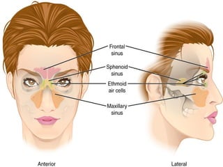
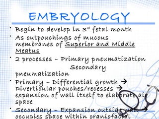
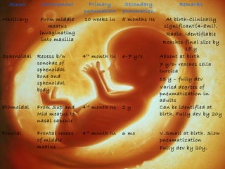
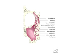
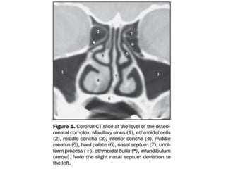
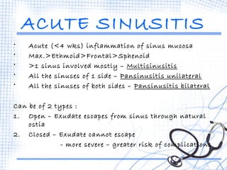
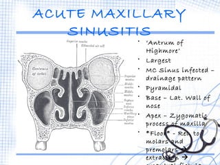
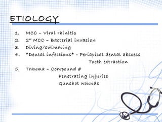
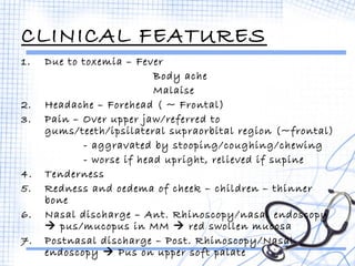
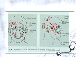
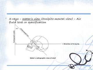
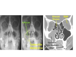
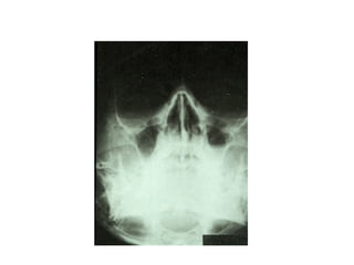
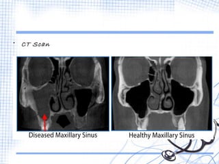
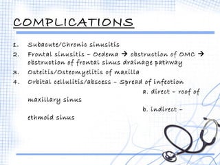
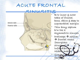
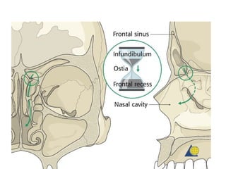
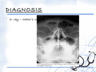
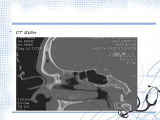
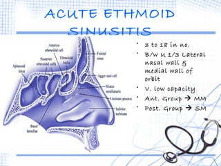
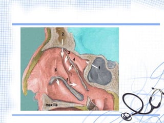
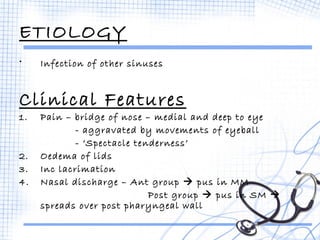
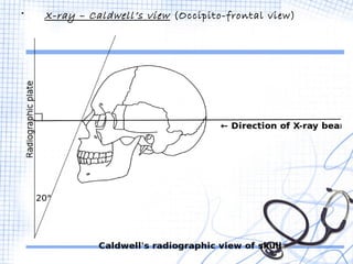
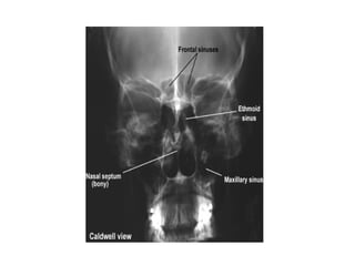
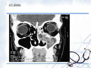
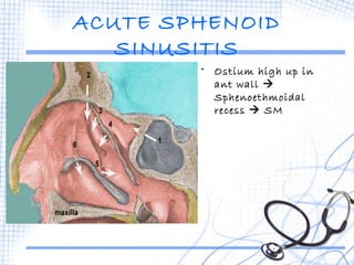
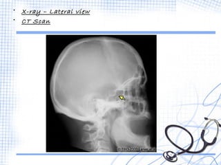
ACUTE 
SINUSITI 
S 
-Anira 
Iqbal 
Batch 2011
EMBRYOLOGY 
• Begin to develop in 3rd fetal month 
• As outpouchings of mucous 
membranes of Superior and Middle 
Meatus 
• 2 processes – Primary pneumatization 
Secondary 
pneumatization 
• Primary – Differential growth  
Diverticular pouches/recesses  
expansion of wall itself to elaborate air 
space 
• Secondary – Expansion outside wall  
occupies space within craniofacial 
bones
Sinus Development Primary 
pneumatizn 
Secondary 
pneumatizn 
Remarks 
Maxillary From middle 
meatus 
invaginating 
into maxilla 
10 weeks iu 5 months IU At birth-Clinically 
significant(4-8ml), 
Radio. Identifiable 
Reaches final size by 
15 y 
Sphenoidal Recess b/w 
conchae of 
sphenoidal 
bone and 
sphenoidal 
body 
4th month IU 6-7 y/o Absent at birth 
7 y/o- reaches sella 
turcica 
15 y – fully dev 
Varied degrees of 
pneumatization in 
adults 
Ethmoidal From Sup and 
Mid meatus to 
nasal capsule 
4th month IU 2 y Can be identified at 
birth. Fully dev by 20y 
Frontal Frontal recess 
of middle 
meatus 
4th month IU 6 mo V.Small at birth. Slow 
pneumatization 
Fully dev by 20y.
OSTEOMEATAL 
COMPLEX 
• Common channel that links frontal sinus, ant. and 
middle ethmoid sinus, and max sinus to middle 
meatus  allows air flow and mucociliary drainage  
needs to be patent for drainage of secretions in 
sinusitis
ACUTE SINUSITIS 
• Acute (<4 wks) inflammation of sinus mucosa 
• Max.>Ethmoid>Frontal>Sphenoid 
• >1 sinus involved mostly – Multisinusitis 
• All the sinuses of 1 side – Pansinusitis unilateral 
• All the sinuses of both sides – Pansinusitis bilateral 
Can be of 2 types : 
1. Open – Exudate escapes from sinus through natural 
ostia 
2. Closed – Exudate cannot escape 
- more severe – greater risk of complications
ETIOLOGY 
EXCITING CAUSES 
1. Nasal infections - Nasal mucosa  Sinus mucosa 
MCC – Viral > 
Bacterial>>Fungal 
2. Swimming/Diving – Infected water  Ostia of sinuses 
- Chlorine  Chemical 
inflammation 
3. Trauma – Compound # or penetrating injuries  
infection 
4. Dental infection – Molar/Premolar infection/extraction 
 Max. sinus
PREDISPOSING CAUSES  Local 
 General 
LOCAL 
1. Obstruction to sinus ventilation and drainage 
• Nasal packing 
• DNS 
• Hypertrophic turbinates 
• Allergy – oedema of sinus ostia 
• Nasal polypi 
• Benign/Malignant neoplasm 
2. Stasis of secretions in nasal cavity 
• Cystic fibrosis – high viscosity of secretions 
• Enlarged adenoids - obstruction 
• Choanal atresia - obstruction
GENERAL 
1. Environment – cold, wet 
- atm. pollution, smoke, dust, 
overcrowding 
2. Poor general health – recurrent attacks of exanth. 
Fevers 
- nutritional deficiencies 
- Systemic disorders (Diabetes) 
- Immunodeficient 
BACTERIOLOGY 
• Streptococcus pneumoniae 
• Hemophilus influenzae 
• Moraxella catarrhalis 
• Streptococcus pyogenes 
• Staphylococcus aureus 
• Klebsiella pneumoniae 
• Anaerobic org. – Dental infections
PATHOLOGY 
Infection  Acute Inflammation of sinus mucosa  
Hyperemia  Exudation (serous  
mucopurulent/purulent)  Outpouring of PMNs  
Increased activity of serous and mucus glands  
severe infection  destruction of mucosal lining 
If failure of ostium to drain  Empyema 
If destruction of bony walls  complications 
• Mild/Non suppurative – less virulent, good immunity, 
drainage 
• Severe/Suppurative
ACUTE MAXILLARY 
SINUSITIS 
• ‘Antrum of 
Highmore’ 
• Largest 
• MC Sinus infected – 
drainage pattern 
• Pyramidal 
• Base – Lat. Wall of 
nose 
• Apex – Zygomatic 
process of maxilla 
• *Floor* - Rel. to 
molars and 
premolars  
extraction  
oroantral fistula
ETIOLOGY 
1. MCC – Viral rhinitis 
2. 2nd MCC – Bacterial invasion 
3. Diving/swimming 
4. *Dental infections* - Periapical dental abscess 
Tooth extraction 
5. Trauma – Compound # 
Penetrating injuries 
Gunshot wounds
CLINICAL FEATURES 
1. Due to toxemia – Fever 
Body ache 
Malaise 
2. Headache – Forehead ( ~ Frontal) 
3. Pain – Over upper jaw/referred to 
gums/teeth/ipsilateral supraorbital region (~frontal) 
- aggravated by stooping/coughing/chewing 
- worse if head upright, relieved if supine 
4. Tenderness 
5. Redness and oedema of cheek – children – thinner 
bone 
6. Nasal discharge – Ant. Rhinoscopy/nasal endoscopy 
 pus/mucopus in MM  red swollen mucosa 
7. Postnasal discharge – Post. Rhinoscopy/Nasal 
endoscopy  Pus on upper soft palate
DIAGNOSIS 
• Transillumination test - Affected sinus  Opaque
• X-rays – Water’s view (Occipito-mental view) – Air 
fluid level or opacification
• CT Scan
COMPLICATIONS 
1. Subacute/Chronic sinusitis 
2. Frontal sinusitis – Oedema  obstruction of OMC  
obstruction of frontal sinus drainage pathway 
3. Osteitis/Osteomyelitis of maxilla 
4. Orbital cellulitis/abscess – Spread of infection 
a. direct – roof of 
maxillary sinus 
b. indirect – 
ethmoid sinus
ACUTE FRONTAL 
SINUSITIS• B/w inner & outer 
table of frontal 
bone, above & deep to 
supraorbital margin 
• Thin bony septum 
b/w the 2 
asymmetric sinuses 
• Drainage  ostium 
 frontal recess 
(hourglass 
structure) 
infundibulum  
Middle meatus 
• Gravity  heals 
faster
ETIOLOGY 
1. Viral rhinitis 
2. Bacterial invasions 
3. Diving/Swimming 
4. Trauma 
5. Ipsilateral Maxillary/Ethmoid sinusitis
CLINICAL FEATURES 
1. Frontal headache – Medial brow area 
‘Office Headache’ – Comes up 
on waking  Gradually increases  Peak at mid-day 
 Subsides 
2. Tenderness – Tapping 
- Pressure upwards on floor of frontal 
sinus 
3. Oedema of upper eyelid 
4. Nasal discharge – Nasal endoscopy – vertical streak 
of mucopus high up in anterior part of middle 
meatus
DIAGNOSIS 
• X- ray – Water’s view
• CT Scan
COMPLICATIONS 
1. Orbital cellulitis 
2. Osteomyelitis of frontal bone and fistula formation 
3. Meningitis 
4. Extradural abscess 
5. Frontal lobe abscess 
6. Chronic frontal sinusitis
ACUTE ETHMOID 
SINUSITIS 
• 3 to 18 in no. 
• B/w U 1/3 Lateral 
nasal wall & 
medial wall of 
orbit 
• V. low capacity 
• Ant. Group  MM 
• Post. Group  SM
ETIOLOGY 
• Infection of other sinuses 
Clinical Features 
1. Pain – bridge of nose – medial and deep to eye 
- aggravated by movements of eyeball 
- ‘Spectacle tenderness’ 
2. Oedema of lids 
3. Inc lacrimation 
4. Nasal discharge – Ant group  pus in MM 
Post group  pus in SM  
spreads over post pharyngeal wall
COMPLICATIONS 
1. Orbital cellulitis and abscess 
2. Optic Nerve  Visual deterioration  blindness 
3. Cavernous sinus thrombosis 
4. Extradural abscess 
5. Meningitis 
6. Brain abscess
• X-ray – Caldwell’s view (Occipito-frontal view)
• CT Scan
ACUTE SPHENOID 
SINUSITIS 
• Ostium high up in 
ant wall  
Sphenoethmoidal 
recess  SM
ETIOLOGY 
• Isolated involvement – rare 
• + Pansinusitis/Post. Ethmoidal sinusitis 
Clinical Features 
1. Headache  occiput/vertex 
- maybe referred to mastoid 
2. Postnasal discharge – Posterior rhinoscopy  pus 
on roof and post. Wall of NP
• X-ray – Lateral view 
• CT Scan
THANK YOU!

Acute Sinusitis

  • 1.
    ACUTE SINUSITI S -Anira Iqbal Batch 2011
  • 3.
    EMBRYOLOGY • Beginto develop in 3rd fetal month • As outpouchings of mucous membranes of Superior and Middle Meatus • 2 processes – Primary pneumatization Secondary pneumatization • Primary – Differential growth  Diverticular pouches/recesses  expansion of wall itself to elaborate air space • Secondary – Expansion outside wall  occupies space within craniofacial bones
  • 4.
    Sinus Development Primary pneumatizn Secondary pneumatizn Remarks Maxillary From middle meatus invaginating into maxilla 10 weeks iu 5 months IU At birth-Clinically significant(4-8ml), Radio. Identifiable Reaches final size by 15 y Sphenoidal Recess b/w conchae of sphenoidal bone and sphenoidal body 4th month IU 6-7 y/o Absent at birth 7 y/o- reaches sella turcica 15 y – fully dev Varied degrees of pneumatization in adults Ethmoidal From Sup and Mid meatus to nasal capsule 4th month IU 2 y Can be identified at birth. Fully dev by 20y Frontal Frontal recess of middle meatus 4th month IU 6 mo V.Small at birth. Slow pneumatization Fully dev by 20y.
  • 6.
    OSTEOMEATAL COMPLEX •Common channel that links frontal sinus, ant. and middle ethmoid sinus, and max sinus to middle meatus  allows air flow and mucociliary drainage  needs to be patent for drainage of secretions in sinusitis
  • 9.
    ACUTE SINUSITIS •Acute (<4 wks) inflammation of sinus mucosa • Max.>Ethmoid>Frontal>Sphenoid • >1 sinus involved mostly – Multisinusitis • All the sinuses of 1 side – Pansinusitis unilateral • All the sinuses of both sides – Pansinusitis bilateral Can be of 2 types : 1. Open – Exudate escapes from sinus through natural ostia 2. Closed – Exudate cannot escape - more severe – greater risk of complications
  • 10.
    ETIOLOGY EXCITING CAUSES 1. Nasal infections - Nasal mucosa  Sinus mucosa MCC – Viral > Bacterial>>Fungal 2. Swimming/Diving – Infected water  Ostia of sinuses - Chlorine  Chemical inflammation 3. Trauma – Compound # or penetrating injuries  infection 4. Dental infection – Molar/Premolar infection/extraction  Max. sinus
  • 11.
    PREDISPOSING CAUSES Local  General LOCAL 1. Obstruction to sinus ventilation and drainage • Nasal packing • DNS • Hypertrophic turbinates • Allergy – oedema of sinus ostia • Nasal polypi • Benign/Malignant neoplasm 2. Stasis of secretions in nasal cavity • Cystic fibrosis – high viscosity of secretions • Enlarged adenoids - obstruction • Choanal atresia - obstruction
  • 12.
    GENERAL 1. Environment– cold, wet - atm. pollution, smoke, dust, overcrowding 2. Poor general health – recurrent attacks of exanth. Fevers - nutritional deficiencies - Systemic disorders (Diabetes) - Immunodeficient BACTERIOLOGY • Streptococcus pneumoniae • Hemophilus influenzae • Moraxella catarrhalis • Streptococcus pyogenes • Staphylococcus aureus • Klebsiella pneumoniae • Anaerobic org. – Dental infections
  • 13.
    PATHOLOGY Infection Acute Inflammation of sinus mucosa  Hyperemia  Exudation (serous  mucopurulent/purulent)  Outpouring of PMNs  Increased activity of serous and mucus glands  severe infection  destruction of mucosal lining If failure of ostium to drain  Empyema If destruction of bony walls  complications • Mild/Non suppurative – less virulent, good immunity, drainage • Severe/Suppurative
  • 14.
    ACUTE MAXILLARY SINUSITIS • ‘Antrum of Highmore’ • Largest • MC Sinus infected – drainage pattern • Pyramidal • Base – Lat. Wall of nose • Apex – Zygomatic process of maxilla • *Floor* - Rel. to molars and premolars  extraction  oroantral fistula
  • 16.
    ETIOLOGY 1. MCC– Viral rhinitis 2. 2nd MCC – Bacterial invasion 3. Diving/swimming 4. *Dental infections* - Periapical dental abscess Tooth extraction 5. Trauma – Compound # Penetrating injuries Gunshot wounds
  • 17.
    CLINICAL FEATURES 1.Due to toxemia – Fever Body ache Malaise 2. Headache – Forehead ( ~ Frontal) 3. Pain – Over upper jaw/referred to gums/teeth/ipsilateral supraorbital region (~frontal) - aggravated by stooping/coughing/chewing - worse if head upright, relieved if supine 4. Tenderness 5. Redness and oedema of cheek – children – thinner bone 6. Nasal discharge – Ant. Rhinoscopy/nasal endoscopy  pus/mucopus in MM  red swollen mucosa 7. Postnasal discharge – Post. Rhinoscopy/Nasal endoscopy  Pus on upper soft palate
  • 19.
    DIAGNOSIS • Transilluminationtest - Affected sinus  Opaque
  • 20.
    • X-rays –Water’s view (Occipito-mental view) – Air fluid level or opacification
  • 23.
  • 24.
    COMPLICATIONS 1. Subacute/Chronicsinusitis 2. Frontal sinusitis – Oedema  obstruction of OMC  obstruction of frontal sinus drainage pathway 3. Osteitis/Osteomyelitis of maxilla 4. Orbital cellulitis/abscess – Spread of infection a. direct – roof of maxillary sinus b. indirect – ethmoid sinus
  • 25.
    ACUTE FRONTAL SINUSITIS•B/w inner & outer table of frontal bone, above & deep to supraorbital margin • Thin bony septum b/w the 2 asymmetric sinuses • Drainage  ostium  frontal recess (hourglass structure) infundibulum  Middle meatus • Gravity  heals faster
  • 27.
    ETIOLOGY 1. Viralrhinitis 2. Bacterial invasions 3. Diving/Swimming 4. Trauma 5. Ipsilateral Maxillary/Ethmoid sinusitis
  • 28.
    CLINICAL FEATURES 1.Frontal headache – Medial brow area ‘Office Headache’ – Comes up on waking  Gradually increases  Peak at mid-day  Subsides 2. Tenderness – Tapping - Pressure upwards on floor of frontal sinus 3. Oedema of upper eyelid 4. Nasal discharge – Nasal endoscopy – vertical streak of mucopus high up in anterior part of middle meatus
  • 29.
    DIAGNOSIS • X-ray – Water’s view
  • 30.
  • 31.
    COMPLICATIONS 1. Orbitalcellulitis 2. Osteomyelitis of frontal bone and fistula formation 3. Meningitis 4. Extradural abscess 5. Frontal lobe abscess 6. Chronic frontal sinusitis
  • 32.
    ACUTE ETHMOID SINUSITIS • 3 to 18 in no. • B/w U 1/3 Lateral nasal wall & medial wall of orbit • V. low capacity • Ant. Group  MM • Post. Group  SM
  • 34.
    ETIOLOGY • Infectionof other sinuses Clinical Features 1. Pain – bridge of nose – medial and deep to eye - aggravated by movements of eyeball - ‘Spectacle tenderness’ 2. Oedema of lids 3. Inc lacrimation 4. Nasal discharge – Ant group  pus in MM Post group  pus in SM  spreads over post pharyngeal wall
  • 35.
    COMPLICATIONS 1. Orbitalcellulitis and abscess 2. Optic Nerve  Visual deterioration  blindness 3. Cavernous sinus thrombosis 4. Extradural abscess 5. Meningitis 6. Brain abscess
  • 36.
    • X-ray –Caldwell’s view (Occipito-frontal view)
  • 38.
  • 39.
    ACUTE SPHENOID SINUSITIS • Ostium high up in ant wall  Sphenoethmoidal recess  SM
  • 40.
    ETIOLOGY • Isolatedinvolvement – rare • + Pansinusitis/Post. Ethmoidal sinusitis Clinical Features 1. Headache  occiput/vertex - maybe referred to mastoid 2. Postnasal discharge – Posterior rhinoscopy  pus on roof and post. Wall of NP
  • 41.
    • X-ray –Lateral view • CT Scan
  • 42.