1. The document summarizes a presentation given by Professor Wade Allison and Professor Akira Tokuhiro arguing that low levels of nuclear radiation are not harmful and current safety regulations are too strict.
2. They claim that fear of radiation causes more harm than the radiation itself through social and economic damage. Regulations have caused unnecessary hardship in Fukushima and Chernobyl.
3. Data from radiation therapy, Hiroshima, Nagasaki, and Chernobyl workers suggests that radiation below certain levels does not increase cancer risks and that current safety levels should be relaxed by a factor of 1000. No deaths are expected from radiation at Fukushima.
This document provides a historical overview of ionizing radiation and its use in cancer treatment from 1895 to the present. Some key events and discoveries discussed include Röntgen's discovery of x-rays in 1895, the early therapeutic uses of radiation in the late 1890s, discoveries of radioactivity and radiation types in the early 1900s, advances in radiation delivery technologies throughout the 20th century including linear accelerators and CT imaging, and the development of concepts in radiobiology from the 1900s-1970s that improved the safety and efficacy of radiation therapy. The document also recognizes the contributions and sacrifices of radiation workers throughout history who advanced the field of radiation oncology but lost their lives from overexposure before safety standards were established.
This document discusses the evolution of radiation therapy from its discovery in the late 19th century to modern techniques. It traces developments such as the discovery of x-rays and radioactivity, early radium and x-ray therapies, and the introduction of cobalt-60 and linear accelerators to improve targeting ability. Modern advances discussed include intensity-modulated radiation therapy (IMRT), image-guided radiation therapy (IGRT), proton beam therapy, and radiosurgery techniques like Gamma Knife and Cyberknife which allow extremely precise high dose radiation treatments.
This document discusses radiation protection and safety criteria related to ionizing radiation. It begins by defining radiation hazards and outlining the biological effects of radiation exposure, which can be either deterministic or probabilistic. Key aspects of radiation protection covered include determining radiation hazards, evaluating radiation doses, and the principles and recommendations established by the International Commission on Radiological Protection. The document then provides examples of calculating radiation intensity and shielding requirements for various radiation sources and energies. It emphasizes protecting workers and the public through principles of time, distance and shielding, and highlights planning considerations for medical x-ray facilities to ensure safe and compliant operation.
This document discusses radiation-induced cell kill and damage. It begins by outlining the topics to be covered, including DNA damage by ionizing radiation, chromosome damage and repair, cell survival curves, and the clinical response of normal tissues to radiation exposure. It then covers various topics in depth, such as the direct and indirect action of ionizing radiation on DNA, the role of linear energy transfer (LET), different types of DNA damage and repair mechanisms, chromosome aberrations, and cell survival curves. The goal of radiotherapy is explained as maximizing tumor kill while minimizing damage to normal tissues.
The document discusses the history and development of brachytherapy for treating cancer of the cervix. It outlines key dates and developments, including the discovery of radium and development of early intracavitary systems like the Paris, Stockholm, and Manchester systems from 1910-1938. It also discusses the MD Anderson system from 1952 and the Mallinckrodt Institute of Radiology system from 1979, as well as requirements, advantages, and dose rates of brachytherapy. The document notes some "caveats" of the Manchester system regarding variable definitions and measurements.
Radioactive Contamination and Procedures of Decontaminationmahbubul hassan
Training Course on Radiation Protection for Radiation Workers and RCOs of BAEC, Medical Facilities and Industries, TI, AERE, BAEC Savar, 27 October 2021
Lanl science 1995 history of radiation standardsJim Werner
2
10
100 millirem (1962)
1
10
0
10
1920
1930
1940
1950
1960
1970
1980
1990
2000
Year
low as reasonably achievable, economic and social factors being taken into account. Limitation requires that no individual shall receive a dose exceeding the recommended limits.
1) Radiation protection standards have evolved over time based on new information about radiation effects and changing attitudes towards risk, beginning with early standards aimed at preventing obvious effects like skin burns and moving to preventing delayed effects like cancer.
2) Early standards in the 1920s-1940
This document provides an overview of radiation hazards and protection. It discusses the different types of radiation including ionizing and non-ionizing radiation. It describes sources of medical radiation exposure like radiography, nuclear imaging, and radiation therapy. The key biological effects of radiation are ionization of atoms which can damage molecules, cells, tissues and organs. The major target of radiation is DNA. Radiation can cause DNA damage, mutations, and altered cell responses. Deterministic effects are harmful tissue reactions that become more serious with increasing dose above a threshold, while stochastic effects like cancer and genetic effects occur probabilistically with no safe threshold. Radiation sensitivity varies between tissues, with rapidly-dividing cells being more sensitive. Proper radiation protection
1. The document discusses the history of various discoveries and technologies in radiation oncology, including the discovery of radium by Marie and Pierre Curie in 1898, the development of the first betatron by Donald Kerst in 1940, and the invention of the gamma knife by Lars Leksell in 1967 to treat small brain tumors.
2. It also covers early researchers like Harold Johns who invented the cobalt bomb for radiation therapy, and the development of the linear accelerator by Henry Kaplan in the 1950s.
3. The document provides brief biographies of several influential figures in the history of radiation oncology such as Theodore Puck, Louis Harold Gray, and Thomas Rockwell Mackie who developed the tom
This document provides an overview of radiation hazards and protection. It begins with definitions of key terms like ionizing radiation and discusses natural and man-made radiation sources. It then examines the biological effects of radiation like deterministic and stochastic effects. Radiation-induced cellular damage and cancer development is explained. Factors affecting radiosensitivity and the relationships between dose and effects are also summarized. Throughout, simple language is used to make radiation concepts accessible to non-experts while still conveying essential scientific information about radiation hazards and safety.
Ppt. on Radiation Hazards by Dr. Brajesh K. Bendr brajesh Ben
This document discusses radiation hazards and provides information about radiation basics. It defines radiation as energy in transit that can be electromagnetic waves or high-speed particles. Ionizing radiation is radiation with sufficient energy to remove electrons from atoms, causing ionization. There are two main types of biological effects from radiation exposure - deterministic effects that occur above a threshold dose and increase in severity with increasing dose, and stochastic effects which have no safe threshold and the probability of damage increases with increasing dose. Some key radiation hazards discussed are acute radiation syndrome, radiation-induced cancer risks particularly for leukemia and thyroid cancer, and fetal radiation risks which are most significant during early stages of pregnancy.
Radiation can cause both deterministic and stochastic biological effects. Deterministic effects occur when radiation doses exceed a threshold and the severity increases with higher doses. These include skin damage, hair loss and cataracts. Stochastic effects like cancer and genetic mutations may occur due to single or multiple low doses and only the probability increases with dose. Cancer is a delayed effect that can take years or decades to appear after radiation exposure. Genetic effects can impact future generations. Both effects have no threshold dose and can potentially occur from even the lowest doses.
The document summarizes the history and development of Boron Neutron Capture Therapy (BNCT) for cancer treatment. It discusses how boron is used to selectively target cancer cells and how neutron capture by boron leads to high-LET particle production within tumor cells. Key developments include the first studies in the 1930s, clinical trials starting in the 1980s focused on glioblastoma, and ongoing research to develop more effective boron delivery agents and dosimetry techniques. BNCT shows potential for treating cancers like brain tumors but challenges remain around boron delivery and normal tissue toxicity.
This document provides an overview of biological effects of ionizing radiation. It defines key terms like dose, exposure, absorbed dose, and effective dose. It describes how radiation can damage cells and cause effects like cancer, and notes some effects are stochastic (probability increases with dose) while others are non-stochastic (above a threshold). Factors like radiation type, dose rate, and exposed area influence effects. The goal is to quantify energy deposition and understand dose-response relationships.
Radiation can damage DNA through ionization, potentially leading to cell death or mutation and increased cancer risk; while high doses cause acute radiation syndromes like hematopoietic syndrome, even low doses slightly increase lifetime cancer risk proportional to dose; medical uses of radiation involve careful consideration of dose required versus risk to maximize benefits like cancer treatment and diagnostics using techniques like x-rays and radiotracers.
This document discusses radiation hazards and includes definitions of radiation, types of radiation like ionizing and non-ionizing, sources of radiation, and effects of radiation on different biological tissues. It covers topics like maximum permissible radiation dosage, acute vs chronic exposure, and effects of radiation on various tissues and whole body irradiation. It also discusses radiation protection principles for patients and clinicians, and concludes with recent advances.
Radiation is energy transmitted through space or matter in the form of waves or particles. It includes visible light, ultraviolet light from the sun, and radio/TV signals. Nuclear radiation comes from unstable atoms undergoing radioactive decay, emitting particles like alpha and beta or electromagnetic waves like gamma rays. Exposure to ionizing radiation can damage living tissue. Natural sources of radiation include cosmic rays, radioactive elements in the earth's crust like radon, and some food/drink. Medical procedures and occupational exposures also contribute. In materials, radiation can cause impurities from nuclear reactions, ionization by charged particles, and displacement of atoms from their normal positions in the crystal structure.
Boron neutron capture therapy (BNCT) is a targeted radiotherapy that uses boron-10 as a delivery agent and neutrons as a source. BNCT selectively kills cancer cells by the high linear energy transfer of alpha particles and lithium ions produced during the nuclear reaction of boron-10 with neutrons. Clinical trials of BNCT have shown effectiveness in treating brain tumors and recurrent head and neck cancers. Further research is still needed to develop more effective boron delivery agents and neutron sources to advance this promising cancer treatment technique.
This document provides an overview of radiation and its effects on the human body. It defines radiation as the process of emitting energy through waves or particles, and identifies ionizing radiation as radiation that can knock electrons out of atoms. The types of ionizing radiation are identified as alpha particles, beta particles, gamma rays, x-rays, and neutrons. Sources of radiation include naturally occurring materials, medical equipment, consumer products, and industrial uses. Exposure to radiation can damage cells and DNA, potentially leading to cell death or cancer development over time. Methods to control radiation exposure include minimizing time spent near sources, maximizing distance, and using shielding to block radiation.
Ionizing radiation has enough energy to remove electrons from atoms, ionizing them. This can cause biological damage by producing free radicals that interact with DNA, RNA, and proteins. The effects of radiation are either deterministic, where severity increases with dose above a threshold, or stochastic, where probability of effects like cancer increases with any dose. Deterministic effects include cell killing while stochastic effects include somatic effects like cancer in exposed individuals and genetic effects that affect future generations. Risk to fetuses depends on gestational period, with early stages most sensitive. Techniques like ALARA, time, distance, shielding and protective materials can minimize radiation exposure.
Ionizing radiation interacts with atoms by removing electrons, leaving unstable molecules that break apart into free radicals. Radiation can cause direct or indirect damage to DNA through these free radicals. Radiation is classified by its linear energy transfer (LET), with high-LET radiation depositing energy densely along its path and more directly damaging DNA, while low-LET radiation interacts more randomly and indirectly through free radicals. Common types of ionizing radiation include alpha particles, beta particles, gamma rays, x-rays, and neutrons. Radiation can damage DNA through base modifications, strand breaks, and chromosome aberrations such as translocations or deletions. Actively dividing cells are generally more radiosensitive than mature cells. Fractionated radiation
The document discusses several key questions about radiation effects: how much radiation is required to increase health risks, what health effects can occur from radiation exposure, and whether any level of radiation can be considered safe. It provides answers to these questions along a continuum, noting that risks depend on factors like radiation type, dose, and location of exposure in the body. While low levels of radiation are generally considered safe and unlikely to cause observable health effects, the linear no-threshold model assumes any dose carries a small risk of effects like cancer.
Laser Therapy, an Emerging Clinical ParadigmPaul Schwen
Laser therapy emerged in the early 20th century and has since grown as a clinical paradigm. Key developments include Finsen using ultraviolet light to treat diseases in 1882, Einstein proposing the theory of lasers in 1916, and Maiman creating the first working laser in 1960. Laser therapy was used widely in Eastern Europe and Asia from the 1970s onward to treat pain, accelerate healing, and reduce inflammation through photobiostimulation. Higher power lasers above 5 watts became more commonly used and effective in the late 20th century. Laser therapy aims to stimulate cells to enhance healing through absorption of infrared light wavelengths.
Boron has potential applications in cancer treatment through two techniques: boron neutron capture therapy (BNCT) and boron nitride nanotube technique (BNNT). BNCT involves selectively delivering boron-10 to tumor cells and then irradiating them with neutrons to trigger fission reactions that destroy nearby tumor cells. BNNTs work by introducing boron nitride nanotubes into cancer cells to trigger apoptosis or cell suicide. Both techniques aim to kill tumor cells while limiting damage to healthy tissue. However, challenges remain such as developing more tumor-selective boron delivery agents and improving neutron sources and dosimetry for BNCT.
This document discusses ionizing radiation, its biological effects, and safety issues. It begins by defining ionizing radiation and its units of measurement. It then describes the mechanisms by which ionizing radiation can damage cells, particularly DNA, and potentially lead to genetic mutations and cancer initiation. Key factors that influence radiosensitivity, such as the cell cycle phase and tissue type, are also covered. The document discusses deterministic effects, which occur above threshold doses, and stochastic effects like cancer that occur probabilistically. Guidelines for radiation protection emphasize justification of exposures and optimizing procedures to minimize risks.
History of radiation therapy and applicationKanhu Charan
1. Radiation oncology has evolved dramatically from the discovery of x-rays in the late 19th century to current technologies.
2. Early radiation treatments used orthovoltage machines which had limitations in treating deep tumors without skin toxicity.
3. Major advances included the development of cobalt-60 teletherapy units and linear accelerators, allowing higher energy penetrating radiation to reach deep tumors.
4. Techniques also advanced from simple external beam radiotherapy to 3D conformal radiation therapy and intensity modulated radiation therapy for improved targeting of tumors and sparing of surrounding healthy tissues.
Radiation can kill or change living cells. The biological effects of radiation depend on the type of radiation, the absorbing tissue, and the total absorbed energy. Different types of radiation have different effects on cells due to their varying abilities to ionize atoms. While natural background radiation exposes people to around 2 millisieverts per year, high doses from events like nuclear accidents or weapons can cause immediate illness and death due to damage to skin, blood, and other tissues. Long-term effects include increased cancer risk believed to be caused by radiation damaging DNA and altering cell reproduction.
The document discusses the evolution of radiation dose standards over time from the discovery of x-rays in 1895. Early researchers did not understand the biological effects of radiation and suffered injuries. Formal standards began in the early 1900s and continued to be refined through the 1900s based on new research findings. The standards set limits on radiation exposure for occupational workers and the general public based on balancing radiation risks and the benefits of practices involving radiation sources.
The document discusses the history of radiation protection, including early pioneers who discovered radiation hazards and effects. It describes some key events like the establishment of the ICRP and AERB, and definitions of key radiation terms. It also outlines the biological effects of radiation exposure, distinguishing between deterministic and stochastic effects. The three principles of radiation protection - justification, optimization and dose limitation - are explained.
Learning more about radioactivity by AREVA - 2005 publicationAREVA
Radioactivity comes from unstable atomic nuclei that spontaneously emit radiation. Some elements like uranium and radium are naturally radioactive, while other radioisotopes have been artificially produced. Radioactivity is measured using units like becquerel (disintegrations per second), gray (energy absorbed), and sievert (biological effects on exposure). Proper shielding, distance, and limiting exposure time can help protect against radiation.
The document discusses various topics related to nuclear technology including nuclear energy, fission, radiation, doses, and effects. It notes that nuclear technology is often misunderstood due to limited knowledge and biased media representations. While high doses of radiation can cause harm, the effects of low doses are unclear and some research has indicated potential health benefits of low radiation exposure.
Ionizing radiation from the Fukushima nuclear accident poses health risks. Exposure to high levels of radiation can cause nausea, hair loss, hemorrhaging and death. While radiation levels near Fukushima are elevated, levels in Tokyo are only 10 times higher than normal which is still considered very low and unlikely to cause immediate health effects. However, Japan will need to monitor long term health consequences from the accident.
This document provides an overview of radiation and its effects on the human body. It defines radiation as the emission of energy through waves or particles. Ionizing radiation like alpha particles, beta particles, gamma rays, x-rays and neutrons have sufficient energy to damage atoms and DNA. Sources include naturally occurring radioactive materials in the environment and man-made sources like nuclear reactors, medical equipment, and consumer products. Exposure to high doses of radiation can damage cells and lead to health issues like cancer, but risks are low from everyday sources like medical x-rays. The document recommends limiting radiation exposure through reducing time spent near sources, increasing distance, and using shielding when possible.
The document discusses various topics related to radiation, including natural and man-made sources of radiation, biological effects, and applications in medicine, power production, agriculture, and industry. It provides an abstract that outlines ionizing radiation exposure to humans, sources of radiation, and protective measures. The presentation aims to describe radiation sources and exposure in simple terms, as well as applications and methods of detection, measurement, and radiation protection.
There are many health risks associated with exposure to radiation from nuclear energy. Short term effects include radiation sickness, while long term effects include increased cancer risks. Children are especially vulnerable, as exposure can lead to thyroid cancer and emotional problems. The Chernobyl disaster caused high rates of thyroid cancer in Ukrainian children decades later. Nuclear accidents can contaminate food and water with radioactive isotopes like iodine-131, posing internal exposure risks.
This document discusses the history and development of radiation protection. Some key points:
- The harmful effects of radiation were initially not well understood after X-rays were discovered in the late 19th century. Several early researchers and technicians suffered health effects.
- Over time, concepts like tolerance doses, maximum permissible doses, and the "as low as reasonably achievable" principle were developed to set safe radiation exposure limits.
- International organizations like the ICRP and IAEA were formed to make recommendations on radiation safety standards and regulation. National bodies like AERB regulate radiation protection in India.
- The principles of justification, optimization and dose limitation form the foundation of modern radiation protection practices and regulation. Exposure
The document discusses various topics relating to nuclear technology including what nuclear technology is, how it produces energy through fission and fusion, common perceptions around nuclear power and weapons, how radiation is portrayed in media, basic facts about radiation and its effects on living things, and the uncertainties around predicting health effects of radiation exposure. It argues that public understanding of nuclear technology is limited and shaped heavily by media and that more complete information is needed for informed decision making.
This document discusses the effects of nuclear radiation on the human body. It defines nuclear radiation and the different types, including alpha particles, beta particles, gamma rays, and neutrons. It explains how radiation is produced through nuclear decay, fission, or fusion and discusses the health impacts of different types of radiation depending on their size and energy. The document provides context on natural sources of radiation and appropriate safety standards to limit health risks from radiation exposure.
Radiation and it’s effect in human lifeMithun Paul
Radiation comes from natural and manmade sources and can be ionizing or non-ionizing. Natural sources include cosmic radiation from space, terrestrial radiation from the Earth's crust, and internal radiation from isotopes inside the human body. Manmade sources include X-rays, nuclear power, and wireless technologies. Radiation can damage human tissue depending on the amount, with effects including hair loss, reduced blood cell count, gastrointestinal issues, and increased cancer risk with higher exposures. Proper shielding, protective equipment, limiting exposure time, and monitoring with dosimeters can help reduce radiation risks.
This document summarizes and rebuts assertions made by Helen Caldicott about radiation risks. The author argues that Caldicott overstates the dangers of internal radiation compared to external radiation. While Chernobyl led to some thyroid cancers due to undigested radiation, precautions are now taken with food safety. The author also argues that Caldicott's claim of 980,000 Chernobyl deaths is an overestimate not supported by official reports from Russia and the UN. In conclusion, the author believes nuclear energy risks are relatively low compared to other energy sources.
001 Efectos Biológicos de las radiaciones ionizantes.pdfcochachi
This document discusses the biological effects of ionizing radiation. It begins by summarizing early discoveries in x-rays and radioactivity in the late 19th century. It then describes some of the first documented cases of radiation injury in the late 1890s, including skin burns and hair loss. The mechanisms of radiation injury are explained, including direct and indirect action and factors that influence radiosensitivity. Both deterministic and stochastic effects are defined. Examples of radiation doses from medical imaging are provided to illustrate radiation risks.
This document discusses the history and current state of radiation therapy. It begins with definitions of radiation oncology and its aims to precisely deliver radiation to tumors while minimizing damage to healthy tissue. It then covers the evolution of the field from early experimentation with X-rays and radium to the development of modern radiation equipment like linear accelerators. The roles of various pioneering physicians are also summarized.
Principles of Radiotherapy1 Darren Fray DM 1 mj.pptxMiguelJohnson8
Radiation therapy uses ionizing radiation to control cancer cells. It has been used for over 100 years since Rontgen discovered x-rays in 1895. Modern radiation therapy employs linear accelerators to deliver targeted photon beams via techniques like external beam radiation therapy, brachytherapy, and stereotactic radiation. The radiation damages cancer cell DNA to cause cell death while sparing normal tissues through fractionated low doses. Side effects depend on treatment area and can include nausea, skin irritation, fatigue, and long term effects like fibrosis and second cancers.
Salesforce Integration for Bonterra Impact Management (fka Social Solutions A...Jeffrey Haguewood
Sidekick Solutions uses Bonterra Impact Management (fka Social Solutions Apricot) and automation solutions to integrate data for business workflows.
We believe integration and automation are essential to user experience and the promise of efficient work through technology. Automation is the critical ingredient to realizing that full vision. We develop integration products and services for Bonterra Case Management software to support the deployment of automations for a variety of use cases.
This video focuses on integration of Salesforce with Bonterra Impact Management.
Interested in deploying an integration with Salesforce for Bonterra Impact Management? Contact us at sales@sidekicksolutionsllc.com to discuss next steps.
TrustArc Webinar - 2024 Global Privacy SurveyTrustArc
How does your privacy program stack up against your peers? What challenges are privacy teams tackling and prioritizing in 2024?
In the fifth annual Global Privacy Benchmarks Survey, we asked over 1,800 global privacy professionals and business executives to share their perspectives on the current state of privacy inside and outside of their organizations. This year’s report focused on emerging areas of importance for privacy and compliance professionals, including considerations and implications of Artificial Intelligence (AI) technologies, building brand trust, and different approaches for achieving higher privacy competence scores.
See how organizational priorities and strategic approaches to data security and privacy are evolving around the globe.
This webinar will review:
- The top 10 privacy insights from the fifth annual Global Privacy Benchmarks Survey
- The top challenges for privacy leaders, practitioners, and organizations in 2024
- Key themes to consider in developing and maintaining your privacy program
HCL Notes und Domino Lizenzkostenreduzierung in der Welt von DLAUpanagenda
Webinar Recording: https://www.panagenda.com/webinars/hcl-notes-und-domino-lizenzkostenreduzierung-in-der-welt-von-dlau/
DLAU und die Lizenzen nach dem CCB- und CCX-Modell sind für viele in der HCL-Community seit letztem Jahr ein heißes Thema. Als Notes- oder Domino-Kunde haben Sie vielleicht mit unerwartet hohen Benutzerzahlen und Lizenzgebühren zu kämpfen. Sie fragen sich vielleicht, wie diese neue Art der Lizenzierung funktioniert und welchen Nutzen sie Ihnen bringt. Vor allem wollen Sie sicherlich Ihr Budget einhalten und Kosten sparen, wo immer möglich. Das verstehen wir und wir möchten Ihnen dabei helfen!
Wir erklären Ihnen, wie Sie häufige Konfigurationsprobleme lösen können, die dazu führen können, dass mehr Benutzer gezählt werden als nötig, und wie Sie überflüssige oder ungenutzte Konten identifizieren und entfernen können, um Geld zu sparen. Es gibt auch einige Ansätze, die zu unnötigen Ausgaben führen können, z. B. wenn ein Personendokument anstelle eines Mail-Ins für geteilte Mailboxen verwendet wird. Wir zeigen Ihnen solche Fälle und deren Lösungen. Und natürlich erklären wir Ihnen das neue Lizenzmodell.
Nehmen Sie an diesem Webinar teil, bei dem HCL-Ambassador Marc Thomas und Gastredner Franz Walder Ihnen diese neue Welt näherbringen. Es vermittelt Ihnen die Tools und das Know-how, um den Überblick zu bewahren. Sie werden in der Lage sein, Ihre Kosten durch eine optimierte Domino-Konfiguration zu reduzieren und auch in Zukunft gering zu halten.
Diese Themen werden behandelt
- Reduzierung der Lizenzkosten durch Auffinden und Beheben von Fehlkonfigurationen und überflüssigen Konten
- Wie funktionieren CCB- und CCX-Lizenzen wirklich?
- Verstehen des DLAU-Tools und wie man es am besten nutzt
- Tipps für häufige Problembereiche, wie z. B. Team-Postfächer, Funktions-/Testbenutzer usw.
- Praxisbeispiele und Best Practices zum sofortigen Umsetzen
leewayhertz.com-AI in predictive maintenance Use cases technologies benefits ...alexjohnson7307
Predictive maintenance is a proactive approach that anticipates equipment failures before they happen. At the forefront of this innovative strategy is Artificial Intelligence (AI), which brings unprecedented precision and efficiency. AI in predictive maintenance is transforming industries by reducing downtime, minimizing costs, and enhancing productivity.
Best 20 SEO Techniques To Improve Website Visibility In SERPPixlogix Infotech
Boost your website's visibility with proven SEO techniques! Our latest blog dives into essential strategies to enhance your online presence, increase traffic, and rank higher on search engines. From keyword optimization to quality content creation, learn how to make your site stand out in the crowded digital landscape. Discover actionable tips and expert insights to elevate your SEO game.
Your One-Stop Shop for Python Success: Top 10 US Python Development Providersakankshawande
Simplify your search for a reliable Python development partner! This list presents the top 10 trusted US providers offering comprehensive Python development services, ensuring your project's success from conception to completion.
How to Interpret Trends in the Kalyan Rajdhani Mix Chart.pdfChart Kalyan
A Mix Chart displays historical data of numbers in a graphical or tabular form. The Kalyan Rajdhani Mix Chart specifically shows the results of a sequence of numbers over different periods.
Ivanti’s Patch Tuesday breakdown goes beyond patching your applications and brings you the intelligence and guidance needed to prioritize where to focus your attention first. Catch early analysis on our Ivanti blog, then join industry expert Chris Goettl for the Patch Tuesday Webinar Event. There we’ll do a deep dive into each of the bulletins and give guidance on the risks associated with the newly-identified vulnerabilities.
Let's Integrate MuleSoft RPA, COMPOSER, APM with AWS IDP along with Slackshyamraj55
Discover the seamless integration of RPA (Robotic Process Automation), COMPOSER, and APM with AWS IDP enhanced with Slack notifications. Explore how these technologies converge to streamline workflows, optimize performance, and ensure secure access, all while leveraging the power of AWS IDP and real-time communication via Slack notifications.
Dive into the realm of operating systems (OS) with Pravash Chandra Das, a seasoned Digital Forensic Analyst, as your guide. 🚀 This comprehensive presentation illuminates the core concepts, types, and evolution of OS, essential for understanding modern computing landscapes.
Beginning with the foundational definition, Das clarifies the pivotal role of OS as system software orchestrating hardware resources, software applications, and user interactions. Through succinct descriptions, he delineates the diverse types of OS, from single-user, single-task environments like early MS-DOS iterations, to multi-user, multi-tasking systems exemplified by modern Linux distributions.
Crucial components like the kernel and shell are dissected, highlighting their indispensable functions in resource management and user interface interaction. Das elucidates how the kernel acts as the central nervous system, orchestrating process scheduling, memory allocation, and device management. Meanwhile, the shell serves as the gateway for user commands, bridging the gap between human input and machine execution. 💻
The narrative then shifts to a captivating exploration of prominent desktop OSs, Windows, macOS, and Linux. Windows, with its globally ubiquitous presence and user-friendly interface, emerges as a cornerstone in personal computing history. macOS, lauded for its sleek design and seamless integration with Apple's ecosystem, stands as a beacon of stability and creativity. Linux, an open-source marvel, offers unparalleled flexibility and security, revolutionizing the computing landscape. 🖥️
Moving to the realm of mobile devices, Das unravels the dominance of Android and iOS. Android's open-source ethos fosters a vibrant ecosystem of customization and innovation, while iOS boasts a seamless user experience and robust security infrastructure. Meanwhile, discontinued platforms like Symbian and Palm OS evoke nostalgia for their pioneering roles in the smartphone revolution.
The journey concludes with a reflection on the ever-evolving landscape of OS, underscored by the emergence of real-time operating systems (RTOS) and the persistent quest for innovation and efficiency. As technology continues to shape our world, understanding the foundations and evolution of operating systems remains paramount. Join Pravash Chandra Das on this illuminating journey through the heart of computing. 🌟
Building Production Ready Search Pipelines with Spark and MilvusZilliz
Spark is the widely used ETL tool for processing, indexing and ingesting data to serving stack for search. Milvus is the production-ready open-source vector database. In this talk we will show how to use Spark to process unstructured data to extract vector representations, and push the vectors to Milvus vector database for search serving.
Digital Marketing Trends in 2024 | Guide for Staying AheadWask
https://www.wask.co/ebooks/digital-marketing-trends-in-2024
Feeling lost in the digital marketing whirlwind of 2024? Technology is changing, consumer habits are evolving, and staying ahead of the curve feels like a never-ending pursuit. This e-book is your compass. Dive into actionable insights to handle the complexities of modern marketing. From hyper-personalization to the power of user-generated content, learn how to build long-term relationships with your audience and unlock the secrets to success in the ever-shifting digital landscape.
Main news related to the CCS TSI 2023 (2023/1695)Jakub Marek
An English 🇬🇧 translation of a presentation to the speech I gave about the main changes brought by CCS TSI 2023 at the biggest Czech conference on Communications and signalling systems on Railways, which was held in Clarion Hotel Olomouc from 7th to 9th November 2023 (konferenceszt.cz). Attended by around 500 participants and 200 on-line followers.
The original Czech 🇨🇿 version of the presentation can be found here: https://www.slideshare.net/slideshow/hlavni-novinky-souvisejici-s-ccs-tsi-2023-2023-1695/269688092 .
The videorecording (in Czech) from the presentation is available here: https://youtu.be/WzjJWm4IyPk?si=SImb06tuXGb30BEH .
Nunit vs XUnit vs MSTest Differences Between These Unit Testing Frameworks.pdfflufftailshop
When it comes to unit testing in the .NET ecosystem, developers have a wide range of options available. Among the most popular choices are NUnit, XUnit, and MSTest. These unit testing frameworks provide essential tools and features to help ensure the quality and reliability of code. However, understanding the differences between these frameworks is crucial for selecting the most suitable one for your projects.
Trusted Execution Environment for Decentralized Process MiningLucaBarbaro3
Presentation of the paper "Trusted Execution Environment for Decentralized Process Mining" given during the CAiSE 2024 Conference in Cyprus on June 7, 2024.
Trusted Execution Environment for Decentralized Process Mining
ACCJ Food Safety
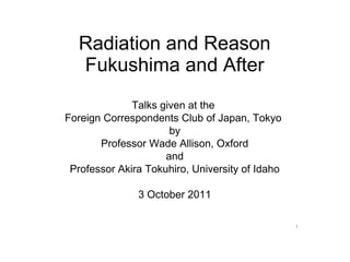
1. Radiation and Reason
Fukushima and After
Talks given at the
Foreign Correspondents Club of Japan, Tokyo
by
Professor Wade Allison, Oxford
and
Professor Akira Tokuhiro, University of Idaho
3 October 2011
1
2. 2006 2009 2011
Wade Allison, Emeritus Professor of
Physics, University of Oxford, UK
Nuclear and medical physicist (no link to the nuclear
industry) http://en.wikipedia.org/wiki/Wade_Allison
Website: http://www.radiationandreason.com
Contact: w.allison@physics.ox.ac.uk
Tokyo, 3 October 2011 slide 2
3. Agenda, points to be explained
1. Low or modest levels of nuclear radiation and radioactivity are not
harmful.
2. Fear of radiation causes personal stress and social damage that is
very harmful.
3. Current food regulations are scientifically unreasonable and cause
hardship, as at Chernobyl.
4. Current evacuation regulations are scientifically unreasonable and
cause hardship, as at Chernobyl.
5. International “safety” levels based on the lowest achievable should be
relaxed upwards by a large factor.
6. Popular clamour in the Cold War era is responsible for this
misunderstanding.
For further detail see http://www.radiationandreason.com
Tokyo, 3 October 2011 slide 3
4. Fear of radiation
Basis:
1. Fear of aftermath of a nuclear holocaust.
?
An effective Cold War message that frightened everybody at the time.
2. You cannot feel nuclear radiation.
But the cells of your body can - and then repair the damage, too.
3. The Regulations warn of radiation dangers.
Misunderstanding here, for which we all share responsibility, in part.
Tokyo, 3 October 2011 slide 4
5. How dangerous is radiation to life? All else follows
What is the effect of radiation on life?
Both data and understanding.
First
Risk assessment.
Public acceptance.
Safety regulations.
Working practices.
Second
Waste. Costs.
Terrorists.
Rogue states.
And finally Dirty bomb threats.
Nuclear blackmail
.
Tokyo, 3 October 2011 slide 5
6. Effect of radiation depends on the dose and the period
Example: for a dose of paracetamol, both the dose and the period
are important.(100 tablets per person at once is fatal, but spread out
regularly over several weeks cures a few headaches.)
For radiation, dose is milli-sievert, mSv, and period, mSv per month.
You can trust radiation doses used in medicine
Today many people benefit from radiation diagnostic scans and some
have radiation therapy for cancer.
A CT scan gives a dose of 5-10 mSv with an external source of
radiation.
PET and SPECT scans give a similar dose from an internal injected
radioactive source.
A screening CT+PET scan gives a whole-body dose of 15mSv.
This radiation and radioactivity, internal and external, are essentially
the same types as that emitted at Fukushima.
Tokyo, 3 October 2011 slide 6
7. Food regulations in error, for example caesium in beef
“Measures against Beef which Exceeds the Provisional Regulation
Values of Radioactive Cesium by the Government to Ensure Safety of
Beef”, issued 27 July 2011
Eating 1 kg of meat with regulation limit of 500Bq/kg gives a dose of
0.008mSv [page 12, section 4. This number has been checked]
Exposure lasts over 4 months while the caesium is excreted
The radioactive caesium dose is evenly spread throughout the body
like the radioactive fluorine in a PET radiation scan which gives 15
mSv all in a couple of hours
Therefore one scan gives the same dose as eating 2000 kg per person
of contaminated meat in 4 months. The Regulation is unreasonable.
After Chernobyl this error was admitted in Norway and Sweden.
The international safety standard (ICRP) underlying such regulations
needs substantial revision.
But 15 mSv is not a dangerous level.
Tokyo, 3 October 2011 slide 7
8. in Norway after Chernobyl
Harbitz, Skuterud and Strand, Norwegian Rad Prot Auth (1998)
Meat at level 6000 Bq per kg and some other food too
- So in Norway they raised the level to factor 12 above the level at Fukushima .
- At this level you can eat 170kg of condemned meat before equivalent to a
CT/PET scan.
- And then the farmers and herders could sell their meat, and nobody suffered.
- Why not at Fukushima?
Tokyo, 3 October 2011 slide 8
10. Real radiation danger levels
Crosses show the mortality of Chernobyl firefighters (curve is for rats).
The numbers show the number who died/total in each dose range.
Above 4,000 mSv 27/42 died from Acute Radiation Syndrome (ARS),
not cancer.
Below 4,000 mSv 1/195 died.
Tokyo, 3 October 2011 slide 10
11. Workers at Chernobyl
- No worker with less than 2000 mSv died from ARS.
Workers at Fukushima
- After six weeks 30 workers had received between 100-250 mSv.
- So there will be no deaths from ARS at Fukushima.
Radiation therapy to cure cancer
- Patients receiving radiotherapy spread over about 6 weeks to cure
cancer get a daily dose of 2000 mSv to the tumour that kills the
cancer cells.
- They also receive daily 1000 mSv to many healthy organs and
tissue that survive -- more than 20,000 mSv per month.
- That is more than 5 X an acute fatal dose (4,000mSv).
- Credible data?
Most people personally know someone who has benefited from such
treatment.
- How? Recovery from radiation damage.
After each daily treatment healthy organs just have time to repair the
radiation damage - and the tumour cells just do not.
Tokyo, 3 October 2011 slide 11
12. Evacuation at Fukushima
- Criterion was set at 20 mSv per year.
- Radiotherapy shows that doses of more than 20,000 mSv per month
are tolerated.
- Radiotherapy equivalent to 1000 years at the evacuation criterion.
This criterion is unreasonable.
- In general, evacuation is at least as traumatic as radiotherapy
treatment.
- The criterion has taken no account of damage to personal and
socio-economic health.
- Radiation safety at the expense of mental health and community well
being is unjustifiable.
Experience from Chernobyl ignored at Fukushima
- The evacuation (and the advice to the population that their health was
threatened by radiation) caused far more damage to public health than
the radiation itself [UN(2011) and IAEA(2006) reports].
- These reports have not been read at Fukushima? Lesson not learnt
and error repeated.
Tokyo, 3 October 2011 slide 12
13. Radiation-induced cancer
There are many overlapping repair methods including immunity.
The immune system may fail (usually with poor health in later years)
resulting in cancer.
Usually it is not possible to distinguish cancers caused by radiation.
Only seen when the lifelong health records of a large population are
compared, those highly irradiated with those not irradiated.
For example, cancer fatalities among the survivors of Hiroshima and
Nagasaki for the period 1950-2000.
The average dose 160 mSv and average cancer risk increased by 1 in 15.
Higher doses show a clear increased risk, but not for those less
than 100 mSv.
Tokyo, 3 October 2011 slide 13
14. Total population 429000 100.00%
What do we know from Known killed or died 1945-1950103000 24.01%
Hiroshima and Nagasaki?
Lost or died 1945-1950 43000 10.02%
Survived to 1950 283000 65.97%
for whom dose known 86955
Died of cancer 1950-2000 32057 7.47%
Died of radiation-induced
cancer 1950-2000 1865 0.44%
Survived Early death
to 1950
and did not
die of Lost
cancer
before 2000
Cancer death 1950-2000
Radiation induced cancer 1950-2000
Tokyo, 3 October 2011 slide 14
15. Solid cancer deaths among Hiroshima and Nagasaki survivors,
1950-2000, separated by dose range (Preston et al., 2004)
Dose range survivor solid cancer survivor deaths1950-2000 extra risk
mSv number actual expected per 1000
less than 5 38507 4270 4282 -2.0 to 1.4
5 to 100 29960 3387 3313 0.0 to 3.5
100 to 200 5949 732 691 3.5 to 12.5
200 to 500 6380 815 736 9 to 18
500 to 1000 3426 483 378 25 to 37
1000 to 2000 1764 326 191 63 to 83
above 2000 625 114 56 72 to 108
all 86611 10127 9647 5.0 to 5.2
“expected” means the number of deaths predicted from those in other cities.
- Doses highlighted have risk compatible with zero, final column.
Tokyo, 3 October 2011 slide 15
16. Why are regulations wrong? Who is to blame?
National regulations are based on advice from the international
committee (ICRP)
ICRP advice is to ignore other risks and to reduce radiation As Low As
Reasonably Achievable (ALARA), close to natural levels. Not for safety
bur for social reasons.
ALARA is what a radiation-phobic world demanded in the Cold War
years. We should correct our mistake.
Safety levels should be As High As Relatively Safe (AHARS), where
“relatively” refers to competing risks.
AHARS levels should take account of recovery from radiation damage
shown by the success of radiotherapy.
What might AHARS safety levels be?
Tokyo, 3 October 2011 slide 16
17. Some monthly doses shown by area with ALARA and AHARS.
Tumour therapy
> 40,000
mSv per month,
death to cell
Healthy tissue
therapy > 20,000
mSv per month,
tolerated dose!
Suggested safe level 100 mSv per month, [conservative by a factor 200]
50 times larger than current evacuation level 2 mSv per month [20 mSv/yr]
ICRP public ALARA level 0.1 mSv per month, [or 1 mSv per yr]
Suggested new safety levels (AHARS): 100 mSv max single dose
100 mSv max in any month
5000 mSv max lifelong
A relaxation by about 1000 times compared to public ALARA, 1 mSv per year .
Tokyo, 3 October 2011 slide 17
18. How many will die from radiation cancer at Fukushima?
It is very unlikely that anyone will die from radiation as a result of
Fukushima, even over the next 50 years. Here is why.....
After six weeks 30 workers had received a radiation dose between 100
and 250 milli-sievert. At Chernobyl no emergency worker who
received less than 2000 milli-sievert died from Acute Radiation
Syndrome, although there were 140 of them.
At Hiroshima and Nagasaki, out of 5949 citizens who received a dose in
this range, 1 in 150 died of radiation-induced cancer in 50 years.
The chance that ANY worker at Fukushima will die of extra cancer is less
than 25%. Doses to the public have been far lower and so without risk.
In Japan seaweed is in the diet and many children received iodine
tablets. Both protect against child thyroid cancer.
At Chernobyl, an iodine-deficient region, 6000 children contracted thyroid
cancer but just 15 died.
No radiation deaths are expected at Fukushima.
Tokyo, 3 October 2011 slide 18
19. Some conclusions
- At Fukushima the mental health, self confidence and livelihood of
hundreds of thousands are put in danger by tight food and evacuation
regulations.
- In fact, at Fukshima as at Chernobyl, appeasing fear of radiation by tight
regulation has had the opposite effect.
- Radiation and radioactivity cure 1000s of cancers a year and are
harmless at low dose.
- Radiation is not a big threat to mankind, unlike geology, climate change,
socio-economic stability, population, water and food supplies.
- Everywhere, new fresh education is needed to explain radiation to more
people in simple terms to remove the stigma.
- Everywhere, we should learn to use nuclear radiation for the benefit of
society with the same care and respect that we already do when it is used
for our personal health.
Tokyo, 3 October 2011 slide 19