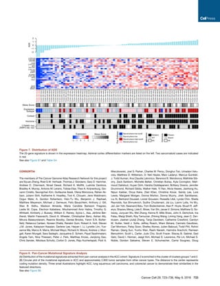
We describe a comprehensive genomic characterization of 91 adrenocortical carcinoma specimens. Analysis identified several new ACC driver genes including PRKAR1A, RPL22, TERF2, CCNE1, and NF1. Genome-wide analysis revealed frequent occurrence of massive DNA loss followed by whole-genome doubling (WGD), associated with aggressive disease. WGD was identified as a hallmark of ACC progression, supported by increased TERT expression, decreased telomere length, and cell-cycle activation. Integrated molecular subtyping identified three ACC subtypes with distinct clinical outcomes and alterations, which could be captured by a 68-CpG DNA-methylation signature for clinical stratification.
Differences in microRNA expression during tumor development in the transition...Enrique Moreno Gonzalez
The prostate is divided into three glandular zones, the peripheral zone (PZ), the transition zone (TZ), and the central zone. Most prostate tumors arise in the peripheral zone (70-75%) and in the transition zone (20-25%) while only 10% arise in the central zone. The aim of this study was to investigate if differences in miRNA expression could be a possible explanation for the difference in propensity of tumors in the zones of the prostate.
The KRAS-Variant and miRNA Expression in RTOG Endometrial Cancer Clinical Tri...UCLA
The KRAS-variant may be a genetic marker of risk for type 2 endometrial cancers. In addition, tumor miRNA expression appears to be associated with patient age, lymphovascular invasion and the KRAS-variant, supporting the hypothesis that altered tumor biology can be measured by miRNA expression, and that the KRAS-variant likely impacts endometrial tumor biology.
Incidence of pneumonia and risk factors among patients with head and neck can...Enrique Moreno Gonzalez
This study investigated the incidence and patient- and treatment-related risk factors related to pneumonia acquired during radiotherapy (PNRT) in head and neck cancer (HNC) patients.
Multicentric and multifocal versus unifocal breast cancer: differences in the...Enrique Moreno Gonzalez
The aim of this study was to evaluate the expression of the cell adhesion-related glycoproteins MUC-1, β-catenin and E-cadherin in multicentric/multifocal breast cancer in comparison to unifocal disease in order to identify potential differences in the biology of these tumor types.
Acute myeloid leukemia (AML) is a hematopoietic malignancy with a dismal outcome in the majority of cases. A detailed understanding of the genetic alterations and gene expression changes that contribute to its pathogenesis is important to improve prognostication, disease monitoring, and therapy. In this context, leukemia-associated misexpression of microRNAs (miRNAs) has been studied, but no coherent picture has emerged yet, thus warranting further investigations.
Clinical and experimental studies regarding the expression and diagnostic val...Enrique Moreno Gonzalez
Carcinoembryonic antigen-related cell adhesion molecule 1 (CEACAM1) is a multifunctional Ig-like cell adhesion molecule that has a wide range of biological functions. According to previous reports, serum CEACAM1 is dysregulated in different malignant tumours and associated with tumour progression. However, the serum CEACAM1 expression in nonsmall-cell lung carcinomas (NSCLC) is unclear. The different expression ratio of CEACAM1-S and CEACAM1-L isoform has seldom been investigated in NSCLC. This research is intended to study the serum CEACAM1 and the ratio of CEACAM1-S/L isoforms in NSCLC.
Thyroid Papillary Carcinoma and Noninvasive Follicular Thyroid Neoplasm with ...CrimsonPublishersGJEM
Papillary thyroid carcinomas (PTC) are well differentiated malignante pithelial tumors with characteristic nuclear features and they originate from epithelial cells of thyroid follicle. Papillary thyroid carcinoma is the most common type of thyroid cancers, which composes 85-90% of all thyroid carcinomas. Number of patients diagnosed with thyroid cancer has significantly increased during the last two decades due to increased awareness of nodular thyroid diseases, developments in diagnostic methods, wide applicability of thyroid fine needle aspiration, new descriptions of histopathology criteria and increased radiation exposure. It is more common in males than females with some ethnic variations. Although it is very rare during early childhood, it is the most common thyroid cancer of this age group. The mean age is 46 years at the time of diagnosis. Tumor has some histologic variants, the most common ones are being classical and follicular variants
https://crimsonpublishers.com/gjem/fulltext/GJEM.000505.php
For more open access journals in Crimson Publishers
Please click on link: https://crimsonpublishers.com
For More Articles on Medical research
Please click on: https://crimsonpublishers.com/gjem/
Overexpression of YAP 1 contributes to progressive features and poor prognosi...Enrique Moreno Gonzalez
Yes-associated protein 1 (YAP 1), the nuclear effector of the Hippo pathway, is a key regulator of organ size and a candidate human oncogene in multiple tumors. However, the expression dynamics of YAP 1 in urothelial carcinoma of the bladder (UCB) and its clinical/prognostic significance are unclear.
hMSH2 Gly322Asp (rs4987188) Single nucleotide polymorphism and the risk of br...Agriculture Journal IJOEAR
Aim: Breast cancer is the most common cancer in women both in the developed and less developed world. The reported study was designed to explore associations between hMSH2 - Gly322Asp (1032G>A, rs4987188) single nucleotide polymorphism (SNP) and the risk of breast carcinoma in the Polish women.
Material and methods: Blood samples were obtained from women with breast cancer (n=225), treated at the Department of Oncological Surgery and Breast Diseases, Polish Mother’s Memorial Hospital – Research Institute between the years 2005 and 2012. A control group included 220 cancer-free women. Genomic DNA was isolated and the SNP Gly322Asp of hMSH2 was determined by High-Resolution Melter method. Odds ratios (ORs) and 95% confidence intervals (CIs) were calculated for each genotype and allele.
Results: This study revealed that single nucleotide polymorphism Gly322Asp of hMSH2 is associated with both breast cancer risk and grading. Moreover, it can be linked with breast carcinoma tumor size and lymph node status. The Asp allele in patients may be a risk factor for breast carcinoma (OR 5.12; 95% CI 3.77 –6.97, p<.0001).
Conclusions: Gly322Asp single nucleotide polymorphism of hMSH2 may be a risk factor of breast cancer in the Polish women.
Recently, a phase II clinical trial in hepatocellular carcinoma (HCC) has suggested that the combination of sorafenib and 5-fluorouracil (5-FU) is feasible and side effects are manageable. However, preclinical experimental data explaining the interaction mechanism(s) are lacking. Our objective is to investigate the anticancer efficacy and mechanism of combined sorafenib and 5-FU therapy in vitro in HCC cell lines MHCC97H and SMMC-7721.
Clinical Genomics for Personalized Cancer Medicine: Recent Advances, Challeng...Yoon Sup Choi
I reviewed recent advances, challenges, and opportunities to implement clinical cancer genomics. Case studies of advanced systems, such as Foundation Medicine, MI-ONCOSEQ are introduced for benchmark. A few fundamental limitations to establish personalized oncology are also discussed.
Cholangiocarcinoma occur in cholangiocytes, either in the intrahepatic, peri-hilar or distal bile duct locations. The three, command distinct attention due to difference in presentation and management. The current 5 years survival for localized intrahepatic cholangiocarcinoma is estimated to be at 24%. With the advent of molecular diagnostics and targeted therapy the field has seen some changes. Surgical resection is still the mainstay of therapy, although transplantation has been proposed in selected patients that have shown disease stability. Localized therapy for intra-hepatic and hilar tumors with neoadjuvant chemotherapy has also been used with some success for smaller tumors.
Objective: The association between telomerase reverse transcriptase (TERT) promoter mutation and outcome of melanoma is unclear and controversial. We aim to conduct a meta-analysis and investigate whether the TERT promoter mutation is a prognostic factor of melanoma.
Study Design: Appropriate studies were searched in 3 databases: PubMed, Web of Science, and Embase. Pooled hazard ratios (HRs) were counted through random effects model.
Results: Heterogeneity was moderate in overall survival (OS) (I2=43.7%, p=0.059) and low in disease-free survival (DFS) (I2=0.0%, p=0.587). Sensitivity analysis indicated that the removal of any of the study did not affect the final results. Evidence for publication bias was not found (Begg’s test, p=0.281; Egger’s test, p=0.078). The pooled OS HRs from combined effects analysis was determined (HR 1.07; 95% CI 0.83–1.39, p=0.585), together with the pooled HRs of DFS (HR 1.65; 95% CI 1.02–2.66, p=0.042). TERT promoter mutation predicted a good outcome in meta-static melanoma patients (HR 0.66; 95% CI 0.46–0.96, p=0.042). The pooled HRs of combined mutation in TERT promoter and BRAF (HR 6.27; 95% CI 2.7–14.58, p=0.000) predicted a bad outcome in melanoma patients.
Conclusion: TERT promoter mutation significantly predicted poor DFS outcome but, on the contrary, predicted a good outcome in metastatic melanoma patients. The combined TERT promoter and BRAF mutation was a significant independent factor of OS in melanoma patients.
Keywords: melanoma; meta-analysis; mutation; prognosis; promoter regions, genetic; skin neoplasms; telomerase; TERT promoter mutation; TERT protein, human
Cord Blood Mesenchymal Stem Cells Conditioned Media Suppress Epithelial Ovari...ijtsrd
Background and objective: Treatment of epithelial ovarian cancer (EOC) is a major challenge with only 30% 5-year survival rate. The outcome of the different therapeutic modalities is still poor, and there is an urgency to find new treatment lines. The effect of mesenchymal stem cells on different tumors is greatly variable. The present work shows the effect of cord blood mesenchymal stem cells conditioned media (MSC CM) in different concentrations on epithelial ovarian cancer stem cells (CD44+ cells) in vitro. Methodology: Ovarian cancer stem cells were subjected to MSC CM of (100%, 75%, 50%, 25%) concentrations for 72 hours followed by investigation of cell morphology, proliferation, apoptosis, cell cycle and expression of certain genes (Oct-4, Sox-2, and Nanog). Results: Cell shrinkage and cell debris was observed with all cancer cell lines by contrast with control. MTT assay showed a reduction in proliferation, in a concentration-dependent manner. The annexin-v results demonstrated a significant early and late apoptosis. There was an increase in the sub-G1 phase of the cell cycles indicating apoptosis. There was a progressive suppression of embryonic stemness genes in all cell lines compared to control. Conclusion: Based on these results, it was concluded that MSC CM may be a potential ovarian cancer inhibitor that may create a new modalities of treatment in ovarian cancer patients. Maher E. Elgaly | Mohamed E. El Ghareeb | Mohamed H. Bedairy | Ahmed M. Badawy | Abeer Shaaban | Farha El shennawy"Cord Blood Mesenchymal Stem Cells Conditioned Media Suppress Epithelial Ovarian Cancer Cells in Vitro" Published in International Journal of Trend in Scientific Research and Development (ijtsrd), ISSN: 2456-6470, Volume-2 | Issue-5 , August 2018, URL: http://www.ijtsrd.com/papers/ijtsrd18182.pdf http://www.ijtsrd.com/other-scientific-research-area/other/18182/cord-blood-mesenchymal-stem-cells-conditioned-media-suppress-epithelial-ovarian-cancer-cells-in-vitro/maher-e-elgaly
A KRAS-variant is a Biomarker of Poor Outcome, Platinum Chemotherapy Resistan...UCLA
Germ-line variants in the 3′ untranslated region (3′UTR) of cancer genes disrupting microRNA (miRNA) regulation have recently been associated with cancer risk. A variant in the 3′UTR of the KRAS oncogene, referred to as the KRAS-variant, is associated with both cancer risk and altered tumor biology. Here we test the hypothesis that the KRAS-variant can act as a biomarker of outcome in epithelial ovarian cancer (EOC), and investigate the cause of altered outcome in KRAS-variant positive EOC patients. As this variant appears to be associated with tumor biology, we additionally test the hypothesis that this variant can be directly targeted to impact cell survival.
EOC patients with complete clinical data were genotyped for the KRAS-variant and analyzed for outcome (n=536), response to neoadjuvant chemotherapy (n=125), and platinum resistance (n=306). Outcome was separately analyzed for women with known BRCA mutations (n=79). Gene expression was analyzed on a subset of tumors with available tissue. Cell lines were employed to confirm altered sensitivity to chemotherapy with the KRAS-variant. The KRAS- variant was directly targeted through siRNA/miRNA oligonucleotides in cell lines and survival was measured.
Post-menopausal EOC patients with the KRAS-variant were significantly more likely to die of ovarian cancer by multivariate analysis (HR=1.67, 95% CI=1.09–2.57, p=0.019, n=279). Possibly explaining this finding, EOC patients with the KRAS-variant were significantly more likely to be platinum resistant (OR=3.18, CI=1.31–7.72, p=0.0106, n=291). Additionally, direct targeting of the KRAS-variant led to a significant reduction in EOC cell growth and survival in vitro.
These findings confirm the importance of the KRAS-variant in EOC, and indicate that the KRAS- variant is a biomarker of poor outcome in EOC likely due to platinum resistance. In addition, this work supports the hypothesis that these tumors have continued dependence on such 3′UTR lesions, and that direct targeting may be a viable future treatment approach.
Breast cancer is the most common cancer among women in different societies. Because life is constructed evolutionary on the gravity of ‘g’, removed gravity as a variable can lead to clarify many biologic questions. Today, the weightlessness is a new method to study cellular changes. Weightlessness leads to metabolic and functional changes of the human body, and studies have shown that weightlessness leads to changes in growth and gene expression in cancer cells. The aim of this study is to evaluate the effect of weightlessness on apoptosis and cellular cycle in breast cancer cells. The tests of Annexin-V, PI-flow cytometer, and MTT have been used here. Cells of the weightlessness group are cultured on Clinostat prepared by the United Nations, and in gravity of 0.001g. The cell death and apoptosis using Annexin kit, and cell cycle using the PI and flow cytometer were investigated. Also, the amount of cell damage was determined by MTT. The apoptosis results showed that weightlessness leads to a reduction of 40% in apoptosis in cell line MCF-7, and the increase in BT-20 cell line for two times. Apoptosis in cell line MDA-MB-468 was not affected, and the results showed that the cell cycle and growth in cell line ZR-75 increased at a rate of five times (35% of weightlessness group versus 7% of control). Cell growth in the other categories showed no significant difference between two groups. Also, no significant difference was observed in the amounts of cell damage in groups of weightlessness and control.
Gene expression mining for predicting survivability of patients in earlystage...ijbbjournal
After numerous breakthroughs in medicine, microbiology, and pathology in the past century, lung cancer
still remains as a leading cause of cancer-related death even in the developed countries. Lung cancer
accounts roughly for 30% of all cancer-related deaths in the world. Diagnosis and treatments are still
based on traditional histopathology. It is of paramount importance to predictthe survivability of patients in
early stages oflung cancer so that specific treatments can be sought. Nonetheless, histopathology has been
shown by previous studies to be inadequate in predicting lung cancerdevelopment and clinical outcome.
The microarray technology allows researchers to examine the expression of thousands of genes
simultaneously. This paper describes a state-of-the-art machine learning based approach called averaged
one-dependence estimators with subsumption resolution to tackle the problem of predictingwhether a
patient in early stages of lung cancer will survive by mining DNA microarray gene expression data. To
lower the computational complexity, we employ an entropy-based geneselection approach to select relevant
genes that are directly responsible for lungcancer survivability prognosis. The proposed system has
achieved an average accuracy of 92.31% in predicting lung cancer survivability over 2 independent
datasets. The experimental results provide confirmation that gene expression mining can be used to predict
survivability of patients in earlystages of lung cancer.
How to deal with complex virus disease problems Senthil Natesan
Group of emerging plant viruses causing economically significant damage to a broad range of field crops, vegetables, ornamentals, fruits, etc.
Viruses can not move by themselves
and they need a “safe” vehicle to spread from plant to plant one such is thrips -Naidu A. Rayapati, Presentation at CPMB, TNAU 19th,August 2009
ANTS - 360 view of your customer - bigdata innovation summit 2016
Dữ liệu lớn, theo nghĩa đen, không phải là một cái gì đó mới, nó đã hiện hữu trong một thời gian dài. Bất kỳ một công ty đã kinh doanh trong một vài năm đều sở hữu một khối lượng dữ liệu từ thông tin khách hàng thông qua hồ sơ giao dịch và sử dụng sản phẩm. Những doanh nghiệp khác nhau sẽ có những khả năng khai thác và sử dụng dữ liệu lớn khác nhau.
Một số doanh nghiệp đã đạt đến độ chín muồi, trong khi đó một số doanh nghiệp khác chỉ vừa bắt đầu cuộc hành trình. Tại Việt Nam các doanh nghiệp viễn thông, các hãng hàng không, các ngân hàng, các tập đoàn bán lẻ, các cơ quan chính phủ đang sở hữu một khối lượng dữ liệu khổng lồ, tuy nhiên, việc xử lý, phân tích dữ liệu lớn còn đang trong giai đoạn rất sơ khai.
Diễn đàn dữ liệu lớn sẽ tập trung chia sẻ những kiến thức thực tế và mang tính chất ứng dụng để người tham dự có thể tích luỹ và áp dụng.
Correlation between vascular endothelial growth factor-A expression and tumor...UniversitasGadjahMada
Vascular endothelial growth factor-A (VEGF-A) has been observed as the predominant angiogenic factor in colorectal cancer (CRC) and the assessment of microvessel density (MVD) has been used to quantify tumor neoangiogenesis. This study aimed to determine clinicopathological and prognostic significance of both angiogenic markers in the local CRC patients. We analyzed tissue samples obtained from 81 cases with CRC. VEGF-A expression and MVD counts were immunohistochemically detected using anti VEGF-A and CD31. The assessments of both markers were classified as low and high. Correlation between VEGF-A expression and MVD value and clinicopathological characteristics were examined using Chi-square test. The overall survival (OS) was plotted using the Kaplan-Meier method. The results indicated a high VEGF-A expression was found more frequently in the rectal location (P=0.042) and T4 tumors (P=0.041) compared to their counterparts. Older patients tended to show a higher MVD value compared to younger cases (P=0.062). In addition, survival analysis showed that males had a worse OS compared to females (P=0.029), and VEGF-A expression and MVD count did not correlate with patients’ survival. In conclusion, there were significant differences of VEGF-A expression according to tumor location and T invasion. Sex, but not angiogenic markers, had an influence on the survival of CRC patients.
Molecular characterization of a patient’s tumor to guide treatment decisions is increasingly being
applied in clinical care and can have a significant impact on disease outcome. These molecular analyses,
including mutation characterization, are typically performed on tissue acquired through a biopsy at diagnosis.
However, tumors are highly heterogeneous and sampling in its entirety is challenging. Furthermore, tumors
evolve over time and can alter their molecular genotype, making clinical decisions based on historical biopsy
data suboptimal. Personalized medicine for cancer patients aims to tailor the best treatment options for the
individual at diagnosis and during treatment. To fully enable personalized medicine it is desirable to have an
easily accessible, minimally invasive way to determine and follow the molecular makeup of a patient’s tumor
longitudinally. One such approach is through a liquid biopsy, where the genetic makeup of the tumor can be
assessed through a bio fluid sample. Liquid biopsies have the potential to help clinicians screen for disease,
stratify patients to the best treatment and monitor treatment response and resistance mechanisms in the tumor. A liquid biopsy can be used for molecular characterization of the tumor and its non-invasive nature
allows repeat sampling to monitor genetic changes over time without the need for a tissue biopsy. This review will summarize three approaches in the liquid biopsy field: circulating tumor cells (CTCs), cell free DNA (cfDNA) and exosomes. We also outline some of the analytical challenges encountered using liquid biopsy techniques to detect rare mutations in a background of wild-type sequences.
Sex-Based Difference in Gene Alterations and Biomarkers in Anal Squamous Cell...semualkaira
anal squamous cell carcinoma (ASCC) is a relatively rare malignancy ac-counting for about 2-3% of all the gastrointestinal tumors. The standard of treatment for localized disease is chemoradiotherapy
Sex-Based Difference in Gene Alterations and Biomarkers in Anal Squamous Cell...semualkaira
anal squamous cell carcinoma (ASCC) is a relatively rare malignancy ac-counting for about 2-3% of all the gastrointestinal tumors. The standard of treatment for localized disease is chemoradiotherapy. Several studies reported a sex disparity
in ASCC prognosis showing a better survival for female compared
to men. Methods: we examined 1,380 patients with ASCC who received comprehensive genomic profiling as part of routine clinical
care and present key
HDAC4 and HDAC7 Promote Breast and Ovarian Cancer Cell Migration by Regulatin...CrimsonpublishersCancer
Breast and ovarian cancer have been remained as a highly malignant tumor among women, posing a serious threat to women health worldwide. In this study, we were aimed to investigate the underlying mechanism of breast and ovarian cancer cell migration. Wound healing assay showed that MDA-MB-231and C13* have higher migration potential compare with MCF-7 and OV2078 cells, as well as regulated epithelial-mesenchymal transition (EMT) marker. We found that HDAC4 and HADC7 mRNA are up regulated in MDA-MB-231 and C13* cells. Moreover, target HDAC4 and HDAC7 by TSA or shRNA block MDA-MB-231and C13* migration. These results reveal a new link between HDACs and EMT in the regulation of breast and ovarian cancer migration.
Presentation by Scott Woodman, MD, PhD. Presented at the 2018 Eyes on a Cure: Patient & Caregiver Symposium, hosted by the Melanoma Research Foundation's CURE OM initiative.
Construction and Validation of Prognostic Signature Model Based on Metastatic...daranisaha
Colorectal Cancer (CRC) is a common malignant cancer with a poor prognosis. Liver metastasis is the dominant cause of death in CRC patients, and it often involves changes in various gene expression profiling. This study proposed to construct and validate a risk model based on differentially expressed genes between the primary and liver metastatic tumors from CRC for prognostic prediction.
1. Article
Comprehensive Pan-Genomic Characterization of
Adrenocortical Carcinoma
Graphical Abstract
Highlights
d Standardized molecular data from 91 cases of adrenocortical
carcinoma
d Driver genes including TP53, ZNFR3, CTNNB1, PRKAR1A,
CCNE1, and TERF2
d Whole-genome doubling event is a marker for ACC
progression
d Three prognostic molecular subtypes captured by a DNA-
methylation signature
Authors
Siyuan Zheng, Andrew D. Cherniack,
Ninad Dewal, ..., Gary D. Hammer,
Thomas J. Giordano,
Roel G.W. Verhaak
Correspondence
giordano@med.umich.edu (T.J.G.),
rverhaak@mdanderson.org (R.G.W.V.)
In Brief
Zheng et al. perform comprehensive
genomic characterization of 91 cases of
adrenocortical carcinoma (ACC). This
analysis expands the list of driver genes
in ACC, reveals whole-genome doubling
as a hallmark of ACC progression, and
identifies three ACC subtypes with
distinct clinical outcome.
Zheng et al., 2016, Cancer Cell 29, 723–736
May 9, 2016 ª2016 Elsevier Inc.
http://dx.doi.org/10.1016/j.ccell.2016.04.002
2. Cancer Cell
Article
Comprehensive Pan-Genomic Characterization
of Adrenocortical Carcinoma
Siyuan Zheng,1 Andrew D. Cherniack,2,3 Ninad Dewal,4 Richard A. Moffitt,5 Ludmila Danilova,6 Bradley A. Murray,2,3,35
Antonio M. Lerario,7,8,9 Tobias Else,8,9 Theo A. Knijnenburg,10 Giovanni Ciriello,11,12 Seungchan Kim,13
Guillaume Assie,14,15,16,17 Olena Morozova,18 Rehan Akbani,1 Juliann Shih,2,3 Katherine A. Hoadley,5 Toni K. Choueiri,3,19
Jens Waldmann,17,20 Ozgur Mete,21 A. Gordon Robertson,22 Hsin-Ta Wu,23 Benjamin J. Raphael,23 Lina Shao,8
(Author list continued on next page)
SUMMARY
We describe a comprehensive genomic characterization of adrenocortical carcinoma (ACC). Using this data-
set, we expand the catalogue of known ACC driver genes to include PRKAR1A, RPL22, TERF2, CCNE1, and
NF1. Genome wide DNA copy-number analysis revealed frequent occurrence of massive DNA loss followed
by whole-genome doubling (WGD), which was associated with aggressive clinical course, suggesting WGD is
a hallmark of disease progression. Corroborating this hypothesis were increased TERT expression,
decreased telomere length, and activation of cell-cycle programs. Integrated subtype analysis identified
three ACC subtypes with distinct clinical outcome and molecular alterations which could be captured by a
68-CpG probe DNA-methylation signature, proposing a strategy for clinical stratification of patients based
on molecular markers.
INTRODUCTION
Adrenocortical carcinoma (ACC) is a rare endocrine malignancy
with an annual incidence of 0.7–2 per million (Bilimoria et al.,
2008; Else et al., 2014a). Current staging schemes such as by
the European Network for the Study of Adrenal Tumors broadly
divides ACCs into four stages (Fassnacht et al., 2009). While
stage I and II tumors are organ confined and potentially curable
by complete resection, advanced stage tumors are invasive
(stage III) and/or metastatic (stage IV), with a dismal 5-year
1Departments of Genomic Medicine, Bioinformatics, and Computational Biology, Endocrine Neoplasia and Hormonal Disorders,
The University of Texas MD Anderson Cancer Center, Houston, TX 77030, USA
2The Eli and Edythe L. Broad Institute of Massachusetts Institute of Technology and Harvard University, Cambridge, MA 02142, USA
3Department of Medical Oncology, Dana-Farber Cancer Institute, Harvard Medical School, Boston, MA 02215, USA
4Human Genome Sequencing Center, Baylor College of Medicine, Houston, TX 77030, USA
5Lineberger Comprehensive Cancer Center, University of North Carolina at Chapel Hill, Chapel Hill, NC 27599, USA
6The Sidney Kimmel Comprehensive Cancer Center at Johns Hopkins University, Baltimore, MD 21287, USA
7Unidade de Suprarrenal, Laborato´ rio de Hormoˆ nios e Gene´ tica Molecular LIM42, Servic¸ o de Endocrinologia e Metabologia,
Hospital das Clı´nicas, Faculdade de Medicina da Universidade de Sa˜ o Paulo, Sa˜ o Paulo 05403-900, Brazil
8Departments of Cell & Developmental Biology, Pathology, Molecular & Integrative Physiology, Internal Medicine, University of Michigan,
Ann Arbor, MI 48109, USA
9University of Michigan Comprehensive Cancer Center, University of Michigan, Ann Arbor, MI 48109, USA
10Institute for Systems Biology, Seattle, WA 98109, USA
11Department of Computational Biology, University of Lausanne, Rue du Bugnon 27, 1005 Lausanne, Switzerland
12Computational Biology Center, Memorial Sloan Kettering Cancer Center, New York, NY 10065, USA
13Translational Genomics Research Institute, Phoenix, AZ 85004, USA
(Affiliations continued on next page)
Significance
Adrenocortical carcinoma (ACC) is a rare endocrine cancer with limited therapeutic options and overall poor outcome. We
comprehensively analyzed 91 ACC specimens from four continents using state-of-the-art genomic technologies and
computational methods. In addition to identification of ACC driver genes, pathways, and refined subtypes, our analysis re-
vealed whole-genome doubling (WGD) as a milestone in disease progression. Our findings suggest that ACC can serve as a
model for how WGD influences disease progression. Our dataset is standardized with other Cancer Genome Atlas (TCGA)
studies, fully available at the TCGA Data Portal, and thus will serve as a valuable research resource.
Cancer Cell 29, 723–736, May 9, 2016 ª 2016 Elsevier Inc. 723
3. survival of 6%–13% for stage IV patients (Else et al., 2014b;
Fassnacht et al., 2009; Fassnacht et al., 2013). Histologic
grading of ACC based on proliferation (Giordano, 2011; Weiss
et al., 1989) refines treatment decisions for specific patient sub-
groups, but is not universally accepted (Miller et al., 2010).
Chemotherapy, radiotherapy, and the adrenolytic agent mito-
tane are the current standard therapeutic modalities for unre-
sectable or metastatic ACC, but all are palliative (Else et al.,
2014a). Our understanding of ACC pathogenesis is incomplete
and additional therapeutic avenues are needed.
Molecular studies have nominated several genes as potential
drivers involved in sporadic adrenocortical tumorigenesis,
including insulin-like growth factor 2 (IGF2), b-catenin (CTNNB1),
and TP53 (Giordano et al., 2003; Tissier et al., 2005). b-Catenin
gain-of-function mutations are evident in approximately 25%
of both benign and malignant sporadic adrenocortical neo-
plasms (Tissier et al., 2005). Recent genomic profiling efforts of
ACC have identified candidate driver genes such as ZNRF3
and TERT, and identified molecular subgroups with variable clin-
ical outcomes (Assie et al., 2014; Pinto et al., 2015). Germline
variants of these genes are also associated with Beckwith-Wie-
demann, familial adenomatous polyposis coli, and Li-Fraumeni
syndromes, in which adrenocortical neoplasia occur.
Comprehensive and integrated genomic characterization of
ACC may identify additional oncogenic alterations, provide a
framework for further research, and guide development of thera-
pies. As one of the rare cancer projects of The Cancer Genome
Atlas (TCGA), we collected the clinical and pathologic features,
genomic alterations, DNA-methylation profiles, and RNA and pro-
teomic signatures of 91 cases of ACC. The comprehensive nature
of the dataset combined with the many easy access routes to the
individualandaggregatedprofileswillserveasapointofreference
for many future ACC and comparative cancer studies. Here, we
report this resource and an integrated analysis of the data.
RESULTS
Patient Cohort, Clinical Annotation, and Analytical
Approach
We analyzed 91 histologically confirmed tumors and matched
blood or normal tissue from a global cohort collected from six
countries including 84 usual type, four oncocytic, two sarcoma-
toid, and one myxoid variant. Median age at diagnosis was
49 years, and female:male ratio was 1.8. While 51 (56%) involved
the left gland and 40 (44%) the right gland, more tumors in the left
gland were diagnosed in males than females (72% versus 47%;
p = 0.03, Fisher’s exact test). The majority of tumors (57%) were
functional; hypercortisolism was the most common endocrinop-
athy. Resected tumors spanned all stages (stage I, n = 9; II,
n = 43; III, n = 19; IV, n = 17; 3 unavailable). Median overall sur-
vival was 78 months with 5-year survival of 59%. Locally invasive
and metastatic tumors (grade III plus IV) had dramatically
Matthew Meyerson,2,3,24 Michael J. Demeure,13,36 Felix Beuschlein,17,25 Anthony J. Gill,26,27 Stan B. Sidhu,26,28
Madson Q. Almeida,7,29 Maria C.B.V. Fragoso,7,29 Leslie M. Cope,6 Electron Kebebew,30 Mouhammed A. Habra,1
Timothy G. Whitsett,13 Kimberly J. Bussey,13,31 William E. Rainey,8,9 Sylvia L. Asa,21 Je´ roˆ me Bertherat,14,15,16,17
Martin Fassnacht,17,32,33 David A. Wheeler,4 The Cancer Genome Atlas Research Network, Gary D. Hammer,8,9,34
Thomas J. Giordano,8,9,34,* and Roel G.W. Verhaak1,34,*
14Inserm U1016, CNRS UMR 8104, Institut Cochin, 75014 Paris, France
15Faculte´ de Me´ decine Paris Descartes, Universite´ Paris Descartes, Sorbonne Paris Cite´ , 75006 Paris, France
16Department of Endocrinology, Referral Center for Rare Adrenal Diseases, Assistance Publique Hoˆ pitaux de Paris, Hoˆ pital Cochin,
75014 Paris, France
17European Network for the Study of Adrenal Tumors, 75014 Paris, France
18University of California Santa Cruz Genomics Institute, University California Santa Cruz, Santa Cruz, CA 95064, USA
19Department of Medicine, Brigham and Women’s Hospital, Boston, MA 02115, USA
20Department of Visceral, Thoracic and Vascular Surgery, University Hospital Giessen and Marburg, Campus Marburg, General Surgery,
Endocrine Center, 34501 Marburg, Germany
21Department of Laboratory Medicine and Pathobiology, University Health Network, Toronto, ON M5G 2C4, Canada
22Canada’s Michael Smith Genome Sciences Centre, BC Cancer Agency, Vancouver, BC V5Z 4S6, Canada
23Department of Computer Science, Brown University, Providence, RI 02906, USA
24Department of Pathology, Harvard Medical School, Boston, MA 02215, USA
25Endocrine Research Unit, Medizinische Klinik und Poliklinik IV, Klinikum der Universita¨ t Mu¨ nchen, 80336 Munich, Germany
26Cancer Diagnosis and Pathology Group and Cancer Genetics Laboratory, Kolling Institute of Medical Research, University of Sydney,
Sydney, NSW 2006, Australia
27Department of Anatomical Pathology, Royal North Shore Hospital, St Leonards, NSW 2065, Australia
28Endocrine Surgical Unit, Royal North Shore Hospital, St Leonards, NSW 2065, Australia
29Instituto do Caˆ ncer do Estado de Sa˜ o Paulo (ICESP), Faculdade de Medicina da Universidade de Sa˜ o Paulo, Sa˜ o Paulo 05403-900, Brazil
30Endocrine Oncology Branch, Center for Cancer Research, National Cancer Institute, National Institutes of Health, Bethesda,
MD 20892, USA
31NantOmics, LLC, The Biodesign Institute, Arizona State University, Tempe, AZ 85287-5001, USA
32Endocrine and Diabetes Unit, Department of Internal Medicine I, University Hospital Wu¨ rzburg, 97080 Wu¨ rzburg, Germany
33Comprehensive Cancer Center Mainfranken, University of Wu¨ rzburg, 97080 Wu¨ rzburg, Germany
34Co-senior author
35Present address: Intellia Therapeutics, Cambridge, MA 02139, USA
36Present address: Ashion Analytics, Phoenix, AZ 85004, USA
*Correspondence: giordano@med.umich.edu (T.J.G.), rverhaak@mdanderson.org (R.G.W.V.)
http://dx.doi.org/10.1016/j.ccell.2016.04.002
724 Cancer Cell 29, 723–736, May 9, 2016
4. reduced median overall survival (18 months), with a 5-year
survival of 22%. Clinicopathologic data are summarized in Table
S1. Patient stage at diagnosis and cortisol hypersecretion were
predictive for both overall survival and disease-free survival
(Else et al., 2014b), but age was only associated with overall
survival. Gender had no clear association with disease stage or
clinical outcome (Table S1).
We generated a comprehensive molecular dataset of the 91
tumors, as follows: whole exome sequence (n = 90), mRNA
sequence (n = 78), miRNA sequence (n = 79), DNA copy number
via SNP arrays (n = 89), DNA methylation via DNA-methylation
arrays (n = 79), and targeted proteome from reverse-phase
protein array (RPPA; n = 45) (Table S1).
Our analytical approach consisted of four main parts. We
began by identifying somatic single-nucleotide variants, gene
fusions, and copy-number alterations. Given the significant
copy-number alterations observed, we performed an extended
bioinformatic analysis that revealed the role of genome doubling
and telomere maintenance in ACC. Using the various molecular
datasets, we derived molecular classifications of ACC and inte-
grated a three-class solution with the somatic genomic land-
scape. We placed ACC in the broader context of cancer by
performing several pan-cancer analyses. Finally, we quantified
adrenal differentiation and tumoral hormone production across
the entire cohort to correlate the genomic results with clinical
1 101 201 301 401 501 601 701 781
CTNNB1
aa
1 101 201 301 393
TAD DBD TMD
Arm Arm Arm Arm
TP53
aa
1 101 201 301 401 501 615 aa
MeninMEN1
1 101 201 301 381
PRKAR1A
aa
Nonsense
Silent
Missense
Splice Site
In-frame indel
Frameshift indel
RIIa cAMP 1 cAMP 2
H
R
D
C
EXOSC10
ENSP00000366135
PI3Kc
ENSP00000354558
MLL
ENSP00000436786 ENST00000524422
ATH
PH
D
SET
A
CB
PP2Ac
PPP1CB
ENSP00000378769
BRE
ENST00000344773
1 101 128
RPL22 Ribosomal_L22e
aa
10-30
10-10
10-3
10-1
0.25
10-30
10-10
10-3
10-1
0.25
TERT (1)
5q35.3 (9)
CDK4 (16)
Xq28 (21)
17q25.3 (3)
BRD4 (35)
CCNE1 (1)
TERF2 (5)
Xp11.22 (5)
ZNRF3 (1)
4q35.1 (6)
LINC00290 (1)
1p36.3 (1)
3q13.31 (1)
CDKN2A (2)
PTPRD (1)
RB1 (2)
PRKAR1A (9)
11p15.5 (12)
RBFOX1 (1)
PARK2 (35)
11q14.1 (6)
17q11.2 (4)
SMAD4 (1)
2
4
6
8
10
12
14
16
18
20
22
1
3
5
7
9
11
13
15
17
19
21
X
Chromosomes
MTOR
ATP5L
Figure 1. Mutational Driver Genes in ACC
(A) Protein domain structure of the five significantly
mutated genes with somatic mutations aligned.
Functional domains and mutation types are indi-
cated in different colors and shapes as shown
in the legend. Arm, Armadillo domain; TAD, tran-
scription-activation domain; DBD, DNA-binding
domain; TMD, tetramerization domain; RIIa, reg-
ulatory subunit of type II PKA R subunit.
(B) Sporadic gene fusions that involve cancer
genes. All exons are represented, with red and blue
indicating high and low expression, respectively.
Lineslinkingtwoexonsindicatethefusionpositions.
Protein domains are related to the exons below
the gene diagram. HRDC, helicase and RNase
D C-terminal; PI3Kc, phosphoinositide 3-kinase,
catalytic domain; ATH, AT-hook motif; PHD, plant
homology domain; SET, Su(var)3–9, enhancer-of-
zeste, trithorax; PP2Ac, protein phosphatase 2A
homologs, catalytic domain.
(C) Focal recurrent amplifications and deletions
in ACCs with the number of genes spanned by
the peak in parentheses. Red and blue indicate
amplification and deletion, respectively. The x axis
at the bottom and top of the figure represents
significance of amplification/deletion per q value.
See also Figure S1 and Table S1.
parameters. All unprocessed data used
in the analysis can be accessed through
the TCGA portals.
Exome and RNA Sequencing
Nominate ACC Driver Events
Somatic single-nucleotide variants and
small indels were detected using five in-
dependent mutation callers, and variants called by at least three
callers or independently validated by RNA sequencing were
included (Figures S1A and S1B). This approach yielded a total
of 8,814 high-confidence mutations (6,664 non-synonymous
and 2,150 synonymous); 3,427 of these were found in two tu-
mors with ultramutator phenotype. Deep-coverage resequenc-
ing (1,5003) achieved a validation rate of 95% (Supplemental
Experimental Procedures and Table S1). The median somatic
mutation density was 0.9 per Mb, similar to pancreatic adeno-
carcinoma but more than twice that of another endocrine tumor,
papillary thyroid cancer (Figure S1C). Similar to other cancers,
ACC demonstrated a marked heterogeneity in mutation density
(range: 0.2–14.0 mutations/Mb, excluding the two ultramuta-
tors). Mutation density was correlated with numerous clin-
icopathologic parameters including overall survival, time to
recurrence, necrosis, stage, Weiss score, and mitotic count
(Figure S1D). Interestingly, these associations remained when
restricting to organ-confined stage I and II diseases.
We used MutSigCV (Lawrence et al., 2013) to identify five
significantly mutated genes (SMGs): TP53, CTNNB1, MEN1,
PRKAR1A, and RPL22. The mutation frequencies ranged from
3.3% to 17.8% of the cohort (Figures 1A and S1E). Mutations
of TP53 and CTNNB1 in ACC are well recognized (Tissier et al.,
2005). As expected, missense mutations in CTNNB1 were
confined to exon 3 (Figure 1A). Six (7%) tumors harbored
Cancer Cell 29, 723–736, May 9, 2016 725
5. inactivating mutations in MEN1, consistent with prior studies
implicating MEN1 in ACC (Assie et al., 2014). While our cohort
is the largest to be sequenced to date, a much larger number
of samples is needed to identify all candidate cancer genes
(Lawrence et al., 2014). To overcome the limitation of sample
size, we compared the mutated genes with the Cancer Gene
Census (Futreal et al., 2004). This approach identified two cancer
genes mutated in more than 5% of the cohort, NF1 and MLL4, in
addition to those nominated by MutSigCV.
Inourcohort,seven(8%)casesharboredinactivatingmutations
in the protein kinase cAMP-dependent regulatory type I alpha
gene (PRKAR1A) (Figure 1A). A homozygous deletion was found
in three additional cases. While inactivating germline PRKAR1A
mutations cause Carney complex and benign primary pigmented
nodular adrenocortical disease (Kirschner et al., 2000), malignant
transformation has been reported in the adrenals of patients with
this rare condition (Anselmo et al., 2012), and sporadic loss-of-
function mutations in PRKAR1A have been found in adrenocor-
tical adenomas and rare carcinomas (Bertherat et al., 2003). Inter-
estingly, DNA sequencing of sporadic adrenocortical adenomas
recently revealed a recurrent activating L206R mutation in the
catalytic subunit of the cAMP-dependent protein kinase A (PKA)
(PRKACA) (Beuschleinetal., 2014;Gohetal., 2014).Thismutation
results in constitutive PKA activity by disrupting the interaction
between PRKACA and the regulatory subunits of PKA including
PRKAR1A (Calebiro et al., 2014; Goh et al., 2014). While we found
no PRKACA mutations in our cohort, we observed decreased
PRKAR1A expression and increased MEK and BRAF protein
expression (Figures S1F and S1G) in mutant cases, suggesting
a potential role for inhibition of the RAF-MEK-ERK cascade in
treatment of some ACCs.
We observed two frameshift mutations in ribosomal protein
L22 (RPL22) and confirmed them by RNA sequencing (Figure 1A).
We detected a third in-frame deletion mutation in a sample with
heterozygous loss of RPL22, and homozygous loss of RPL22 in
three ACCs. These findings suggest a role for somatic alteration
of RPL22 in 7% of ACC, which has previously been related to
MDM2-mediated p53 ubiquitination and degradation (Zhang
and Lu, 2009).
Using two complementary analytical methods to detect fusion
transcripts in mRNA-sequencing data (McPherson et al., 2011;
Torres-Garcia et al., 2014), we identified 156 singleton but no
recurrent gene-fusion events in 48 of 78 (62%) tumors (range:
1–16) (Figure S1H and Table S1). Gene fusions occur at much
lower frequencies than mutations and copy-number variants
(Yoshihara et al., 2015), and a larger cohort is needed to deter-
mine the frequency of fusions reported here. However, we did
identify private in-frame fusions involving known cancer genes
(Figure 1B). A highly expressed EXOSC10-MTOR fusion retained
the mTOR catalytic domain and resulted in elevated levels of
total and phosphorylated mTOR protein in this tumor (Figure S1I).
The fusion point of a MLL-ATP5L fusion fell within the MLL break-
point cluster region associated with acute myeloid leukemia
(Krivtsov and Armstrong, 2007). Both fusion cases lacked muta-
tions in SMGs. A fusion involving the gene BRE, reported to pro-
mote tumor cell growth and adrenal neoplasia (Chan et al., 2005;
Miao et al., 2001), was identified in one tumor. While more data
are required to ascertain their roles in ACC, these private fusions
may represent functional transcripts.
Whole-Genome Doubling Is a Common Event in ACC
We assessed somatic copy-number alterations and loss of het-
erozygosity (LOH) in 89 tumors. Using GISTIC2 (Mermel et al.,
2011), we identified recurrent focal amplifications of TERT
(5p15.33), TERF2 (16q22.1), CDK4 (12q14.1), and CCNE1
(19q12) and deletions of RB1 (13q14.2), CDKN2A (9p21.2), and
ZNRF3 (22q12.1) (q % 0.01; Figure 1C and Table S1). A focal
deletion peak around 4q34.3-4q35.1 centered on a long noncod-
ing RNA LINC00290, which has been reported as a deletion
target in pediatric ACCs and other cancers (Letouze et al.,
2012; Zack et al., 2013). ZNRF3 homozygous deletions ap-
peared in 16% (n = 14) of tumors assayed; by including non-
silent mutations, 19.3% of ACCs harbored alterations in this
gene. TERT and TERF2, two telomere-maintenance-related
genes (Blasco, 2005), were focally amplified in 15% and 7%
of cases, respectively. TERT promoter hotspot mutations have
been recently discovered in human cancers (Huang et al.,
2013). We resequenced the TERT promoter region of all 91
tumors. In agreement with a recent study (Liu et al., 2014), we
identified four cases with the C228T mutation, but no C250T
mutations.
Arm-level copy-number changes were frequent in ACC (Fig-
ures 2A and S2A). Clustering of 89 tumors based on their arm-
level alterations produced three groups with striking differences:
chromosomal (n = 54; 61%); noisy (n = 27; 30%); and quiet (n = 8;
9%) (Figure S2A). The chromosomal group showed the highest
frequency of whole-chromosome arm gains and losses. The
noisy group was characterized by a significantly higher number
of chromosomal breaks as well as frequent loss of 1p with 1q
intact. Tumors in the quiet group had few large copy-number
alterations. Kaplan-Meier analysis demonstrated a significant
decrease in survival in the noisy group relative to the chromo-
somal and quiet subtypes, suggesting that this copy-number
phenotype is characteristic of aggressive disease (Figure S2A).
To validate these subtypes, we analyzed an independent dataset
of ACC tumors (n = 119) profiled on different versions of the
Illumina BeadArray platform (Assie et al., 2014). Based on the
chromosome 1p/1q pattern, the noisy group and its survival
association were recovered in this independent cohort (Fig-
ure S2B), suggesting that the group distinctions are robust. We
did not observe copy-number-quiet ACC in the independent
cohort.
We next determined tumor purity, ploidy, and whole-genome
doubling (WGD) by integrating allelic copy-number profiles and
DNA mutation data using the previously validated ABSOLUTE al-
gorithm (Carter et al., 2012). ACC samples were pure relative to
other tumor types (tumor purity 0.82 ± 0.15; Figure 2B). Consis-
tently, fractions of tumor infiltrated stromal cells estimated from
gene expression signatures (Yoshihara et al., 2013) were low
relative to a panel of 14 other cancer types (Figure S2C). Immune
scores were lower in cortisol-secreting ACCs (p = 0.015), consis-
tent with the suppression of T cell activity by glucocorticoids
(Palacios and Sugawara, 1982). We found that hypodiploid
karyotypes (ploidy %1.6) occurred more frequently in ACC
than in 11 other tumor types (31% versus 1%; Figure 2B), a
frequency that was only matched by chromophobe renal cell
carcinoma. WGD occurred in 68% of the noisy subtype, 51%
of the chromosomal subtype, and none of the diploid-quiet sub-
type cases. We inferred the temporal order of somatic mutations
726 Cancer Cell 29, 723–736, May 9, 2016
6. for genes implicated in ACC based on variant allele fractions
(VAF) and genotype (Figures 2C and S2D). Mutations in TP53
(n = 7), MEN1 (n = 3), RPL22 (n = 2), and ZNRF3 (n = 2) were
predicted to have occurred prior to WGD. Only 4/9 CTNNB1 mu-
tations were predicted to be pre-doubling, while three were post-
doubling and the remaining two showed further reduced VAFs
suggestive of subclonality. PRKAR1A mutations split evenly as
having occurred before WGD, after WGD or being subclonal.
We observed that LOH patterns were nearly identical between
hypodiploid and hyperdiploid cases in the chromosomal group
(rho = 0.96, p < 10À5
) (Figures S3A and S3B). We hypothesized
that the undoubled chromosomal ACCs were precursors to the
chromosomal ACC that underwent WGD. This model is corrob-
orated by the difference in outcome and absolute copy number
(Figures 3A, S3C, and S3D). We did not find a survival difference
between non-WGD and WGD noisy samples suggesting that
additional factors may contribute to disease course or that we
were underpowered to detect a statistically significant signal.
Mutation density further corroborated the WGD hypothesis as
the higher mutation frequency in WGD cases was eliminated
after normalization by ploidy (Figure S3E).
We employed gene-set-enrichment analysis using gene
expression data to uncover differences between WGD and
non-WGD tumors. We identified significantly enhanced path-
ways in WGD tumors including telomere regulation, cell-cycle
regulation, and DNA replication repair (Figures S3F and S3G).
The identification of cell-cycle regulation was verified by an inde-
pendent algorithm, Evaluation of Dependency DifferentialitY
(Jung and Kim, 2014), which detected enrichment of the PARKIN
pathway (Figure S3F) including PARK2, a recently reported
master regulator of G1/S cyclins (Gong et al., 2014).
Inthetelomereregulationpathway,TERTexpressionwassignif-
icantly higher in the WGD group (false discovery rate [FDR] = 0.05)
(Figure 3B). Given the role of telomerase in maintaining telomere
length (Blasco, 2005), we used a spill-over sequence generated
by exome sequencing to infer the telomere length of tumors and
normal samples (Ding et al., 2014). Most tumors (73%) exhibited
shorter telomeres than their matched normal samples (Figure 3C).
WGD cases harbored shorter telomeres than non-WGD cases
(FigureS3H).TheassociationbetweenWGDandTERTexpression
may suggest that increased TERT was required as a compensa-
tory mechanism for telomere maintenance. Alternatively, TERT
may have been non-functional with eroding telomeres as a conse-
quence. This confirms previous observations that the majority of
ACCs show telomerase activity, particularly in those with relatively
short telomeres, while only a minority uses alternative telomere
lengthening (ALT) as a telomere-maintenance mechanism (Else
et al., 2008). Telomere crisis is thought to drive tetraploidization
A
B
C
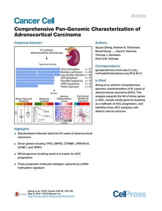
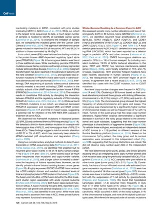
Figure 2. Landscape of DNA Copy-Number Alteration in ACC
(A) Three major copy-number patterns in ACC. Unsupervised clustering divided the cohort (n = 89) into quiet, chromosomal and noisy subtypes.
(B) Pan-cancer purity and ploidy including ACC. Sample sizes are indicated on top. Average tumor purity is plotted as a gray line for each cancer type. The
percentages of whole-genome doubling and hypodiploidy (ploidy %1.6) are listed in red and blue, respectively. LUAD, lung adenocarcinoma; LUSC, lung
squamous; HNSC, head and neck; KIRC, clear cell renal cell; BRCA, breast; BLCA, bladder; CRC, colorectal; THCA, thyroid papillary; UCEC, endometrial; GBM,
glioblastoma; OV, ovarian; KICH, kidney chromophobe.
(C) Purity-adjusted variant allele fraction in genome-doubled and -undoubled tumors. Only tumors with high purity (R0.8) and mutation density less than 5 were
included. Cutoffs labeled in the figure are recognized as turning points in the density distributions of variant allele fractions.
See also Figure S2.
Cancer Cell 29, 723–736, May 9, 2016 727
7. (Davoli and de Lange, 2012) and may be directly associated with
WGD events in these tumors. Similar to pediatric ACC and other
cancers (Heaphy et al., 2011; Pinto et al., 2015), mutations in
ATRX and DAXX (n = 7) were associated with longer telomeres
(p = 5.4 3 10À5
, Fisher’s exact test; Figure 3C). We did not find sig-
nificant associationsbetween telomere lengthand TERTpromoter
mutations or TERT amplification. Most cancers express TERT
in the context of decreased telomere length (Maser and DePinho,
2002), suggesting that increased TERT maintains telomere
homeostasis to a level that is sufficient for cancer cells to survive
crisis. TERF2 was amplified in 7% of the cohort but similarly did
notcorrelatewithtelomerelength.TERF2servesasananchorpro-
tein of the shelterin complex which binds to telomeric DNA, but
its direct involvement in telomere elongation has not been deter-
mined (Blasco, 2005). It has been documented that TERF2 might
have telomere-independent functions (Stewart and Weinberg,
2006), raising interesting hypotheses about its role in adrenal
tumorigenesis.
Molecular Classes of ACC Are Captured by DNA-
Methylation Signatures
To derive a robust molecular classification, we used unsupervised
clustering to analyze the genomic and transcriptomic datasets
(Figures S4 and S2A). This approach yielded four mRNA-expres-
sion groups (Figure S4A-B), six microRNA-expression groups
A B
C
Figure 3. Comparison of WGD and Non-
WGD ACCs
(A) Event-free survival of copy-number subtypes
and whole-genome doubling groups. p value rep-
resents the statistical significance of event-free
survival differences between the five groups.
(B) TERT expression in genome-undoubled
and -doubled tumors. Boxplot shows median and
interquartile range of TERT expressions, with
whiskers extending to extreme values within 1.5
interquartile ranges from the upper and lower
quartiles. Each dot corresponds to a tumor.
(C) Telomere length was estimated using off-
target exome-sequencing data corrected for
tumor purity and ploidy. Top panel shows TERT
expression with ‘‘x’’ representing missing values.
TERT promoter C228T mutation (black), amplifi-
cation (red), and TERF2 amplification (red) are
noted in the second panel. The bottom panel
represents ATRX and DAXX mutations in light blue
and dark red, respectively.
See also Figure S3.
(Figures S4C–S4K), three DNA-methyl-
ation groups (Figure S4L), three copy-
number groups (Figure S2A), and three
protein-expression groups (Figures S4M
and S4N). Except for microRNA-based
clustering, the individual cluster analyses
all resulted in classifications with signifi-
cant differences in outcome. These sub-
types are summarized in Table S2, and
the characteristics of each molecular clas-
sificationaredescribedindetailintheSup-
plemental Information. We integrated the
ACC subsets identified across the DNA copy-number, DNA-
methylation, mRNA-expression, and miRNA-expression plat-
forms through a Cluster of Cluster (CoC) analysis (Figure 4).
Combined, the molecular classifications converged into three
CoC subtypes (Figures 4A and S4O). Transcriptome clustering
in a non-overlapping ACC sample set has previously identified
an aggressive C1A subtype and an indolent C1B subtype (de Rey-
nies et al., 2009; Giordano et al., 2009). A comparison between
CoC and C1A/C1B showed that the majority of CoC I were classi-
fied as C1B, while most CoC II and CoC III were predicted as C1A
(Figure 4A). Pan-cancer pathway-enrichment analyses showed
significant up-regulation of genes in immune-mediated pathways
in CoC I tumors and mitotic pathways in CoC III tumors (Fig-
ure S4P). We compared the clinical outcome of the three CoC
clusters. Disease progression rates of the three CoCs were 7%,
56%, and 96%, respectively. Survival analysis showed a dismal
median event-free survival of 8 months for CoC III (Figure 4B),
while median event-free survival time was not reached in CoC I.
CoC II was more heterogeneous in outcome with an event-free
survival of 38 months. Stage III/IV tumors represented 25%,
47%, and 52% of CoC I, II, and III, respectively, and stage I/II
cases in CoC III showed MKI67 values in accordance with their
grade classification (Figure 5A).
While the CoC analysis showed that molecular data can deter-
mine outcome with high significance, implementing four parallel
728 Cancer Cell 29, 723–736, May 9, 2016
8. profiling platforms poses a clinical challenge. The four expres-
sion subtypes and three methylation subtypes rendered discrim-
inative representations of each CoC group (Figure 4A), offering
plausible routes for clinical implementation. Given the poten-
tial of the methylation platform to provide accurate data on
formalin-fixed, paraffin-embedded tumor samples (de Ruijter
et al., 2015; Thirlwell et al., 2010), we derived a methylation
signature consisting of 68 probes that were included in both Illu-
mina Human Methylation 450K and 27K arrays and tested its
performance. This signature robustly classified our cohort into
three ACC survival groups with 92.4% accuracy (Table S2). Clas-
sification of 51 methylation profiles from an independent cohort
(Assie et al., 2014) accurately validated the prognostic power of
the methylation signature (Figure S4Q).
Integrated Genomic Landscape of ACC
We generated a genomic landscape of ACC by further integrating
multimodal mutational and epigenetic data with the CoC three-
class solution (Figure 5A). In addition to DNA copy number and
mutations, we assessed epigenetic silencing and germline muta-
tions of 177 manually selected genes (Table S3). We found 9
germline mutations, including two TP53R337H
in two Brazilian
patients, two MSH6, one MSH2 and one NF1R1534X
mutations.
The MSH6 and MSH2 mutations support recent observations
that ACC is a Lynch syndrome-associated cancer (Raymond
et al., 2013). The two TP53R337H
mutated patients were younger
than the rest of the cohort (23 and 30; median age of cohort:
49). We also found a single TINF2S245Y
variant that is associated
with dyskeratosis congenita. The likely benign variant APCE1317Q
was observed in two cases (Rozek et al., 2006).
Collectively the genes altered most frequently by somatic mu-
tations, DNA copy-number alterations, and epigenetic silencing
were TP53 (21%), ZNRF3 (19%), CDKN2A (15%), CTNNB1
(16%), TERT (14%), and PRKAR1A (11%). The majority of
gene alterations were either mutation or copy-number changes,
except for CDKN2A, which was targeted by both deletion and
epigenetic silencing through promoter DNA methylation. Alter-
ations of ZNRF3, CTNNB1, APC, and MEN1 resulted in modifica-
tion of the Wnt/b-catenin pathway in 41% of cases (Figure 5B).
Wnt-pathway activity, as measured by expression of canonical
Wnt target genes, was increased in ACC with Wnt-pathway
alterations relative to Wnt wild-type samples (Figure S5A). So-
matic alterations in TP53, CDKN2A, RB1, CDK4, and CCNE1
emphasize the importance of the p53 apoptosis/Rb1 cell-cycle
pathway, and were altered in 44.9% of the cases (Figure 5B).
Finally, histone modification genes (MLL, MLL2, and MLL4)
and chromatin remodeling genes (ATRX and DAXX) were collec-
tively altered in 22% of cases, suggesting a role for epigenetic
deregulation in ACC tumorigenesis (Figure 5C). Unsupervised
HotNet2 analysis of the protein-protein interaction network iden-
tified four significant subnetworks containing at least four genes
(Figure S5B, p < 0.0001) (Leiserson et al., 2015). In addition to
p53, Rb-cell-cycle, and Wnt-alteration subnetworks, HotNet2
showed a PKA network affecting 16 samples in total.
Combining somatic mutations, copy-number alterations, and
epigenetic modification, we found at least one alteration of po-
tential driver genes in 69% of tumors (Figure 5A). The majority
of CoC I tumors did not harbor a driver alteration that we could
detect. We also examined the expression of IGF2 and an estab-
lished proliferation marker, MKI67. MKI67 expression was lower
in CoC I tumors, consistent with the indolent phenotype of this
subtype. IGF2 expression was unanimously high independent
of ACC classification. Expression of IGF2 was relatively high in
67 of 78 (86%) ACCs (cutoff log2(RSEM) = 14 determined by
expression distribution), with no association to either promoter
DNA methylation or genome rearrangement.
Based on existing clinical trials and FDA-approved drugs for
cancers, we found 51 potentially actionable alterations, including
both mutations and copy-number alterations, in 22 ACCs using
precision heuristics for interpreting the actionable landscape
(Van Allen et al., 2014), from cyclin-dependent kinases to
DNA repair protein poly ADP-ribose polymerase (Figure S5C
and Table S3).
Pan-Cancer Analyses Provide Context to ACC
Pan-cancer analysis of genomic data has provided insights on a
wide range of cancer-related questions. In an attempt to under-
stand the driving mechanisms of ACC, we grouped our cohort
on the basis of recurrent cancer-driving alterations using
OncoSign (Ciriello et al., 2013). Our results broadly recapitulated
the C class (OSC1-3, copy-number driven) and M class (OSC
4–5, mutation driven) that have been described as a general
trend across cancers (Ciriello et al., 2013) (Figure S6A).
The accumulation of somatic mutations in cancer is caused by
mutational processes that can be deconvoluted as mutational
signatures (Alexandrov et al., 2013). To examine mutational pro-
cesses in ACC, we extracted six mutational signatures from
85 ACCs (mutation number R10) and about 2,900 other can-
cers using non-negative matrix factorization (Figure S6B). We
compared the six signatures with the 22 signatures from an
A B Figure 4. Cluster of Clusters
(A) CoCs from four platforms (DNA copy number,
black; mRNA expression, red; DNA methylation,
blue; miRNA expression, purple) divided the cohort
into three groups. Presence or absence of mem-
bership for each sample is represented by black or
light blue bars, respectively. Sample parameters
are aligned on top of the heatmap. White bars
indicate data not available.
(B) Event-free survival of the three CoC groups.
Pairwise log-rank test p values are shown.
See also Figure S4 and Table S2.
Cancer Cell 29, 723–736, May 9, 2016 729
9. independent study (Alexandrov et al., 2013). Signature 1 resem-
bled the age- and DNA-mismatch repair-deficiency signatures,
all featuring C > T substitution in the CG context. Signature 2
resembled the smoking signature featuring C > A substitution
in the CG context. Signature 5 resembled UV and APOBEC
signatures featuring C > G, C > T, and C > T substitutions in
the contexts of TC, CC, and TC, respectively. We did not find
an association between signatures and sample country of origin
(p = 0.9, Fisher’s exact test). The majority of ACCs exhibited sig-
natures 1, 2 and 4 (Figure 6A). Signature 1 captured the majority
of gastrointestinal cancers (stomach, esophageal, colorectal)
but also four ACC with a relatively high mutation frequency,
all of which harbored mutations in the DNA-mismatch repair
pathway (Figure 6B). One case with a germline MSH6 muta-
tion but relatively modest mutation density (0.51 mutations/Mb)
also clustered with this signature. The smoking-associated
A
B C
Figure 5. Genomic Landscape of ACC
(A) The ensemble of mutations, copy-number alterations, methylations, subtypes, and clinicopathologic parameters.
(B) Aggregated alterations of the p53/Rb pathway and the Wnt pathways. Activating and deactivating alterations are indicated in red and blue, respectively.
(C) Mutations in epigenetic regulatory genes.
See also Figure S5 and Table S3.
730 Cancer Cell 29, 723–736, May 9, 2016
10. adenocarcinoma and squamous cell lung cancers in signature 2
also featured four high mutation frequency ACCs. Smoking has
been listed as an ACC risk factor (Hsing et al., 1996). A set of
human papillomavirus (HPV)-driven cancers, including cervical,
bladder, and head and neck cancers, clustered together but
did not capture any ACC. We analyzed RNA and exome-
sequencing data for microbial sequence reads (Chu et al.,
2014) and identified human herpes virus sequences in nine
exomes but not HPV sequence reads. De novo assembly of
herpes virus sequences confirmed their presence but returned
no evidence for viral genomic integration (Figure S6C).
Molecular Correlates of ACC Pathology and
Adrenocortical Differentiation
We evaluated adrenocortical differentiation by using 25 genes
with very high expression levels in the adult adrenal cortex and
that are of importance for adrenal function, including steroido-
genic enzymes, cholesterol transporters, and their transcriptional
regulator, SF1 (NR5A1) (Figure S7A and Table S4). We derived a
single metric, termed Adrenocortical Differentiation Score (ADS),
to measure adrenocortical differentiation (Figure 7). Sorting the
tumors according to their ADS values we observed that functional
tumors showed higher ADS values, which did not correlate with
Weiss histopathology score (p = 0.41, ANOVA). TP53 mutations
were present across the spectrum of ADS values (p = 0.56,
Fisher’s exact test), whereas Wnt-related mutations appeared
to be enriched among tumors with higher ADS values (p =
0.0091, Fisher’s exact test). Not surprisingly, the two sarcoma-
toid tumors had the lowest ADS values with very low expression
of NR5A1 and steroidogenic enzymes (Figure S7B). One such
patient had elevated serum cortisol levels, suggesting a mixed
histology in which a differentiated component expressed
steroidogenic enzymes and an undifferentiated component did
not. The other tumor was histologically mixed with separate com-
ponents of usual and sarcomatoid ACC. We suggest that these
sarcomatoid, NR5A1-negative tumors do indeed represent
dedifferentiated ACCs, rather than retroperitoneal sarcomas.
DISCUSSION
As part of TCGA, we present an integrated molecular character-
ization of a large cohort of ACC. The tumors were derived from
four continents and thus represent a near-global sampling of
this disease. The data presented here represent a resource for
future investigations to facilitate ACC research across myriad
avenues, including via pan-cancer analysis.
Our study builds on prior efforts on adult ACC from European
(Assie et al., 2014) and North American patients (Juhlin et al.,
2015), as well as pediatric ACC patients from North America
and Brazil (Pinto et al., 2015). Using high-quality multidimen-
sional genomic data, we confirm many alterations as essential
for ACC development and progression but also expand the so-
matic genetic landscape of ACC to nearly double the known
ACC driver genes. The high frequency of PRKAR1A mutations
expands the role of PKA signaling in ACC and is consistent
with PRKACA somatic mutations being the founder lesion of
benign adrenal tumor associated with endocrinopathies such
as Cushing syndrome (Beuschlein et al., 2014). By integrating
data from multiple platforms, we demonstrated the critical role
of WGD. While WGD is common across cancer (Zack et al.,
2013), an association between WGD and patient outcome has
only been reported in ovarian cancer (Carter et al., 2012). The
exceptional high level of tumor purity and the clear evidence
that WGD is a marker of tumor progression make ACC a model
disease for better understanding of the mechanisms that result
in doubling and the molecular context needed to sustain it.
Weiss et al. (1989) first proposed that ACC consists of two
pathologic classes that have different mitotic rates and distinct
clinical outcomes. This proliferation-based two-grade (low
and high) system has been confirmed by several transcriptome
studies (de Reynies et al., 2009; Giordano et al., 2009) and
has been clinically implemented by some centers (Giordano,
2011). We confirmed the two-grade classification with mRNA-
sequencing data and, importantly, used multidimensional data
to extend the molecular classification to three classes that
have markedly distinct biological properties and significantly
different patient outcomes. Clinical implementation of this
three-class grading system using DNA-methylation profiles
could facilitate improved patient care, although additional trans-
lational efforts are needed for its implementation. Despite these
uncertainties, our results represent an extensive ensemble of
molecular ACC subtypes and thus are likely to be useful in direct-
ing the future development of clinically applicable classifiers.
Moreover, our results illustrate how molecular data, combined
with traditional clinicopathologic assessment, might inform ther-
apeutic decisions and lead to advances in patient outcomes. The
diversity of genomic alterations, especially the significant copy-
number changes seen in the majority of ACC, suggests that
combined inhibition of disease pathways, however challenging,
likely holds the key to successful targeted therapy for ACC.
EXPERIMENTAL PROCEDURES
Tumor and normal samples were obtained from patients after informed consent
and with approval from local Institutional Review Boards (IRB). Details on all
contributingcenters and their IRB approval canbe found in theSupplemental In-
formation. DNA, RNA, and protein were purified and distributed throughout the
TCGA network. In total, 91 primary tumors with associated clinicopathologic
data were assayed on at least one molecular-profiling platform. Platforms
included exome sequencing, mRNA sequencing, miRNA sequencing, SNP ar-
rays, DNA-methylation arrays, and reverse-phase protein arrays. Mutation
calling was performed by five independent callers, and a voting mechanism
was used to generate the final mutation set. MutSigCV (version 1.4) was used
to determine significantly mutated genes (Lawrence et al., 2013). GISTIC2.0
was used to identify recurrent deletion and amplification peaks (Mermel et al.,
2011). Consensus clustering was used to derive miRNA, mRNA, methylation,
and protein subtypes. Tumor purity, ploidy, and WGD were determined
by ABSOLUTE (Carter et al., 2012). The data and analysis results can be
explored through the TCGA Data Portal (https://tcga-data.nci.nih.gov/
tcga/tcgaCancerDetails.jsp?diseaseType=ACC), the Broad Institute GDAC
FireBrowse portal (http://firebrowse.org/?cohort=ACC), Memorial Sloan Ketter-
ing Cancer Center cBioPortal (http://www.cbioportal.org/public-portal/study.
do?cancer_study_id=acc_tcga), and Regulome Explorer (http://explorer.
cancerregulome.org/all_pairs/?dataset=TCGA_ACC). See also the Supple-
mental Experimental Procedures and the ACC publication page (https://
tcga-data.nci.nih.gov/docs/publications/acc_2016/).
SUPPLEMENTAL INFORMATION
Supplemental Information includes Supplemental Experimental Procedures,
seven figures, and four tables and can be found with this article online at
http://dx.doi.org/10.1016/j.ccell.2016.04.002.
Cancer Cell 29, 723–736, May 9, 2016 731
12. CONSORTIA
The members of The Cancer Genome Atlas Research Network for this project
are Siyuan Zheng, Roel G.W. Verhaak, Thomas J. Giordano, Gary D. Hammer,
Andrew D. Cherniack, Ninad Dewal, Richard A. Moffitt, Ludmila Danilova,
Bradley A. Murray, Antonio M. Lerario, Tobias Else, Theo A. Knijnenburg, Gio-
vanni Ciriello, Seungchan Kim, Guillaume Assie´ , Olena Morozova, Rehan Ak-
bani, Juliann Shih, Katherine A. Hoadley, Toni K. Choueiri, Jens Waldmann,
Ozgur Mete, A. Gordon Robertson, Hsin-Tu Wu, Benjamin J. Raphael,
Matthew Meyerson, Michael J. Demeure, Felix Beuschlein, Anthony J. Gill,
Stan B. Sidhu, Madson Almeida, Maria Candida Barisson Fragoso,
Leslie M. Cope, Electron Kebebew, Mouhammed Amir Habra, Timothy G.
Whitsett, Kimberly J. Bussey, William E. Rainey, Sylvia L. Asa, Je´ roˆ me Ber-
therat, Martin Fassnacht, David A. Wheeler, Christopher Benz, Adrian Ally,
Miruna Balasundaram, Reanne Bowlby, Denise Brooks, Yaron S.N. Butter-
field, Rebecca Carlsen, Noreen Dhalla, Ranabir Guin, Robert A. Holt, Steven
J.M. Jones, Katayoon Kasaian, Darlene Lee, Haiyan I. Li, Lynette Lim, Yus-
sanne Ma, Marco A. Marra, Michael Mayo, Richard A. Moore, Andrew J. Mun-
gall, Karen Mungall, Sara Sadeghi, Jacqueline E. Schein, Payal Sipahimalani,
Angela Tam, Nina Thiessen, Peter J. Park, Matthias Kroiss, Jianjiong Gao,
Chris Sander, Nikolaus Schultz, Corbin D. Jones, Raju Kucherlapati, Piotr A.
Mieczkowski, Joel S. Parker, Charles M. Perou, Donghui Tan, Umadevi Velu-
volu, Matthew D. Wilkerson, D. Neil Hayes, Marc Ladanyi, Marcus Quinkler,
J. Todd Auman, Ana Claudia Latronico, Berenice B. Mendonca, Mathilde Sib-
ony, Zack Sanborn, Michelle Bellair, Christian Buhay, Kyle Covington, Mah-
moud Dahdouli, Huyen Dinh, Harsha Doddapaneni, Brittany Downs, Jennifer
Drummond, Richard Gibbs, Walker Hale, Yi Han, Alicia Hawes, Jianhong Hu,
Nipun Kakkar, Divya Kalra, Ziad Khan, Christine Kovar, Sandy Lee, Lora
Lewis, Margaret Morgan, Donna Morton, Donna Muzny, Jireh Santibanez,
Liu Xi, Bertrand Dousset, Lionel Groussin, Rossella Libe´ , Lynda Chin, Sheila
Reynolds, Ilya Shmulevich, Sudha Chudamani, Jia Liu, Laxmi Lolla, Ye Wu,
Jen Jen Yeh, Saianand Balu, Tom Bodenheimer, Alan P. Hoyle, Stuart R. Jeff-
erys, Shaowu Meng, Lisle E. Mose, Yan Shi, Janae V. Simons, Matthew G. So-
loway, Junyuan Wu, Wei Zhang, Kenna R. Mills Shaw, John A. Demchok, Ina
Felau, Margi Sheth, Roy Tarnuzzer, Zhining Wang, Liming Yang, Jean C. Zen-
klusen, Jiashan (Julia) Zhang, Tanja Davidsen, Catherine Crawford, Carolyn
M. Hutter, Heidi J. Sofia, Jeffrey Roach, Wiam Bshara, Carmelo Gaudioso,
Carl Morrison, Patsy Soon, Shelley Alonso, Julien Baboud, Todd Pihl, Rohini
Raman, Qiang Sun, Yunhu Wan, Rashi Naresh, Harindra Arachchi, Rameen
Beroukhim, Scott L. Carter, Juok Cho, Scott Frazer, Stacey B. Gabriel, Gad
Getz, David I. Heiman, Jaegil Kim, Michael S. Lawrence, Pei Lin, Michael S.
Noble, Gordon Saksena, Steven E. Schumacher, Carrie Sougnez, Doug
Figure 6. Pan-Cancer Mutational Signature Analysis
(A) Distribution of the mutational signatures extracted from pan-cancer analysis in the ACC cohort. Signature 2 is enriched in the cluster of clusters groups 1 and 2.
(B) Circular plot of the mutational signatures in ACC and approximately 2,900 tumor samples from other cancer types. The distance to the center represents
coding mutation density. Three small illustrations highlight ACC, lung squamous cell carcinoma, and colorectal cancer to demonstrate their similarities in the
featured directions.
See also Figure S6.
−2
−1
0
1
2
TCGA−OR−A5JB
TCGA−OR−A5J8
TCGA−OR−A5JO
TCGA−OR−A5LK
TCGA−PK−A5HA
TCGA−OR−A5LT
TCGA−OR−A5JD
TCGA−OR−A5JT
TCGA−OR−A5J5
TCGA−OR−A5JQ
TCGA−OR−A5JV
TCGA−OR−A5KZ
TCGA−PK−A5H9
TCGA−OR−A5J2
TCGA−PA−A5YG
TCGA−OR−A5JI
TCGA−OR−A5LJ
TCGA−OR−A5LP
TCGA−OR−A5JJ
TCGA−OR−A5L9
TCGA−OR−A5K4
TCGA−OR−A5L6
TCGA−OR−A5LO
TCGA−OR−A5JZ
TCGA−OR−A5KX
TCGA−PK−A5HB
TCGA−OR−A5LN
TCGA−OR−A5LD
TCGA−P6−A5OF
TCGA−OR−A5K3
TCGA−OR−A5LR
TCGA−OR−A5K1
TCGA−OR−A5KU
TCGA−OR−A5JR
TCGA−OR−A5J1
TCGA−OR−A5JA
TCGA−OR−A5LA
TCGA−OR−A5J9
TCGA−OR−A5JK
TCGA−OR−A5L5
TCGA−OR−A5JY
TCGA−OU−A5PI
TCGA−OR−A5KY
TCGA−OR−A5K8
TCGA−OR−A5LM
TCGA−OR−A5JX
TCGA−OR−A5KO
TCGA−OR−A5KW
TCGA−PK−A5H8
TCGA−OR−A5LS
TCGA−OR−A5L8
TCGA−OR−A5LH
TCGA−OR−A5LE
TCGA−OR−A5K5
TCGA−OR−A5JG
TCGA−OR−A5KV
TCGA−OR−A5JL
TCGA−OR−A5JP
TCGA−OR−A5LB
TCGA−OR−A5LC
TCGA−OR−A5KT
TCGA−OR−A5JC
TCGA−OR−A5K0
TCGA−OR−A5L4
TCGA−OR−A5JM
TCGA−OR−A5JF
TCGA−OR−A5JW
TCGA−OR−A5J7
TCGA−OR−A5K2
TCGA−OR−A5J6
TCGA−OR−A5JE
TCGA−OR−A5LL
TCGA−OR−A5J3
TCGA−OR−A5JS
TCGA−OR−A5LG
TCGA−OR−A5K6
TCGA−OR−A5K9
TCGA−OR−A5L3
NR5A1
MC2R
DLK1
FDX1
INHA
CYP11B2
SCARB1
SOAT1
FDXR
CYP11A1
SULT2A1
NOV
CYP17A1
STAR
HSD3B2
CYP11B1
CYP21A2
2 9
Weiss Score
ExpSubtype
Steroid-pheno-low+prolif
Steroid-pheno-low
Steroid-pheno-high+prolif
Steroid-pheno-low
Expression
−2 −1 0 1 2
Alterations
Mutation
WT
Hormone
Present
Absent
ADS
NANA
Cortisol
Weiss Score
Expression
Subtype
Hormone
CTNNB1 mut
ZNRF3 del, mut
Figure 7. Distribution of ADS
The 25-gene signature is shown in the expression heatmap. Adrenal cortex differentiation markers are listed on the left. Two sarcomatoid cases are indicated
in red.
See also Figure S7 and Table S4.
Cancer Cell 29, 723–736, May 9, 2016 733
13. Voet, Hailei Zhang, Jay Bowen, Sara Coppens, Julie M. Gastier-Foster, Mark
Gerken, Carmen Helsel, Kristen M. Leraas, Tara M. Lichtenberg, Nilsa C.
Ramirez, Lisa Wise, Erik Zmuda, Stephen Baylin, James G. Herman, Janine
LoBello, Aprill Watanabe, David Haussler, Amie Radenbaugh, Arjun Rao,
Jingchun Zhu, Detlef K. Bartsch, Silviu Sbiera, Bruno Allolio, Timo Deutsch-
bein, Cristina Ronchi, Victoria M. Raymond, Michelle Vinco, Lina Shao, Linda
Amble, Moiz S. Bootwalla, Phillip H. Lai, David J. Van Den Berg, Daniel J. Wei-
senberger, Bruce Robinson, Zhenlin Ju, Hoon Kim, Shiyun Ling, Wenbin Liu,
Yiling Lu, Gordon B. Mills, Kanishka Sircar, Qianghu Wang, Kosuke Yoshi-
hara, Peter W. Laird, Yu Fan, Wenyi Wang, Eve Shinbrot, Martin Reincke,
John N. Weinstein, Sam Meier, and Timothy Defreitas.
AUTHOR CONTRIBUTIONS
The TCGA consortium contributed collectively to this study. Project activities
were coordinated by the National Cancer Institute and National Human
Genome Research Institute Project Teams. Initial guidance in the project
design was provided by the Disease Working Group. We also acknowledge
the following TCGA investigators of the Analysis Working Group, who contrib-
uted substantially to the analysis and writing of this manuscript: project
leaders: R.G.W.V., T.J.G., and G.D.H.; data coordinator: S.Z.; manuscript
coordinator: S.Z., T.J.G., and R.G.W.V.; analysis coordinator: S.Z.; writing
team: S.Z., G.D.H, T.J.G., and R.G.W.V. DNA sequence analysis: N.D.,
D.A.W., S.Z., and R.G.W.V.; mRNA analysis: R.A.M., K.A.H., S.Z., and O.M.;
miRNA analysis: G.A.R.; DNA copy-number analysis: A.D.C., B.A.M., S.Z.,
J.S., and R.G.W.V.; DNA-methylation analysis: L.D. and L.M.C.; pathway
analysis: S.K., T.A.K., B.J.R., S.Z., and T.G.W.; clinical data pathology and
disease expertise: T.J.G., G.D.H., T.E., A.M.L., M.F., J.B., W.E.R., K.J.B.,
and S.L.A.
ACKNOWLEDGMENTS
We are grateful to all the patients and families who contributed to this study, to
Ina Felau, Margi Sheth, and Jiashan (Julia) Zhang for project management.
Supported by the following grants from the United States NIH:
5U24CA143799, 5U24CA143835, 5U24CA143840, 5U24CA143843,
5U24CA143845, 5U24CA143848, 5U24CA143858, 5U24CA143866,
5U24CA143867, 5U24CA143882, 5U24CA143883, 5U24CA144025,
U54HG003067, U54HG003079, and U54HG003273, P30CA16672. G.D.H.
has equity interest in, is a consultant for, and scientific advisory board director
of Millendo Therapeutics. R.B. is a consultant for and received grant funding
from Novartis. J.S., B.A.M., A.D.C., and M.M. received grant support from
Bayer. S.L.A. is a member of the Medical Advisory Board of Leica Aperio.
D.J.W. is a consultant for Zymo Research Corporation.
Received: August 11, 2015
Revised: December 8, 2015
Accepted: April 5, 2016
Published: May 9, 2016
REFERENCES
Alexandrov, L.B., Nik-Zainal, S., Wedge, D.C., Aparicio, S.A., Behjati, S.,
Biankin, A.V., Bignell, G.R., Bolli, N., Borg, A., Borresen-Dale, A.L., et al.
(2013). Signatures of mutational processes in human cancer. Nature 500,
415–421.
Anselmo, J., Medeiros, S., Carneiro, V., Greene, E., Levy, I., Nesterova, M.,
Lyssikatos, C., Horvath, A., Carney, J.A., and Stratakis, C.A. (2012). A large
family with Carney complex caused by the S147G PRKAR1A mutation shows
a unique spectrum of disease including adrenocortical cancer. J. Clin.
Endocrinol. Metab. 97, 351–359.
Assie, G., Letouze, E., Fassnacht, M., Jouinot, A., Luscap, W., Barreau, O.,
Omeiri, H., Rodriguez, S., Perlemoine, K., Rene-Corail, F., et al. (2014).
Integrated genomic characterization of adrenocortical carcinoma. Nat.
Genet. 46, 607–612.
Bertherat, J., Groussin, L., Sandrini, F., Matyakhina, L., Bei, T., Stergiopoulos,
S., Papageorgiou, T., Bourdeau, I., Kirschner, L.S., Vincent-Dejean, C., et al.
(2003). Molecular and functional analysis of PRKAR1A and its locus
(17q22-24) in sporadic adrenocortical tumors: 17q losses, somatic muta-
tions, and protein kinase A expression and activity. Cancer Res. 63, 5308–
5319.
Beuschlein, F., Fassnacht, M., Assie, G., Calebiro, D., Stratakis, C.A.,
Osswald, A., Ronchi, C.L., Wieland, T., Sbiera, S., Faucz, F.R., et al. (2014).
Constitutive activation of PKA catalytic subunit in adrenal Cushing’s syn-
drome. N. Engl. J. Med. 370, 1019–1028.
Bilimoria, K.Y., Shen, W.T., Elaraj, D., Bentrem, D.J., Winchester, D.J.,
Kebebew, E., and Sturgeon, C. (2008). Adrenocortical carcinoma in the
United States: treatment utilization and prognostic factors. Cancer 113,
3130–3136.
Blasco, M.A. (2005). Telomeres and human disease: ageing, cancer and
beyond. Nat. Rev. Genet. 6, 611–622.
Calebiro, D., Hannawacker, A., Lyga, S., Bathon, K., Zabel, U., Ronchi, C.,
Beuschlein, F., Reincke, M., Lorenz, K., Allolio, B., et al. (2014). PKA catalytic
subunit mutations in adrenocortical Cushing’s adenoma impair association
with the regulatory subunit. Nat. Commun. 5, 5680.
Carter, S.L., Cibulskis, K., Helman, E., McKenna, A., Shen, H., Zack, T., Laird,
P.W., Onofrio, R.C., Winckler, W., Weir, B.A., et al. (2012). Absolute quantifi-
cation of somatic DNA alterations in human cancer. Nat. Biotechnol. 30,
413–421.
Chan, B.C., Li, Q., Chow, S.K., Ching, A.K., Liew, C.T., Lim, P.L., Lee, K.K.,
Chan, J.Y., and Chui, Y.L. (2005). BRE enhances in vivo growth of tumor cells.
Biochem. Biophys. Res. Commun. 326, 268–273.
Chu, J., Sadeghi, S., Raymond, A., Jackman, S.D., Nip, K.M., Mar, R.,
Mohamadi, H., Butterfield, Y.S., Robertson, A.G., and Birol, I. (2014).
BioBloom tools: fast, accurate and memory-efficient host species sequence
screening using bloom filters. Bioinformatics 30, 3402–3404.
Ciriello, G., Miller, M.L., Aksoy, B.A., Senbabaoglu, Y., Schultz, N., and
Sander, C. (2013). Emerging landscape of oncogenic signatures across human
cancers. Nat. Genet. 45, 1127–1133.
Davoli, T., and de Lange, T. (2012). Telomere-driven tetraploidization occurs in
human cells undergoing crisis and promotes transformation of mouse cells.
Cancer Cell 21, 765–776.
de Reynies, A., Assie, G., Rickman, D.S., Tissier, F., Groussin, L., Rene-Corail,
F., Dousset, B., Bertagna, X., Clauser, E., and Bertherat, J. (2009). Gene
expression profiling reveals a new classification of adrenocortical tumors
and identifies molecular predictors of malignancy and survival. J. Clin.
Oncol. 27, 1108–1115.
de Ruijter, T.C., de Hoon, J.P., Slaats, J., de Vries, B., Janssen, M.J., van
Wezel, T., Aarts, M.J., van Engeland, M., Tjan-Heijnen, V.C., Van Neste, L.,
and Veeck, J. (2015). Formalin-fixed, paraffin-embedded (FFPE) tissue epige-
nomics using Infinium HumanMethylation450 BeadChip assays. Lab. Invest.
95, 833–842.
Ding, Z., Mangino, M., Aviv, A., Spector, T., and Durbin, R.; Consortium, U. K
(2014). Estimating telomere length from whole genome sequence data.
Nucleic Acids Res. 42, e75.
Else, T., Giordano, T.J., and Hammer, G.D. (2008). Evaluation of telomere
length maintenance mechanisms in adrenocortical carcinoma. J. Clin.
Endocrinol. Metab. 93, 1442–1449.
Else, T., Kim, A.C., Sabolch, A., Raymond, V.M., Kandathil, A., Caoili, E.M.,
Jolly, S., Miller, B.S., Giordano, T.J., and Hammer, G.D. (2014a).
Adrenocortical carcinoma. Endocr. Rev. 35, 282–326.
Else, T., Williams, A.R., Sabolch, A., Jolly, S., Miller, B.S., and Hammer, G.D.
(2014b). Adjuvant therapies and patient and tumor characteristics associated
with survival of adult patients with adrenocortical carcinoma. J. Clin.
Endocrinol. Metab. 99, 455–461.
Fassnacht, M., Johanssen, S., Quinkler, M., Bucsky, P., Willenberg, H.S.,
Beuschlein, F., Terzolo, M., Mueller, H.H., Hahner, S., Allolio, B., et al.
(2009). Limited prognostic value of the 2004 International Union against
Cancer staging classification for adrenocortical carcinoma: proposal for a
Revised TNM Classification. Cancer 115, 243–250.
734 Cancer Cell 29, 723–736, May 9, 2016
14. Fassnacht, M., Kroiss, M., and Allolio, B. (2013). Update in adrenocortical
carcinoma. J. Clin. Endocrinol. Metab. 98, 4551–4564.
Futreal, P.A., Coin, L., Marshall, M., Down, T., Hubbard, T., Wooster, R.,
Rahman, N., and Stratton, M.R. (2004). A census of human cancer genes.
Nat. Rev. Cancer 4, 177–183.
Giordano, T.J. (2011). The argument for mitotic rate-based grading for
the prognostication of adrenocortical carcinoma. Am. J. Surg. Pathol. 35,
471–473.
Giordano, T.J., Thomas, D.G., Kuick, R., Lizyness, M., Misek, D.E., Smith, A.L.,
Sanders, D., Aljundi, R.T., Gauger, P.G., Thompson, N.W., et al. (2003).
Distinct transcriptional profiles of adrenocortical tumors uncovered by DNA
microarray analysis. Am. J. Pathol. 162, 521–531.
Giordano, T.J., Kuick, R., Else, T., Gauger, P.G., Vinco, M., Bauersfeld, J.,
Sanders, D., Thomas, D.G., Doherty, G., and Hammer, G. (2009). Molecular
classification and prognostication of adrenocortical tumors by transcriptome
profiling. Clin. Cancer Res. 15, 668–676.
Goh, G., Scholl, U.I., Healy, J.M., Choi, M., Prasad, M.L., Nelson-Williams, C.,
Kunstman, J.W., Korah, R., Suttorp, A.C., Dietrich, D., et al. (2014). Recurrent
activating mutation in PRKACA in cortisol-producing adrenal tumors. Nat.
Genet. 46, 613–617.
Gong, Y., Zack, T.I., Morris, L.G., Lin, K., Hukkelhoven, E., Raheja, R., Tan, I.L.,
Turcan, S., Veeriah, S., Meng, S., et al. (2014). Pan-cancer genetic analysis
identifies PARK2 as a master regulator of G1/S cyclins. Nat. Genet. 46,
588–594.
Heaphy, C.M., de Wilde, R.F., Jiao, Y., Klein, A.P., Edil, B.H., Shi, C.,
Bettegowda, C., Rodriguez, F.J., Eberhart, C.G., Hebbar, S., et al. (2011).
Altered telomeres in tumors with ATRX and DAXX mutations. Science
333, 425.
Hsing, A.W., Nam, J.M., Co Chien, H.T., McLaughlin, J.K., and Fraumeni, J.F.,
Jr. (1996). Risk factors for adrenal cancer: an exploratory study. Int. J. Cancer
65, 432–436.
Huang, F.W., Hodis, E., Xu, M.J., Kryukov, G.V., Chin, L., and Garraway, L.A.
(2013). Highly recurrent TERT promoter mutations in human melanoma.
Science 339, 957–959.
Juhlin, C.C., Goh, G., Healy, J.M., Fonseca, A.L., Scholl, U.I., Stenman, A.,
Kunstman, J.W., Brown, T.C., Overton, J.D., Mane, S.M., et al. (2015).
Whole-exome sequencing characterizes the landscape of somatic mutations
and copy number alterations in adrenocortical carcinoma. J. Clin. Endocrinol.
Metab. 100, E493–E502.
Jung, S., and Kim, S. (2014). EDDY: a novel statistical gene set test method to
detect differential genetic dependencies. Nucleic Acids Res. 42, e60.
Kirschner, L.S., Sandrini, F., Monbo, J., Lin, J.P., Carney, J.A., and Stratakis,
C.A. (2000). Genetic heterogeneity and spectrum of mutations of the
PRKAR1A gene in patients with the carney complex. Hum. Mol. Genet. 9,
3037–3046.
Krivtsov, A.V., and Armstrong, S.A. (2007). MLL translocations, histone
modifications and leukaemia stem-cell development. Nat. Rev. Cancer 7,
823–833.
Lawrence, M.S., Stojanov, P., Polak, P., Kryukov, G.V., Cibulskis, K.,
Sivachenko, A., Carter, S.L., Stewart, C., Mermel, C.H., Roberts, S.A., et al.
(2013). Mutational heterogeneity in cancer and the search for new cancer-
associated genes. Nature 499, 214–218.
Lawrence, M.S., Stojanov, P., Mermel, C.H., Robinson, J.T., Garraway, L.A.,
Golub, T.R., Meyerson, M., Gabriel, S.B., Lander, E.S., and Getz, G. (2014).
Discovery and saturation analysis of cancer genes across 21 tumour types.
Nature 505, 495–501.
Leiserson, M.D., Vandin, F., Wu, H.T., Dobson, J.R., Eldridge, J.V., Thomas,
J.L., Papoutsaki, A., Kim, Y., Niu, B., McLellan, M., et al. (2015). Pan-cancer
network analysis identifies combinations of rare somatic mutations across
pathways and protein complexes. Nat. Genet. 47, 106–114.
Letouze, E., Rosati, R., Komechen, H., Doghman, M., Marisa, L., Fluck, C., de
Krijger, R.R., van Noesel, M.M., Mas, J.C., Pianovski, M.A., et al. (2012). SNP
array profiling of childhood adrenocortical tumors reveals distinct pathways of
tumorigenesis and highlights candidate driver genes. J. Clin. Endocrinol.
Metab. 97, E1284–E1293.
Liu, T.T., Brown, T.C., Juhlin, C.C., Andreasson, A., Wang, N., Backdahl, M.,
Healy, J.M., Prasad, M.L., Korah, R., Carling, T., et al. (2014). The activating
TERT promoter mutation C228T is recurrent in subsets of adrenal tumors.
Endocr. Relat. Cancer 21, 427–434.
Maser, R.S., and DePinho, R.A. (2002). Connecting chromosomes, crisis, and
cancer. Science 297, 565–569.
McPherson, A., Hormozdiari, F., Zayed, A., Giuliany, R., Ha, G., Sun, M.G.,
Griffith, M., Heravi Moussavi, A., Senz, J., Melnyk, N., et al. (2011). deFuse:
an algorithm for gene fusion discovery in tumor RNA-Seq data. PLoS
Comput. Biol. 7, e1001138.
Mermel, C.H., Schumacher, S.E., Hill, B., Meyerson, M.L., Beroukhim, R., and
Getz, G. (2011). GISTIC2.0 facilitates sensitive and confident localization of the
targets of focal somatic copy-number alteration in human cancers. Genome
Biol. 12, R41.
Miao, J., Panesar, N.S., Chan, K.T., Lai, F.M., Xia, N., Wang, Y., Johnson, P.J.,
and Chan, J.Y. (2001). Differential expression of a stress-modulating gene,
BRE, in the adrenal gland, in adrenal neoplasia, and in abnormal adrenal tis-
sues. J. Histochem. Cytochem. 49, 491–500.
Miller, B.S., Gauger, P.G., Hammer, G.D., Giordano, T.J., and Doherty, G.M.
(2010). Proposal for modification of the ENSAT staging system for adre-
nocortical carcinoma using tumor grade. Langenbecks Arch. Surg. 395,
955–961.
Palacios, R., and Sugawara, I. (1982). Hydrocortisone abrogates prolifer-
ation of T cells in autologous mixed lymphocyte reaction by rendering
the interleukin-2 Producer T cells unresponsive to interleukin-1 and un-
able to synthesize the T-cell growth factor. Scand. J. Immunol. 15,
25–31.
Pinto, E.M., Chen, X., Easton, J., Finkelstein, D., Liu, Z., Pounds, S.,
Rodriguez-Galindo, C., Lund, T.C., Mardis, E.R., Wilson, R.K., et al. (2015).
Genomic landscape of paediatric adrenocortical tumours. Nat. Commun. 6,
6302.
Raymond, V.M., Everett, J.N., Furtado, L.V., Gustafson, S.L., Jungbluth, C.R.,
Gruber, S.B., Hammer, G.D., Stoffel, E.M., Greenson, J.K., Giordano, T.J., and
Else, T. (2013). Adrenocortical carcinoma is a lynch syndrome-associated
cancer. J. Clin. Oncol. 31, 3012–3018.
Rozek, L.S., Rennert, G., and Gruber, S.B. (2006). APC E1317Q is not associ-
ated with Colorectal Cancer in a population-based case-control study in
Northern Israel. Cancer Epidemiol. Biomarkers Prev. 15, 2325–2327.
Stewart, S.A., and Weinberg, R.A. (2006). Telomeres: cancer to human aging.
Annu. Rev. Cell Dev. Biol. 22, 531–557.
Thirlwell, C., Eymard, M., Feber, A., Teschendorff, A., Pearce, K., Lechner,
M., Widschwendter, M., and Beck, S. (2010). Genome-wide DNA methyl-
ation analysis of archival formalin-fixed paraffin-embedded tissue using
the Illumina Infinium HumanMethylation27 BeadChip. Methods 52,
248–254.
Tissier, F., Cavard, C., Groussin, L., Perlemoine, K., Fumey, G., Hagnere, A.M.,
Rene-Corail, F., Jullian, E., Gicquel, C., Bertagna, X., et al. (2005). Mutations of
beta-catenin in adrenocortical tumors: activation of the Wnt signaling pathway
is a frequent event in both benign and malignant adrenocortical tumors.
Cancer Res. 65, 7622–7627.
Torres-Garcia, W., Zheng, S., Sivachenko, A., Vegesna, R., Wang, Q., Yao, R.,
Berger, M.F., Weinstein, J.N., Getz, G., and Verhaak, R.G. (2014). PRADA:
pipeline for RNA sequencing data analysis. Bioinformatics 30, 2224–2226.
Van Allen, E.M., Wagle, N., Stojanov, P., Perrin, D.L., Cibulskis, K., Marlow, S.,
Jane-Valbuena, J., Friedrich, D.C., Kryukov, G., Carter, S.L., et al. (2014).
Whole-exome sequencing and clinical interpretation of formalin-fixed,
paraffin-embedded tumor samples to guide precision cancer medicine. Nat.
Med. 20, 682–688.
Weiss, L.M., Medeiros, L.J., and Vickery, A.L., Jr. (1989). Pathologic features
of prognostic significance in adrenocortical carcinoma. Am. J. Surg. Pathol.
13, 202–206.
Cancer Cell 29, 723–736, May 9, 2016 735
15. Yoshihara, K., Shahmoradgoli, M., Martinez, E., Vegesna, R., Kim, H., Torres-
Garcia, W., Trevino, V., Shen, H., Laird, P.W., Levine, D.A., et al. (2013).
Inferring tumour purity and stromal and immune cell admixture from expres-
sion data. Nat. Commun. 4, 2612.
Yoshihara, K., Wang, Q., Torres-Garcia, W., Zheng, S., Vegesna, R., Kim, H.,
and Verhaak, R.G. (2015). The landscape and therapeutic relevance of can-
cer-associated transcript fusions. Oncogene 34, 4845–4854.
Zack, T.I., Schumacher, S.E., Carter, S.L., Cherniack, A.D., Saksena, G.,
Tabak, B., Lawrence, M.S., Zhang, C.Z., Wala, J., Mermel, C.H., et al.
(2013). Pan-cancer patterns of somatic copy number alteration. Nat. Genet.
45, 1134–1140.
Zhang, Y., and Lu, H. (2009). Signaling to p53: ribosomal proteins find their
way. Cancer Cell 16, 369–377.
736 Cancer Cell 29, 723–736, May 9, 2016