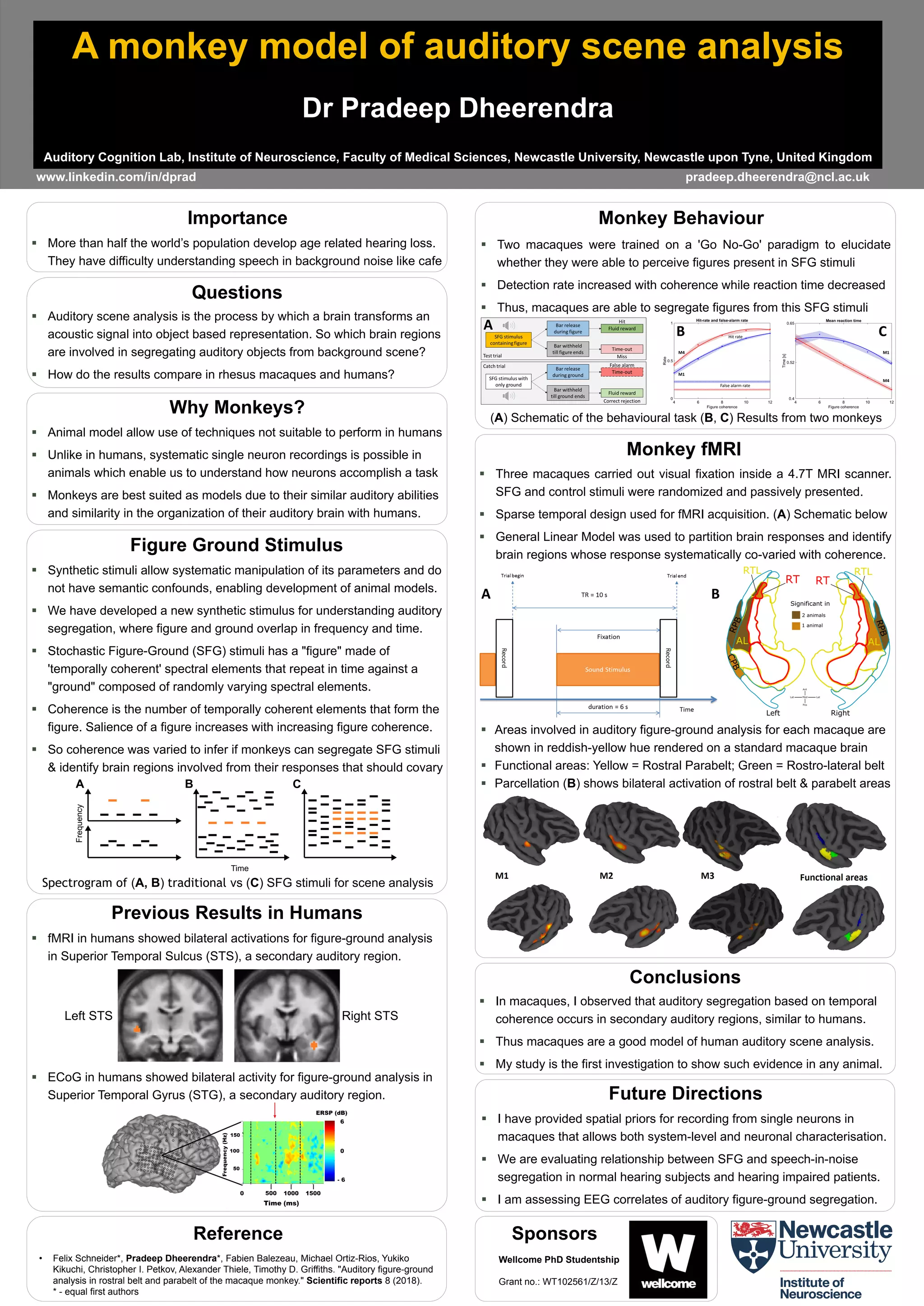
A monkey model of auditory scene analysis
Dr Pradeep Dheerendra
Auditory Cognition Lab, Institute of Neuroscience, Faculty of Medical Sciences, Newcastle University, Newcastle upon Tyne, United Kingdom
 More than half the world’s population develop age related hearing loss.
They have difficulty understanding speech in background noise like cafe
Importance
 Auditory scene analysis is the process by which a brain transforms an
acoustic signal into object based representation. So which brain regions
are involved in segregating auditory objects from background scene?
 How do the results compare in rhesus macaques and humans?
Questions
www.linkedin.com/in/dprad pradeep.dheerendra@ncl.ac.uk
 I have provided spatial priors for recording from single neurons in
macaques that allows both system-level and neuronal characterisation.
 We are evaluating relationship between SFG and speech-in-noise
segregation in normal hearing subjects and hearing impaired patients.
 I am assessing EEG correlates of auditory figure-ground segregation.
Future Directions
Figure Ground Stimulus
 Synthetic stimuli allow systematic manipulation of its parameters and do
not have semantic confounds, enabling development of animal models.
 We have developed a new synthetic stimulus for understanding auditory
segregation, where figure and ground overlap in frequency and time.
 Stochastic Figure-Ground (SFG) stimuli has a "figure" made of
'temporally coherent' spectral elements that repeat in time against a
"ground" composed of randomly varying spectral elements.
 Coherence is the number of temporally coherent elements that form the
figure. Salience of a figure increases with increasing figure coherence.
 So coherence was varied to infer if monkeys can segregate SFG stimuli
& identify brain regions involved from their responses that should covary
Spectrogram of (A, B) traditional vs (C) SFG stimuli for scene analysis
Reference
• Felix Schneider*, Pradeep Dheerendra*, Fabien Balezeau, Michael Ortiz-Rios, Yukiko
Kikuchi, Christopher I. Petkov, Alexander Thiele, Timothy D. Griffiths. "Auditory figure-ground
analysis in rostral belt and parabelt of the macaque monkey." Scientific reports 8 (2018).
* - equal first authors
 In macaques, I observed that auditory segregation based on temporal
coherence occurs in secondary auditory regions, similar to humans.
 Thus macaques are a good model of human auditory scene analysis.
 My study is the first investigation to show such evidence in any animal.
Conclusions
 Animal model allow use of techniques not suitable to perform in humans
 Unlike in humans, systematic single neuron recordings is possible in
animals which enable us to understand how neurons accomplish a task
 Monkeys are best suited as models due to their similar auditory abilities
and similarity in the organization of their auditory brain with humans.
Why Monkeys?
 fMRI in humans showed bilateral activations for figure-ground analysis
in Superior Temporal Sulcus (STS), a secondary auditory region.
Previous Results in Humans
 ECoG in humans showed bilateral activity for figure-ground analysis in
Superior Temporal Gyrus (STG), a secondary auditory region.
Left STS Right STS
Sponsors
Wellcome PhD Studentship
Grant no.: WT102561/Z/13/Z
 Areas involved in auditory figure-ground analysis for each macaque are
shown in reddish-yellow hue rendered on a standard macaque brain
 Functional areas: Yellow = Rostral Parabelt; Green = Rostro-lateral belt
 Parcellation (B) shows bilateral activation of rostral belt & parabelt areas
 Three macaques carried out visual fixation inside a 4.7T MRI scanner.
SFG and control stimuli were randomized and passively presented.
 Sparse temporal design used for fMRI acquisition. (A) Schematic below
 General Linear Model was used to partition brain responses and identify
brain regions whose response systematically co-varied with coherence.
Monkey fMRI
A B
(A) Schematic of the behavioural task (B, C) Results from two monkeys
 Two macaques were trained on a 'Go No-Go' paradigm to elucidate
whether they were able to perceive figures present in SFG stimuli
 Detection rate increased with coherence while reaction time decreased
 Thus, macaques are able to segregate figures from this SFG stimuli
Monkey Behaviour
False alarm rate
Hit rate
M4
M1
M1
M4
A B C

A monkey model of auditory scene analysis

  • 1.
    A monkey modelof auditory scene analysis Dr Pradeep Dheerendra Auditory Cognition Lab, Institute of Neuroscience, Faculty of Medical Sciences, Newcastle University, Newcastle upon Tyne, United Kingdom  More than half the world’s population develop age related hearing loss. They have difficulty understanding speech in background noise like cafe Importance  Auditory scene analysis is the process by which a brain transforms an acoustic signal into object based representation. So which brain regions are involved in segregating auditory objects from background scene?  How do the results compare in rhesus macaques and humans? Questions www.linkedin.com/in/dprad pradeep.dheerendra@ncl.ac.uk  I have provided spatial priors for recording from single neurons in macaques that allows both system-level and neuronal characterisation.  We are evaluating relationship between SFG and speech-in-noise segregation in normal hearing subjects and hearing impaired patients.  I am assessing EEG correlates of auditory figure-ground segregation. Future Directions Figure Ground Stimulus  Synthetic stimuli allow systematic manipulation of its parameters and do not have semantic confounds, enabling development of animal models.  We have developed a new synthetic stimulus for understanding auditory segregation, where figure and ground overlap in frequency and time.  Stochastic Figure-Ground (SFG) stimuli has a "figure" made of 'temporally coherent' spectral elements that repeat in time against a "ground" composed of randomly varying spectral elements.  Coherence is the number of temporally coherent elements that form the figure. Salience of a figure increases with increasing figure coherence.  So coherence was varied to infer if monkeys can segregate SFG stimuli & identify brain regions involved from their responses that should covary Spectrogram of (A, B) traditional vs (C) SFG stimuli for scene analysis Reference • Felix Schneider*, Pradeep Dheerendra*, Fabien Balezeau, Michael Ortiz-Rios, Yukiko Kikuchi, Christopher I. Petkov, Alexander Thiele, Timothy D. Griffiths. "Auditory figure-ground analysis in rostral belt and parabelt of the macaque monkey." Scientific reports 8 (2018). * - equal first authors  In macaques, I observed that auditory segregation based on temporal coherence occurs in secondary auditory regions, similar to humans.  Thus macaques are a good model of human auditory scene analysis.  My study is the first investigation to show such evidence in any animal. Conclusions  Animal model allow use of techniques not suitable to perform in humans  Unlike in humans, systematic single neuron recordings is possible in animals which enable us to understand how neurons accomplish a task  Monkeys are best suited as models due to their similar auditory abilities and similarity in the organization of their auditory brain with humans. Why Monkeys?  fMRI in humans showed bilateral activations for figure-ground analysis in Superior Temporal Sulcus (STS), a secondary auditory region. Previous Results in Humans  ECoG in humans showed bilateral activity for figure-ground analysis in Superior Temporal Gyrus (STG), a secondary auditory region. Left STS Right STS Sponsors Wellcome PhD Studentship Grant no.: WT102561/Z/13/Z  Areas involved in auditory figure-ground analysis for each macaque are shown in reddish-yellow hue rendered on a standard macaque brain  Functional areas: Yellow = Rostral Parabelt; Green = Rostro-lateral belt  Parcellation (B) shows bilateral activation of rostral belt & parabelt areas  Three macaques carried out visual fixation inside a 4.7T MRI scanner. SFG and control stimuli were randomized and passively presented.  Sparse temporal design used for fMRI acquisition. (A) Schematic below  General Linear Model was used to partition brain responses and identify brain regions whose response systematically co-varied with coherence. Monkey fMRI A B (A) Schematic of the behavioural task (B, C) Results from two monkeys  Two macaques were trained on a 'Go No-Go' paradigm to elucidate whether they were able to perceive figures present in SFG stimuli  Detection rate increased with coherence while reaction time decreased  Thus, macaques are able to segregate figures from this SFG stimuli Monkey Behaviour False alarm rate Hit rate M4 M1 M1 M4 A B C