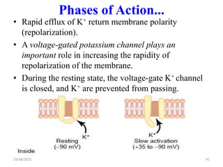
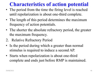
The document discusses the physiology of nerve cells. It covers the structures and functions of neurons and neuroglia. Neurons generate and propagate action potentials for communication. The resting membrane potential is maintained by ion gradients established by the sodium-potassium pump and leak channels. When stimulated, voltage-gated sodium channels open, causing rapid depolarization. Then voltage-gated potassium channels open to repolarize the membrane back to the resting potential.
The document discusses the nervous system, including neurons, the central nervous system, and peripheral nervous system. It describes the structure and function of neurons, including different parts like the cell body, dendrites, and axon. It discusses how neurons transmit electrical signals via action potentials and communicate via synapses. The central nervous system contains the brain and spinal cord, which control and coordinate the body. The peripheral nervous system includes nerves that connect the central nervous system to the rest of the body.
This document provides an overview of neurons and synapses in 3 chapters:
1) It describes the basic anatomy and types of neurons and glial cells in the brain. Neurons have dendrites, a cell body, an axon, and axon terminals. There are sensory, motor and interneurons.
2) It explains how chemical signaling works at synapses, including the release and uptake of neurotransmitters and the effects of excitatory and inhibitory postsynaptic potentials.
3) It covers how electrical signaling in the form of action potentials is generated and propagated in neurons and how this leads to the release of neurotransmitters and communication between neurons. Population and local coding of information is also discussed.
1) Neurons communicate with each other via synapses, transmitting signals chemically or electrically. At chemical synapses, an action potential causes neurotransmitter release, generating excitatory or inhibitory postsynaptic potentials.
2) The membrane potential of neurons is maintained by ion gradients. At rest it is negative, around -70mV. An action potential is a brief reversal of the potential triggered when the membrane reaches threshold.
3) Glial cells support and protect neurons. The main types are astrocytes, oligodendrocytes, and microglia. Astrocytes help form the blood-brain barrier and regulate the extracellular environment.
This document provides an overview of nerve tissue physiology. It discusses the two principal cell types in the nervous system - neurons and neuroglial cells. Neurons are specialized for signal conduction while neuroglial cells provide support and protection. The document then examines the structure and function of neurons, including their cell body, dendrites, axon, and synaptic transmission. It also explores concepts such as membrane potentials, action potentials, refractory periods, and the mechanisms of electrical and chemical synaptic transmission.
The nervous system consists of the central nervous system (brain and spinal cord) and peripheral nervous system. It functions to communicate and coordinate the body's activities, act as the site of reasoning in the brain, and adapt and respond to changes inside and outside the body. Neurons are the basic functional units and come in three types: sensory, motor, and interneurons. Neurons connect via synapses and transmit electrochemical signals through the body. The signals allow for coordination of muscles, glands, and organs. Diseases and disorders can disrupt the nervous system's functioning.
This document provides an overview of nerve-muscle physiology. It discusses the structure and types of neurons, including their classification based on number of poles and function. It also describes the structure and types of muscle, including skeletal, cardiac and smooth muscle. Additionally, it explains the electrophysiology of nerves and muscle, including the properties of electrical excitability, refractory period, and accommodation. The document outlines the process of the action potential in nerves and muscle cells. It concludes with a brief description of electrophysiology in the central nervous system.
Neurons are the basic building blocks of the nervous system and communicate via electrical and chemical signals. There are three main types of neurons - sensory neurons that receive information, interneurons that process information within the central nervous system, and motor neurons that send signals to muscles and glands. Glial cells provide support and insulation for neurons and are involved in nutrient supply, debris removal, and synaptic function. Neurons and glial cells work cooperatively to carry out the vital functions of the nervous system.
This document provides an overview of a lecture on nervous tissue that will take place on November 21st from 10:30 am to 12:30 pm at the KILIMANJARO CHRISTIAN MMEDICAL UNIVERSITY. The objectives are to learn about the cellular and structural components of nervous tissue, the structure and types of neurons, glial cells, and healing and repair of neurons. The main topics that will be covered include the cells of the nervous tissue, the components and classifications of neurons, myelination of axons, synapses, neuroglia, and regeneration of nerves after injury.
This document provides an overview of a lecture on nervous tissue that will take place on November 21st from 10:30 am to 12:30 pm at the KILIMANJARO CHRISTIAN MMEDICAL UNIVERSITY. The objectives are to learn about the cellular and structural components of nervous tissue, the structure and types of neurons, glial cells, and healing and repair of neurons. The lecture will cover the two main cell types - neurons and glial cells, the parts of a neuron including the cell body, dendrites and axon, myelination, synapses, and the roles of different glial cells.
This document provides an overview of a lecture on nervous tissue that will take place on November 21st from 10:30 am to 12:30 pm at the KILIMANJARO CHRISTIAN MMEDICAL UNIVERSITY. The objectives are to learn about the cellular and structural components of nervous tissue, the structure and types of neurons, glial cells, and healing and repair of neurons. The lecture will cover the cells of the nervous tissue including neurons and glial cells, the components of neurons, classifications of neurons, structures of the neuron cell body, dendrites, axons, synapses, myelination, neuroglia, and the relationship between neurons, glia cells and blood vessels.
The document discusses the cells of the nervous system. It describes two main types of cells - non-excitable neuroglial cells and excitable neuron cells. Neuroglial cells make up over 50% of the nervous system and include ependymal, microglial, astrocyte, Schwann and oligodendrocyte cells. Neurons are the excitable cells that transmit nerve impulses. The document then provides details about the structure and function of neurons, types of neurons, generation and conduction of nerve impulses, and the synapse. It also includes information about the meninges and major parts of the human brain.
Neurons are electrically excitable cells that communicate with each other and the body. The human nervous system contains around 100 billion neurons. There are three main types of neurons - sensory neurons relay signals from sense organs to the central nervous system, motor neurons relay signals from the CNS to effector organs, and interneurons connect sensory and motor neurons. Each neuron has a cell body, dendrites that receive signals, and an axon that transmits signals. When a neuron is stimulated, it generates an action potential down its axon via changes in membrane potential. Neurotransmitters are released at synapses to transmit signals between neurons.
Nerve cells, Nervous communication & its link to the celllular signallingSabahat Ali
The document discusses the structure and function of neurons. It notes that neurons are specialized cells that communicate via electrical and chemical signals. They contain dendrites that receive signals, a cell body, and an axon that transmits signals. At synapses, chemical neurotransmitters transmit signals between neurons or to other cell types. Neurons form circuits that allow for complex coordinated responses. The action potential involves changes in ion channel permeability that propagate electrical signals rapidly along axons. Calcium acts as an important intracellular messenger in neurons and other cell types, often working through the calcium sensor protein calmodulin.
The nervous system allows animals to detect and communicate information about the external and internal environment. It has two main parts: the central nervous system (CNS) and peripheral nervous system (PNS).
Neurons are the basic functional units and carry electrical signals through their cell body, dendrites, and axon. The axon is covered by a myelin sheath that insulates and speeds up signal transmission. At nodes of Ranvier, gaps in the myelin allow signals to travel faster.
The resting membrane potential of neurons is around -70mV, created by ion concentration gradients and selective permeability to ions like potassium and sodium. When stimulated, neurons generate action potentials through changes in sodium and potassium permeability that
The human neural system is divided into the central nervous system (CNS) and peripheral nervous system (PNS). The CNS includes the brain and spinal cord for information processing and control, while the PNS comprises nerves associated with the CNS. Nerve fibers of the PNS are afferent (transmit impulses to the CNS) and efferent (transmit impulses from the CNS). The PNS is further divided into the somatic and autonomic systems. Neurons are the basic structural and functional units, composed of a cell body, dendrites, and axon. Impulses are generated and conducted along neurons when the membrane becomes permeable to ions, then restored to its
The nervous system consists of the brain, spinal cord and nerves. It detects changes inside and outside the body and responds through electrical signals called nerve impulses. Neurons conduct these impulses while neuroglia provide support. There are two main types of synapses - electrical and chemical. At chemical synapses, a neurotransmitter is released from the presynaptic neuron and binds to receptors on the postsynaptic neuron.
These slides contain the basic information and principle of nervous transduction, It also includes the information about the type of the neurons, structure of the neuron, resting and active membrane potential, synapes and events occurring in it, and introduction to the neurotransmitters.
Lecture 5 Functions of Body Functions and Biophysics final_560db1d02acd3c34c3...jrdys25ycm
This document provides information about the neuronal membrane and resting membrane potential in neurons. It discusses the structure and types of neurons and neuroglial cells, and their functions in supporting and protecting neurons. It defines the resting membrane potential as the potential difference between the inside and outside of the nerve fiber during rest, which is typically -70 mV. It explains that the resting membrane potential is caused by the distribution of ions like sodium, potassium, and chloride inside and outside the cell, the selective permeability of the cell membrane to different ions, and the sodium-potassium pump, which helps maintain ion concentration gradients.
This document provides an overview of the central nervous system. It discusses the main components and functions.
The central nervous system consists of the brain and spinal cord. The brain is made up of the cerebrum, diencephalon, brainstem and cerebellum. The spinal cord contains ascending and descending tracts that transmit sensory and motor signals between the brain and body.
The brain and spinal cord contain grey matter with neuron cell bodies and white matter with myelinated axons. Neuroglia provide support to neurons. The brain and spinal cord are protected by meninges and cerebrospinal fluid.
Neurons are the basic functional units and come in different types. They transmit signals through electrical
The document provides information on neurons and supporting cells in the nervous system. It discusses the key parts of neurons including the cell body, dendrites and axon. It describes how neurons are classified based on their structure and function. The roles of supporting cells like astrocytes and oligodendrocytes are also summarized. The document then explains the ion channels that underlie electrical signaling in neurons and discusses graded and action potentials.
types of neurons, structure and functions, types of glia cells, their structure and function, functioning of a neuron - resting potential, action potential, graded potential, absolute and relative refractory period.
Neurons are the basic structural and functional units of the nervous system. They transmit electrical and chemical signals and have three main parts - the cell body, dendrites, and axon. The cell body contains the nucleus. Dendrites receive signals and the long axon conducts signals away from the cell body. Neurons communicate with each other via synapses, where neurotransmitters are released by the presynaptic neuron and bind to receptors on the postsynaptic cell. This allows signals to be transmitted electrically along neurons and chemically between neurons.
This document provides an overview of neurophysiology and the nervous system. It begins by outlining the learning objectives, which are to describe the organization of the nervous system, types of cells and their functions, different neurotransmitters and their roles, and functions of the spinal cord and brain. It then introduces neurophysiology and the components of the nervous system. The rest of the document discusses the structure and function of neurons, synaptic transmission, and various neurotransmitters like acetylcholine, glutamate, dopamine, norepinephrine, and epinephrine. It also mentions some clinical correlates related to different neurotransmitters.
Neurochemistry involves the study of neurochemicals that influence neuronal function and neural networks. Key developments include the discovery that messages are sent chemically between neurons rather than electrically. Molecular neuroscience applies concepts of molecular biology to the nervous system. Main components of neuroanatomy include the brain, neurons, synapses, and neuroglia. Neurons have a cell body, dendrites that receive inputs, and an axon that transmits signals. Synapses allow communication between neurons. Neurotransmitters are the chemical messengers released at synapses to affect another neuron.
1. The nucleus contains DNA which provides instructions for protein synthesis. The nucleolus is involved in ribosome synthesis and RNA transport.
2. The Golgi apparatus packages proteins and neurotransmitters into vesicles. Polyribosomes have multiple ribosomes attached to mRNA for protein copying.
3. Mitochondria produce energy in the form of ATP which powers biochemical reactions like ion pumping across membranes.
Indian Dental Academy: will be one of the most relevant and exciting training center with best faculty and flexible training programs for dental professionals who wish to advance in their dental practice,Offers certified courses in Dental implants,Orthodontics,Endodontics,Cosmetic Dentistry, Prosthetic Dentistry, Periodontics and General Dentistry.
TEST BANK For Accounting Information Systems, 3rd Edition by Vernon Richardso...rightmanforbloodline
TEST BANK For Accounting Information Systems, 3rd Edition by Vernon Richardson, Verified Chapters 1 - 18, Complete Newest Version
Unlocking the Secrets to Safe Patient Handling.pdfLift Ability
Furthermore, the time constraints and workload in healthcare settings can make it challenging for caregivers to prioritise safe patient handling Australia practices, leading to shortcuts and increased risks.
MBC Support Group for Black Women – Insights in Genetic Testing.pdfbkling
Christina Spears, breast cancer genetic counselor at the Ohio State University Comprehensive Cancer Center, joined us for the MBC Support Group for Black Women to discuss the importance of genetic testing in communities of color and answer pressing questions.
DECODING THE RISKS - ALCOHOL, TOBACCO & DRUGS.pdfDr Rachana Gujar
Introduction: Substance use education is crucial due to its prevalence and societal impact.
Alcohol Use: Immediate and long-term risks include impaired judgment, health issues, and social consequences.
Tobacco Use: Immediate effects include increased heart rate, while long-term risks encompass cancer and heart disease.
Drug Use: Risks vary depending on the drug type, including health and psychological implications.
Prevention Strategies: Education, healthy coping mechanisms, community support, and policies are vital in preventing substance use.
Harm Reduction Strategies: Safe use practices, medication-assisted treatment, and naloxone availability aim to reduce harm.
Seeking Help for Addiction: Recognizing signs, available treatments, support systems, and resources are essential for recovery.
Personal Stories: Real stories of recovery emphasize hope and resilience.
Interactive Q&A: Engage the audience and encourage discussion.
Conclusion: Recap key points and emphasize the importance of awareness, prevention, and seeking help.
Resources: Provide contact information and links for further support.
The best massage spa Ajman is Chandrima Spa Ajman, which was founded in 2023 and is exclusively for men 24 hours a day. As of right now, our parent firm has been providing massage services to over 50,000+ clients in Ajman for the past 10 years. It has about 8+ branches. This demonstrates that Chandrima Spa Ajman is among the most reasonably priced spas in Ajman and the ideal place to unwind and rejuvenate. We provide a wide range of Spa massage treatments, including Indian, Pakistani, Kerala, Malayali, and body-to-body massages. Numerous massage techniques are available, including deep tissue, Swedish, Thai, Russian, and hot stone massages. Our massage therapists produce genuinely unique treatments that generate a revitalized sense of inner serenely by fusing modern techniques, the cleanest natural substances, and traditional holistic therapists.
R3 Stem Cell Therapy: A New Hope for Women with Ovarian FailureR3 Stem Cell
Discover the groundbreaking advancements in stem cell therapy by R3 Stem Cell, offering new hope for women with ovarian failure. This innovative treatment aims to restore ovarian function, improve fertility, and enhance overall well-being, revolutionizing reproductive health for women worldwide.
Can coffee help me lose weight? Yes, 25,422 users in the USA use it for that ...nirahealhty
The South Beach Coffee Java Diet is a variation of the popular South Beach Diet, which was developed by cardiologist Dr. Arthur Agatston. The original South Beach Diet focuses on consuming lean proteins, healthy fats, and low-glycemic index carbohydrates. The South Beach Coffee Java Diet adds the element of coffee, specifically caffeine, to enhance weight loss and improve energy levels.
The facial nerve, also known as cranial nerve VII, is one of the 12 cranial nerves originating from the brain. It's a mixed nerve, meaning it contains both sensory and motor fibres, and it plays a crucial role in controlling various facial muscles, as well as conveying sensory information from the taste buds on the anterior two-thirds of the tongue.
Comprehensive Rainy Season Advisory: Safety and Preparedness Tips.pdfDr Rachana Gujar
The "Comprehensive Rainy Season Advisory: Safety and Preparedness Tips" offers essential guidance for navigating rainy weather conditions. It covers strategies for staying safe during storms, flood prevention measures, and advice on preparing for inclement weather. This advisory aims to ensure individuals are equipped with the knowledge and resources to handle the challenges of the rainy season effectively, emphasizing safety, preparedness, and resilience.
Gemma Wean- Nutritional solution for Artemiasmuskaan0008
GEMMA Wean is a high end larval co-feeding and weaning diet aimed at Artemia optimisation and is fortified with a high level of proteins and phospholipids. GEMMA Wean provides the early weaned juveniles with dedicated fish nutrition and is an ideal follow on from GEMMA Micro or Artemia.
GEMMA Wean has an optimised nutritional balance and physical quality so that it flows more freely and spreads readily on the water surface. The balance of phospholipid classes to- gether with the production technology based on a low temperature extrusion process improve the physical aspect of the pellets while still retaining the high phospholipid content.
GEMMA Wean is available in 0.1mm, 0.2mm and 0.3mm. There is also a 0.5mm micro-pellet, GEMMA Wean Diamond, which covers the early nursery stage from post-weaning to pre-growing.
Exploring the Benefits of Binaural Hearing: Why Two Hearing Aids Are Better T...Ear Solutions (ESPL)
Binaural hearing using two hearing aids instead of one offers numerous advantages, including improved sound localization, enhanced sound quality, better speech understanding in noise, reduced listening effort, and greater overall satisfaction. By leveraging the brain’s natural ability to process sound from both ears, binaural hearing aids provide a more balanced, clear, and comfortable hearing experience. If you or a loved one is considering hearing aids, consult with a hearing care professional at Ear Solutions hearing aid clinic in Mumbai to explore the benefits of binaural hearing and determine the best solution for your hearing needs. Embracing binaural hearing can lead to a richer, more engaging auditory experience and significantly improve your quality of life.
LGBTQ+ Adults: Unique Opportunities and Inclusive Approaches to CareVITASAuthor
This webinar helps clinicians understand the unique healthcare needs of the LGBTQ+ community, primarily in relation to end-of-life care. Topics include social and cultural background and challenges, healthcare disparities, advanced care planning, and strategies for reaching the community and improving quality of care.
2. Outline
• Introduction
• Functional structures of neurons and neuroglia
• Membrane Potential
• Resting membrane potential
• Ion channels
• Action potential
• Initiation of Action Potentials
• Propagation of Action Potential
• Synaptic transmission
• Classification of Neurotransmitters
29/08/2023 2
3. Objectives
At the end of this session the students will expected to:
• Describe functional structures of nerve cells
• Clearly state the causes of resting membrane
potential.
• List types of ion channel with their functions.
• Describe phases of action potential and its
propagation.
• Describe mechanisms of electrical and chemical
synapstic transmission. 3
4. Introduction
• In order to coordinate the human body functions,
mainly two control systems exist.
• One, the endocrine system, is a collection of blood-
borne messengers that works slowly.
• The other is the nervous system, a rapid control
system.
• Nerve and muscle cells respond to stimulus through
excitation.
• Excitation is the ability a cell to generate and
propagate action potential.
• The stimulus for excitation can be mechanical,
chemical or electrical.
29/08/2023 4
5. Introduction...
• Two types of physicochemical disturbances are
produced during excitation of nerve cells:
Local, non propagated potentials known as graded
potentials, and
Propagated potentials i.e. action potentials (AP).
• Generation and propagation of AP in excitable tissues
is the means of intercellular communication.
• The nervous system has two principal cell types:
neurons and neuroglial cells.
29/08/2023 5
6. Neuroglia
• Neuroglial cells are non excitable cells that support
the functions of neurons.
• Neuroglia are smaller than neurons, but they are 10 to
50 times more numerous than neurons.
• Unlike neurons, they can multiply in the mature NS
• Potential causes of glioma (brain tumour)
• Glial cells physically and metabolically support
neurons
29/08/2023 6
7. Neuroglia...
• They include: Astrocytes, microglia,
oligodendrocytes, ependymal cells, schwann cells &
Satellite cells.
A. ASTROCYTES:
• Star shaped cells having many processes
• The largest and the most numerous of the neuroglia.
Functions of astrocytes:-
o Form blood-brain barrier, by wrapped around blood
capillaries and isolate neurons of the CNS.
o In the embryo, astrocytes secrete chemicals that
appear to regulate the growth and interconnection
among neurons in the brain.
29/08/2023 7
8. Neuroglia...
o Provide nourishment for CNS neurons
o Maintain the appropriate chemical environment for
the generation of nerve impulses.
o E.g. regulate the conc. K+; take up excess
neurotransmitters.
B. Microglia:- small cells used as tissue macrophages
in the CNS.
C. Oligodendrocytes: - form myelin sheath around
axons in the CNS.
D. Ependymal cells: - cells line the ventricles of the
brain and central canal of the spinal cord.
• Produce and assist in the flow of cerebrospinal fluid.
29/08/2023 8
9. Neuroglia...
E. Schwann cells: - form the myelin sheath around
axons of PNS.
• Participate in axon regeneration
– Secrete growth-promoting factors that promote axonal
growth toward the distal stump.
• The amount of myelin increases from birth to
maturity
F. Satellite cells: - Surround the cell bodies of neurons
of PNS ganglia.
• Regulate the exchanges of materials between
neuronal cell bodies and interstitial fluid.
9
10. Neurons
• Functional and structural units of nervous system.
• Specialized for transmission of information from one
part of the body to other as nerve impulse.
• Have three distinct parts:-
1. Cell body/ soma:
Contains nucleus and other cell organelles in the
cytoplasm.
The cell body maintains the functional and structural
integrity of the axon.
2. Axon:
Is a long, thin, cylindrical projection from cell body
at axon hillock (used as trigger zone to generate
action potential). 10
11. Neurons...
The cytoplasm of an axon is axoplasm that contains:
– Mitochondria
– Neurofibrils
– Microtubules (important for axonal transport)
Axonal transport
• Transport of substances along microtubules that run
along the length of the axon.
• Slow axonal transport occurs at 0.5 to 10 mm/day.
– Supplies new axoplasm to developing or regenerating
axons.
• Fast axonal transport occurs at about 400 mm/day.
– Transport synaptic vesicles and materials that maintain
axolemma & synaptic end bulbs.
29/08/2023 11
13. Neurons...
Surrounded by a plasma membrane known as the
axolemma.
Propagates nerve impulses toward another neurons,
muscles or glands.
At the end, axon dividing into many fine processes,
axon terminals.
The tips of axon terminals swell into bulb-shaped
structures, synaptic end bulbs.
Synaptic end bulbs contain many synaptic vesicles
that store a chemical neurotransmitter.
29/08/2023 13
14. Neurons...
3. Dendrites:
Short,thin and
highly branched.
Are the receiving or
input portions of a
neuron.
Changes in
dendritic spines
have been
implicated in
learning and long-
term memory.
14
29/08/2023
15. Neurons...
Structurally, neurons are
classified as:-
I. Multipolar neurons:
• Have several dendrites
and one axon.
• Common in the brain
and spinal cord (motor
& interneurons)
II. Bipolar neurons
• Have one main dendrite
and one axon.
• Found in the special
sense.eg:-retina of eye ,
tongue, ear etc
29/08/2023 15
16. Neurons...
III. Unipolar neurons
• Have dendrites and one axon that are fused together.
• Sensory neurons in function.
Functionally, neurons are classified as:
1. Sensory or afferent neurons-
Convey action potential from receptors to CNS
2. Motor or efferent neurons-
Convey action potentials away from the CNS to
effectors
3. Interneurons or association neurons-
Mainly located within the CNS between sensory and
motor neurons.
29/08/2023 16
18. Ion Equilibrium Potential
• There are two independent forces acting on a
particular ion across the membrane:
Concentration Force
• Determined by the concentration difference across the
membrane.
• The greater the concentration difference, the greater
the concentration force.
Electrical Force
• The size of this force is determined by the electrical
difference across the membrane also known as
membrane potential (in mV).
29/08/2023 18
19. Ion Equilibrium Potential
• Equilibrium potential is the membrane potential at which
no net movement of ion across the membrane.
– i.e concentration gradient force is equal in magnitude but
opposite in direction to the electrical force.
• Given the concentration gradient for any ion, the
equilibrium potential for that ion can be calculated by
means of the Nernst equation.
• EMF (Elec.Mot.Force) (mV) = 61.5 logc1/c2
Z
– Z- valence of the ion
– C1 concentration outside the cell,
– C2 concentration inside the cell.
• The main function of the Nernst equation is to calculate
the ion's equilibrium potential at a given concentration
gradient.
29/08/2023 19
20. Ion Equilibrium Potential
• Assume an initial membrane conductance of zero for both
ions.
1. What is the direction of the sodium ion concentration force?
2. What are the direction of the
sodium ion electrical force?
3. What is the direction of net
force on the sodium ions?
4. If the membrane conductance
to sodium ion increases (sodium channels are opened),
what will be the directional change in the electrical potential in
B? That is, will it move in a positive or negative direction?
20
21. Membrane Potential
• All cells have a potential difference across their plasma
membranes which is known as membrane potential.
Ionic hypothesis
• In order for a potential difference to occur across a
membrane, there must be an unequal distribution of ions
across the membrane (i.e., a concentration gradient).
• Results in unequal distribution of positive and negative
charges across the cell membrane (electrical gradient).
29/08/2023 21
22. Membrane Potential cont’d
During at rest or at
equilibrium in the ICF
there are:
• High conc. of K +
(140mEq/L) and
• -vely charged non
diffusible proteins and
Phosphate compounds.
In the ECF there are:
• High conc. of Na+
and chloride ions.
29/08/2023 22
23. Membrane Potential cont’d
• Separation of charges (+,-) create an electrical
potential difference across the cell membrane (the
membrane potential).
• The membrane potential at rest is resting membrane
potential (RMP).
• The RMP exists because there are excess of negative
ions inside the cell and an excess of positive ions
outside.
• The RMP of a typical neuron is -90mv.
• What makes inside the membrane –ve and outside
+ve?
29/08/2023 23
24. Resting membrane potential
• The resting membrane potential arises from three
major factors:-
1. The Na+/K+ ATPase constantly pumping 3 Na+ ions
out ward for every 2 K+ ions imported.
• Thus more positive charge is leaving than entering.
• The activity of Na+/K+ ATPase is the non stop
process as long as the cell supply ATP.
2. Out ward diffusion of K+ through K+ leak channel.
29/08/2023 24
25. Resting membrane...
• There are Na+-K+ leak channels that continuously
leak Na+ in ward and K+ out ward, but K+ leak
channels are abundant in number & more permeable
to K+ more K+ are leaving than Na+ .
• As a result, inside of the membrane becomes
increasingly negative, and the outside of the
membrane becomes increasingly positive.
29/08/2023 25
26. Resting membrane...
3. There are negatively charged non diffusible proteins
with in the ICF that can not travel across the
membrane.
• Negatively charged organic
molecules, such as proteins
and phosphate compounds
account for negativity inside
the cell.
• These make up the interior has
less +ve than the exterior.
29/08/2023 26
27. Resting membrane...
o The resting membrane potential represents an
equilibrium situation at which:
– The concentration and electrical forces are equal and
opposite.
• If they are not equal, there will be a net diffusion of the
ion across the membrane.
o A cell's resting membrane potential is very sensitive to
changes in the extracellular K+ conc. but not for sodium.
– Because sodium channels are less leaky & few in number.
29/08/2023 27
28. Measuring the Membrane Potential
• Microelectrode is
impaled through the cell
membrane to the
interior of the fiber.
• Then another electrode
is placed in the
extracellular fluid
• The potential difference
between the inside and
outside of the fiber is
measured using an
appropriate voltmeter.
29/08/2023 28
29. Ion Channels
• Since ions are charged elements, they need membrane
proteins that form channels (passages) to cross cell mm.
• The transmission of electrical signals produced by
neurons and muscle fibers depend on two types of ion
channels:
I. Leak channels
• These channels have no gates, they are always open
permit specific ions to pass.
• E.g. Na+ leak channels ,K+ leak channels (they are
more leaky & numerous than Na+ leak channels)
II. Gated channels
• Ion channels that open and close due to the presence of
“gates.”
29/08/2023 29
30. Ion Channels cont’d
• The gate is a part of the channel protein that can seal
the channel pore or move aside to open the pore.
Depending on the stimulus that cause to open or close
the gate, gated ion channels can be classified as:
o Ligand-gated channels,
o Mechanically gated channels, and
o Voltage-gated channels.
I. Ligand-gated ion channels
• Opens and closes in response to a specific chemical
stimulus.
• Chemical ligands can be- neurotransmitters,
hormones, and particular ions.
29/08/2023 30
31. Ion Channels cont’d
II. Mechanically gated channel
• Open or close in response to mechanical stimulation in the
form of vibration (such as sound waves), touch, pressure, or
tissue stretching.
• The force distorts the channel from its resting position,
opening the gate.
• Common in receptors of special senses (auditory receptors )
and somato-sensory receptors.
III.voltage-gated channel
• Opens in response to a change in membrane potential.
29/08/2023 31
32. Ion Channels cont’d
• Voltage-gated channels participate in the generation and
conduction of action potentials.
• Common in membrane of excitable tissues.
A particular type of ion may pass through several different types
of channels.
• The following are common types of ion channels for specific
ions:
Na+ - channels:-
There are three major types of Na+ - channels
Na+ leak channel –found in almost all cells
-allow inflow Na+
Voltage gate- Na+ - channels
-Common in excitable cells
-Participate in depolarization phase of action potential
29/08/2023 32
33. Ion Channels cont’d
Liganed -gated Na+ - channels
-Respond to neurotransmitters (like Ach)
-Mostly found on the postsynaptic cell
membrane
K+ -channels
• There are four major classes:
K+ leak channel
-found in almost all cells and allow K+ outflow
-more leaky & numerous in number than Na+ leak
channel
Voltage-gated K+ channel
-Respond to membrane potential change & increase
rapidity of membrane repolarisation.
29/08/2023 33
34. Ion Channels cont’d
Ligand-gated K+ channel
-Respond to chemical ligands
-Mostly found on postsynaptic membrane to
inhibit synaptic transmission.
G-protein-gated K+ channel
-Function in the acetylcholine-dependent slowing
of the sinoatrial node pacemaker.
-In neurons, a number of transmitters can activate
it to hyperpolarize the membrane.
Ca2+ -channels:
Voltage gated Ca2+ -channels with subtypes: L,T and
N-type voltage gated Ca2+ -channels.
29/08/2023 34
35. Ion Channels cont’d
L-type(long-lasting) calcium channels
– Mostly found in muscle cells
– Remain open for longer time (responsible for
prolonged action potential in cardiac muscle)
N-type (Neural-Type) calcium channels
– Found primarily at presynaptic terminals and are
involved in neurotransmitter release.
T-type(transient) calcium channel
• Open and close in short time
• Respond to low voltage change
• Found in pace maker cells (neuronal rhythmicity)
29/08/2023 35
36. Ion Channels cont’d
Ligand-gated Ca2+ -channels
– Respond to chemical molecules or Ca2+
– Function to release Ca2+ from internal stores
G- protein-gated Ca2+ -channels
Chloride channels
• Ligand gated Cl- channels
– GABA receptors
– Glycine receptors
29/08/2023 36
37. Action potential
• AP is rapid change in the membrane potential that
spread rapidly along the nerve fiber membrane.
• Rapid membrane depolarization followed by a
repolarization back to the normal negative membrane
potential.
29/08/2023 37
38. Action potential...
Phases of the action potential:-
1. Resting phase- polarized state before the action
potential begins.
2. Depolarization phase-
Stimulus causes the membrane of the axon to
depolarize to threshold (–55mv).
Voltage-gated Na+ channels open rapidly rapid
influx of Na+ →depolarization (-90mv to zero -
+35mv)→ inactivation gates of Na+ channels closed.
29/08/2023 38
39. Action potential...
Voltage-gated Na+ channels:-
This channel has two gates,
the activation gate
(outside the membrane),
and the inactivation gate
(inside).
• During resting membrane
potential( –90 mv) the
activation gate is closed
and the inactivation gate is
remain in open state.
29/08/2023 39
40. Phases of Action...
• Inactivation gate of sodium channel will not reopen
until the membrane potential returns to or near
RMP level.
• So once closed, the voltage-gated Na+ channels will
not respond to a second stimulus until the cell almost
completely repolarizes.
3. Repolarizing Phase- is the return of membrane
polarity lost by depolarization.
• The same threshold level for depolarization also
opens voltage-gated K+ channels.
• But voltage-gated K+ channels are slow to open than
voltage-gated Na+ channels. That is why they open
just at the time voltage-gated Na+ channels are
beginning to close.
40
41. Phases of Action...
29/08/2023 41
• Rapid efflux of K+ return membrane polarity
(repolarization).
• A voltage-gated potassium channel plays an
important role in increasing the rapidity of
repolarization of the membrane.
• During the resting state, the voltage-gate K+ channel
is closed, and K+ are prevented from passing.
42. Phases of Action...
• Thus, in Na+ entry to the cell and simultaneous in
K+ exit from the cell combine to speed repolarization
process.
• The original gradients (RMP) are reestablished via
the (active) Na/K-ATPase pump.
If the voltage-gated K+ channels do not open, what
happens to the cell? Repolarization slows. Is there a
possibility for the cell to repolarize?yes.Na/K
ATPase.
What if the voltage-gated Na+ channels are
inactivated by medication? Does the depolarization
29/08/2023 42
43. Phases of Action...
• When the inside of a cell becomes more positive
relative to the outside, a phenomenon known as
overshoot occurs which is the reversal of the
membrane potential polarity.
• When the membrane potential is more negative than
the resting level, the membrane is said to be
hyperpolarized.
29/08/2023 43
44. Phases of Action...
• The following shows the action potential in three
types of excitable cells.
What differences do you observe?fast,slower,...of AP.
29/08/2023 44
45. Characteristics of action potential
All-or-none law
• If the stimulus is at or above threshold intensity, the action
potential occurs with constant amplitude regardless of the
strength of the stimulus.
• Once membrane is depolarized to threshold level, membrane
events are no longer dependent upon stimulus strength.
Refractoriness
• Is period of unresponsiveness of membrane to the second
excitatory signals. It can be absolute or relative:
1. Absolute Refractory period
• It is the period during which no matter how strong the
stimulus, it cannot induce a second action potential.
29/08/2023 45
46. Characteristics of action potential
• The period from the time the firing level is reached
until repolarization is about one-third complete.
• The length of this period determines the maximum
frequency of action potentials.
• The shorter the absolute refractory period, the greater
the maximum frequency.
2. Relative Refractory Period
• Is the period during which a greater than normal
stimulus is required to induce a second AP.
• Starts when repolarization is about one-third
complete and ends just before RMP is maintained.
46
29/08/2023
47. Characteristics of action potential
• It coincides with the period when the voltage-gated K
channels are still open after inactivated Na channels
have returned to their resting state.
29/08/2023 47
48. Initiation of Action Potentials
• Appropriate stimulus that brings membrane potential
to the threshold can initiate AP.
Graded Potentials
• Graded means magnitude of change in membrane
potential depends on the strength of the stimulus.
• A graded potential is a small deviation of resting
membrane potential that makes the membrane either
more polarized or less polarized.
• Depending on their location, graded potential can be
classified as: -Receptor potential (in the sensory
receptors)
-Generator (pacemaker) potential
-Synaptic potential
29/08/2023 48
50. Initiation of Action...
• A very weak stimulus at point
A causes the membrane
potential to change from –90 to
–85 mv, but cannot produce
AP.
• At point B, the stimulus is
greater, but again, the intensity
is still not enough to generate
AP.
• At point C the stimulus is
stronger that reaches
threshold level to generate AP.
50
29/08/2023
51. Initiation of Action...
• Graded potentials can become stronger and last
longer by summation to reach the threshold level.
• Summation is the process by which graded potentials
add together.
• If two depolarizing graded potentials summate, the
net result is a larger depolarizing graded potential
AP.
Summation
• There are two types of summation:
• Spatial summation and
• Temporal summation.
29/08/2023 51
52. Initiation of Action...
1. Spatial summation is
summation of postsynaptic
potentials in response to
stimuli that occur at
different locations in the
membrane of a
postsynaptic cell at the
same time.
• Results from the build up of
neurotransmitter released
simultaneously by several
presynaptic end bulbs.
29/08/2023 52
53. Initiation of Action...
2. Temporal summation is
summation of
postsynaptic potentials in
response to stimuli that
occur at the same
location in the membrane
of the postsynaptic cell
but at different times.
• Results from stimulation
of a single postsynaptic
membrane two or more
times in rapid succession.
29/08/2023 53
54. Comparison of Graded Potentials and Action Potentials in Neurons
Graded Potential Action Potential
Amplitude varies with conditions of the initiating
event
All-or-none once membrane is depolarized to
threshold, event amplitude is independent of
initiating
Can be summed Cannot be summed
Has no threshold Has a threshold that is usually about -55 mV
depolarized relative to the resting potential
Has no refractory period Has a refractory period
Is conducted decrementally; that is, amplitude
decreases with distance
Is conducted without decrement; the
depolarization is amplified to a constant value at
each point along the membrane
Duration varies with initiating conditions Duration constant for a given cell type under
constant conditions
Can be a depolarization or a hyperpolarization Always consists of depolarizing phase followed
by repolarizing phase and return to resting
membrane potential.
Initiated by environmental stimulus (receptor),
by neurotransmitter (synapse), or spontaneously
Initiated by a graded potential
Mechanism depends on ligand-sensitive channels or
other chemical or physical changes
Mechanism depends on voltage-gated channels
54
29/08/2023
55. Propagation of Action Potential
• When sodium ions flow in, they
cause voltage-gated Na-ion
channels in adjacent segments
of the membrane to open.
• The AP regenerates over and over
at adjacent regions of membrane
from the trigger zone to the axon
terminals.
• This process is a ve-feedback
vicious cycle that, once it starts
continues until all the voltage-
gated Na+ channels have
become activated (opened). 55
56. Propagation of Action...
Local anesthesia that block pain and other somatic
sensations act by blocking the opening of voltage-
gated Na+ channels.
There are two types of propagation:
I. Continuous conduction:- involves step by-step
depolarization and repolarization of each adjacent
segments of the plasma membrane.
• Occurs in unmyelinated axons and in muscle fibers.
• Ionic currents flow across each adjacent segment of
the membrane.
• Slow type of AP propagation.
29/08/2023 56
58. Propagation of Action...
II. Saltatory conduction:-action potentials jump from
one node to the next as they propagate along a
myelinated fiber.
• Few voltage-gated channels are present in regions
where a myelin sheath covers the axolemma.
• By contrast, at the nodes of Ranvier, the axolemma
has many voltage-gated channels & other channels.
• AP conducts by opening a smaller number of
channels only at the nodes, rather than many channels
in each adjacent segment of membrane.
• Much faster than continuous conduction
• More energy-efficient mode of conduction WHY?
29/08/2023 58
60. Propagation of Action...
Orthodromic vs antidromic conduction
• Action potential travels in all directions away from the
stimulus
• When an action potential is initiated in the middle of
axon, two impulses traveling in opposite directions.
o Orthodromic conduction:- impulse from
receptors/synapes to axon terminal.
o Antidromic conduction:- from axonsoma then die at
synapse.
29/08/2023 60
61. Factors that affect the speed of propagation
• The speed of propagation of an action potential is
affected by various factors:
1. Myelination: action potentials propagate more
rapidly along myelinated axons than along
unmyelinated axons.
2. Axon diameter: larger-diameter axons propagate
action potentials faster than smaller ones due to their
larger surface areas.
3. Temperature: axons propagate action potentials at
lower speeds when cooled (cold press used as pain
killer).
29/08/2023 61
62. Factors That Affect the Speed...
4. Conc. of ions in the ECF:
Hyperkalemia (K+ )- prolong depolarization slow
transmission
Hypokalemia (K+)- hyperpolarization cell
excitibility
Hypocalcemia (Ca2+): excitability by increasing the
opening of sodium channels in excitable membranes.
• Conversely, an increase in extracellular Ca2+ conc. can
stabilize the membrane by excitability.
29/08/2023 62
63. Types of Nerve Fibers
• Collection of axons are called nerve fibers.
• Axons can be classified into three major groups based on the
amount of myelination, their diameters, and their propagation
speeds:
1. Type A fibers
• Are the largest-diameter axons (5–20m) and myelinated.
• Has ,, and subtypes.
Type A fibers:-
• Motor to the skeletal muscle
• Sensory from muscle spindles and golgi tendon organs
• Diameter – 5-10m
• Conduction velocity 60-120m/s
29/08/2023 63
64. Types of Nerve...
Type A fibers: - both motor and sensory in function.
• 5-15m in diameter
• Conduction velocity 30-80m/s
Type A and A fibers:- both motor and sensory in function
• 1-10m in diameter
• Conduction velocity 6-30m/s
2. Type B fibers
• Myelineted fibers with diameters of 2–3m.
• Conduction velocity 15 m/sec
• Both motor and sensory in function- motors are all
preganglionic autonomic fibers
29/08/2023 64
65. Types of Nerve...
• B fibers also conduct sensory nerve impulses from
the viscera to the brain and spinal cord.
3. Type C fibers
• All are unmyelinated
• 0.5 to 2 micrometers in diameter
• Nerve impulse propagation ranges from 0.5 to 2
m/sec
• Both motor and sensory in function:- motor post
ganglionic fibers
• Sensory :-unmyelinated fibers carrying pain, itch,
temperature, and crude touch sensations.
29/08/2023 65
66. Synaptic transmission
• Synapse is the junction b/n two cells in which one
must be a neuron.
• It is the site of transmission from one neuron to the
next.
Depending on the chemical nature of messenger ,
synapses can be classified as:-
I. Electrical synapses- action potentials (impulses)
conduct directly between adjacent cells through
flow of ions in gap junctions.
Common in visceral smooth muscle, cardiac
muscle, and the developing embryo.
29/08/2023 66
67. Synaptic transmission...
• Electrical synapses can coordinate the activity of a
group of neurons or muscle fibers
• i.e large number of neurons or muscle fibers can produce
action potentials in one pitch.
2. Chemical synapses contain:
– Presynaptic axon consists of neurotransmitter,
– Synaptic cleft containing ECF with enzymes and
– Postsynaptic cell consists of receptors for
neurotransmitters that produce postsynaptic
potential.
• Chemical synapses relay signals more slowly than
electrical synapses.
29/08/2023 67
68. Synaptic transmission...
Depending on the post synaptic cell type, there are 3
types of synapses:-
I. Neuroneuronal junction
II. Neuromuscular junction
III. Neuroglandular junction
There are 3 types of neuroneuronal junctions
(Axo-dendritic, axosomatic and axoaxonic)
29/08/2023 68
70. Mechanism of Chemical Synapse
Transmission
1. A nerve impulse arrives at a synaptic end bulb of a
presynaptic axon.
2. Depolarizing of the axon terminals opens voltage-gated
Ca2+channels.
3. In flow of Ca2+ triggers exocytosis of the synaptic vesicles to
release neurotransmitter.
4. Neurotransmitter molecules diffuse across the synaptic cleft
and bind to their receptors in the postsynaptic neuron’s
plasma membrane.
5. Opening of ligand-gated channels allows particular ions to
flow across the membrane postsynaptic potential (EPSP /
IPSP).
29/08/2023 70
72. Mechanism of Chemical Synapse...
6.When a depolarizing postsynaptic potential reaches threshold,
it triggers an action potential in the axon of the postsynaptic
neuron.
Neurotransmitter binds directly to ligand-gated ion channels
(produce EPSP/IPSP) or G-protein channels( usually
hyperpolarization of the mm).
EPSPs
• Transient depolarization
• Excitatory because membrane potential moves closer to
threshold
• Increase in conductance to Na+
• Na+ influx causes depolarization.
IPSPs
• Transient hyperpolarizations
29/08/2023 72
73. Mechanism of Chemical Synapse...
• Inhibitory because membrane potential moves farther
away from its threshold .
• Increased conductance to CI-
• CI- influx causes hyperpolarization
• Also can be produced by increased K+ conductance
Neurotransmitters removed from the synaptic cleft
via:
Enzymatic degradation in the synaptic cleft
Diffuse to blood stream (very small amount)
Reuptake by presynaptic neuron
Removed by glial cells
29/08/2023 73
74. Properties of synaptic transmission
I. Unidirectional conduction
II. Synaptic delay (0.5-1 ms in chemical synapses)
III. Fatigue- in response of postsynapic neurons as repetitive
stimulation by presynaptic neurons.
IV. Synaptic potentiation (facilitation)- persistence of out put
signal after the stoppage of input signal.
V. Affected by blood pH & tissue oxygenation
-alkalosis synaptic transmission
-acidosis synaptic transmission
-hypoxia synaptic transmission
VI. Synapse is the site for the action of different types of drugs.
29/08/2023 74
75. Neurotransmitters
Classes Neurotransmitters receptors Distribution & role
Monoamines Acetylcholine Nicotinic &
muscarinic
Excitatory in CNS
and PNS
Serotonin 5HT1,2,3,4 Excitatory in CNS
Catecholami
nes
Dopamine Dopaminergic
receptors A,B
Inhibitory in the
Basal ganglia
(BG).
Norepinephrine & epinephrine &
adrenoreceptores
Excitatory in PNS
Histamine Histaminergic Rs:-
H1, H2
Excitatory
Amino Acids Glutamate NMDA* receptores Excitatory
Glycine Glycine receptors inhibitory
GABA GABAA &B Inhibitory in BG
Neuropeptid
e
Enkephalin, neurotensin,
somatostatin, substance P,
Cholecystokinin, hypothalamic
hormones.
In the CNS and
glands pituitary.
*N-methyl-D-aspartate
29/08/2023 75
This results from growth-promoting factors secreted by Schwann cells that attract axons toward the distal stump. Adhesion molecules of the immunoglobulin superfamily (eg, NgCAM/L1) promote axon growth along cell membranes and extracellular matrices
The main reason for this point of origin of the action potential is that the soma has relatively few voltage-gated sodium channels in its membrane, which makes it difficult for the EPSP to open the required number of sodium channels to elicit an action potential. Plays a major role in “summation” of signals entering the neuron from multiple sources.
At physiological pH (around 7.4) the carboxyl group will be unprotonated and the amino group will be protonated. An amino acid with no ionizable R-group would be electrically neutral at this pH.
They are activated by neurotransmitters such as acetylcholine, dopamine, serotonin, opioids, somatostatin, adenosine and
GABA which act through the pertussis toxin-sensitive
heterotrimeric G proteins Gαi and Gαo (Module 2: Figure
heterotrimeric G protein signalling). It is the βγ subunit released from these G proteins that is responsible for activating the GIRKs. The GIRKs have multiple
functions:
• GIRKs function in the acetylcholine-dependent slowing
of the sinoatrial node pacemaker (Module 7: Figure
cardiac pacemaker).
• In neurons, a number of transmitters can activate GIRKs to hyperpolarize the membrane giving rise to
slow inhibitory postsynaptic potentials that reduces neuronal activity as has been described during the
synchronization of oxytocin neurons (Module 10: Figure
oxytocin neurons)
These channels/transporters have two main functions. First, they carry charge and thus contribute to the regulation of membrane potential and thus cell excitability. Secondly, the bulk flow of Cl− contributes to cell volume regulation and the flow of ions and water
across transporting epithelial. From a signalling perspective, attention has focused on those channels and transporters
that contribute to the control of cell function:
• Ca2+ -sensitive Cl− channels (CLCAs)
• Cation-chloride cotransporters
• CLC chloride channels and transporters
• Cystic fibrosis transmembrane conductance regulator
(CFTR)
• GABA receptors
• Glycine receptors (GlyRs)
In addition to actions on GABAA chloride channels, inhaled anesthetics have been reported to cause membrane hyperpolarization (an inhibitory action) via their activation of ligand-gated potassium channels.
The same increase in voltage that opens the activation gate also closes the inactivation gate. The inactivation gate, however, closes a few 10,000ths of a second after the activation gate opens.
To say that these electrical signals are graded means that they vary in amplitude (size), depending on the strength of the stimulus
But the changed postsynaptic potential lasts up to 15 milliseconds after the synaptic membrane channels have already closed. Therefore, a second opening of the same channels can increase the postsynaptic potential to a still greater level, and the more rapid the rate of stimulation, the greater the postsynaptic potential becomes. Thus, successive discharges from a single presynaptic terminal, if they occur rapidly enough, can add to one another; that is, they can “summate.”This type of summation is called temporal summation.
An antidromic impulse in an axon refers to conduction opposite of the normal (orthodromic) direction. That is, it refers to conduction along the axon away from the axon terminal(s) and towards the soma
Encounters the much larger membrane area, and thus capacitance, of the larger axon. The time course is faster for the action potential due to the faster conduction velocity in the larger axon. When the axon's diameter is changed, its area per unit length is changed. This results in two electrical changes: (1) its capacitance per unit length is changed proportionally, and (2) its conductance per unit length is changed proportionally (its resistance is increased)
When the extracellular fluid concentration of calcium ions falls below normal, the nervous system becomes progressively more excitable, because this causes increased neuronal membrane permeability to sodium ions, allowing easy initiation of action potentials. At plasma calcium ion concentrations about 50 per cent below normal, the peripheral nerve fibers become so excitable that they begin to discharge spontaneously, initiating trains of nerve impulses that pass to the peripheral skeletal muscles to elicit tetanic muscle contraction. Consequently, hypocalcemia causes tetany
K+ affect membrane potential (conc. gradient), as conc. gradient decrease rate of change in membrane potential (Vmax) prolong depolarization slower propagation of AP