Steps of VirusInfection
A virus must use cell processes to replicate. The viral replication cycle can produce
dramatic biochemical and structural changes in the host cell, which may cause cell
damage. These changes, called cytopathic (causing cell damage) effects, can change
cell functions or even destroy the cell. Some infected cells, such as those infected by the
common cold virus known as rhinovirus, die through lysis (bursting) or apoptosis
(programmed cell death or "cell suicide"), releasing all progeny virions at once. The
symptoms of viral diseases result from the immune response to the virus, which
attempts to control and eliminate the virus from the body and from cell damage caused
by the virus. Many animal viruses, such as HIV (Human Immunodeficiency Virus), leave
the infected cells of the immune system by a process known as budding, where virions
leave the cell individually. During the budding process, the cell does not undergo lysis
and is not immediately killed. However, the damage to the cells that the virus infects
may make it impossible for the cells to function normally, even though the cells remain
alive for a period of time.
There is a period between infection of a cell and the appearance of new infectious virus that is known
as the latent period. During this time, several different stages in the virus life cycle are occurring.
2.
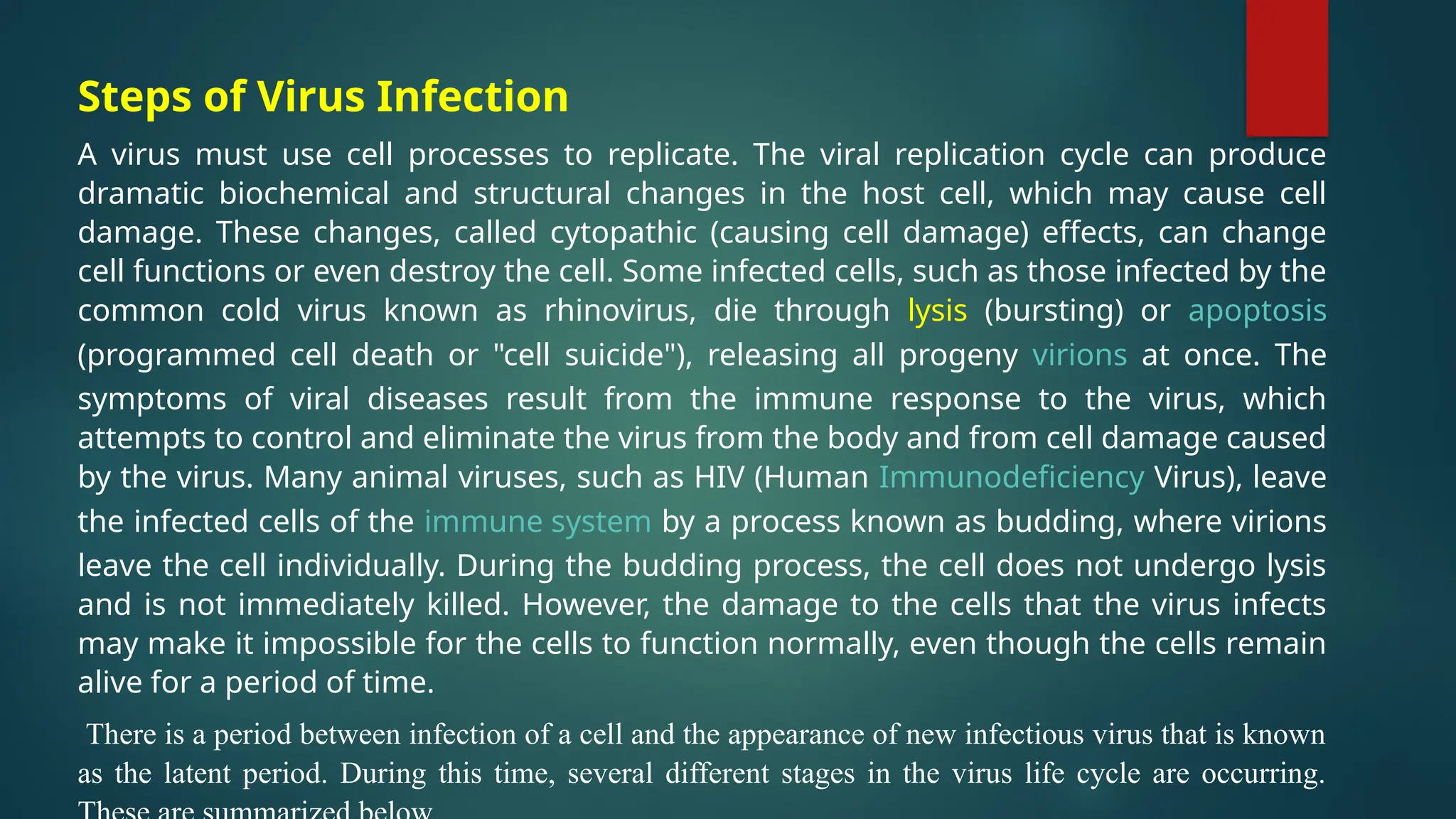
Viral particles disappearupon penetration, none are seen during biosynthesis and
assembly, and eventually all cells die so no new virions can be produced.
The eclipse period is the period when all viral particles are present but before they are
assembled.
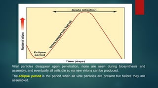
3.
Burst time isthe time from phage adsorption to release.
Burst size is the number of newly synthesized phages produced from one
infected cell.
4.
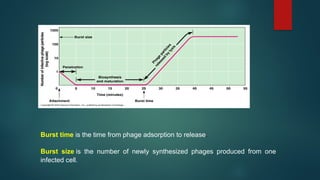
Steps in a"Model" Viral Life Cycle:
1) Attachment (Adsorption)
2) Penetration
3) Uncoating
4) Targeting
5) Gene expression.
-synthesis of viral mRNA (transcription)
-synthesis of viral proteins (translation)
6) Genome replication
7) Virion assembly/maturation
8) Release of new infectious virus
-lysis : breakdown of cell membrane and release of virus
-budding: viruses "bud" through cell membrane and are released without
necessarily killing the cell. Viruses acquire envelopes (membranes)
during this process.
6.
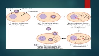
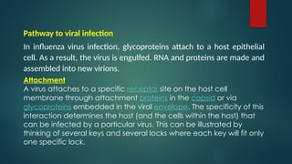
Pathway to viralinfection
In influenza virus infection, glycoproteins attach to a host epithelial
cell. As a result, the virus is engulfed. RNA and proteins are made and
assembled into new virions.
Attachment
A virus attaches to a specific receptor site on the host cell
membrane through attachment proteins in the capsid or via
glycoproteins embedded in the viral envelope. The specificity of this
interaction determines the host (and the cells within the host) that
can be infected by a particular virus. This can be illustrated by
thinking of several keys and several locks where each key will fit only
one specific lock.
7.
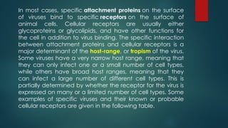
In most cases,specific attachment proteins on the surface
of viruses bind to specific receptors on the surface of
animal cells. Cellular receptors are usually either
glycoproteins or glycolipids, and have other functions for
the cell in addition to virus binding. The specific interaction
between attachment proteins and cellular receptors is a
major determinant of the host-range, or tropism of the virus.
Some viruses have a very narrow host range, meaning that
they can only infect one or a small number of cell types,
while others have broad host ranges, meaning that they
can infect a large number of different cell types. This is
partially determined by whether the receptor for the virus is
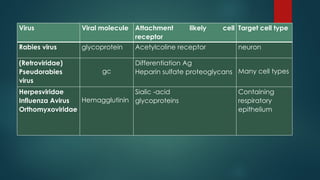
expressed on many or a limited number of cell types. Some
examples of specific viruses and their known or probable
cellular receptors are given in the following table.
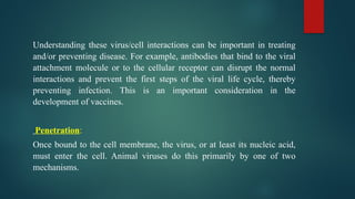
Understanding these virus/cellinteractions can be important in treating
and/or preventing disease. For example, antibodies that bind to the viral
attachment molecule or to the cellular receptor can disrupt the normal
interactions and prevent the first steps of the viral life cycle, thereby
preventing infection. This is an important consideration in the
development of vaccines.
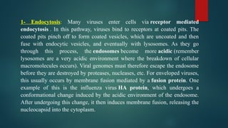
Penetration:
Once bound to the cell membrane, the virus, or at least its nucleic acid,
must enter the cell. Animal viruses do this primarily by one of two
mechanisms.
10.
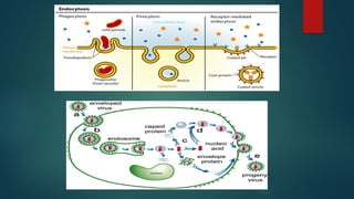
1- Endocytosis: Manyviruses enter cells via receptor mediated
endocytosis . In this pathway, viruses bind to receptors at coated pits. The
coated pits pinch off to form coated vesicles, which are uncoated and then
fuse with endocytic vesicles, and eventually with lysosomes. As they go
through this process, the endosomes become more acidic (remember
lysosomes are a very acidic environment where the breakdown of cellular
macromolecules occurs). Viral genomes must therefore escape the endosome
before they are destroyed by proteases, nucleases, etc. For enveloped viruses,
this usually occurs by membrane fusion mediated by a fusion protein. One
example of this is the influenza virus HA protein, which undergoes a
conformational change induced by the acidic environment of the endosome.
After undergoing this change, it then induces membrane fusion, releasing the
nucleocapsid into the cytoplasm.
11.
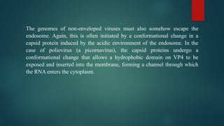
The genomes ofnon-enveloped viruses must also somehow escape the
endosome. Again, this is often initiated by a conformational change in a
capsid protein induced by the acidic environment of the endosome. In the
case of poliovirus (a picornavirus), the capsid proteins undergo a
conformational change that allows a hydrophobic domain on VP4 to be
exposed and inserted into the membrane, forming a channel through which
the RNA enters the cytoplasm.
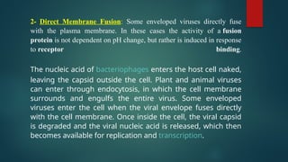
13.
2- Direct MembraneFusion: Some enveloped viruses directly fuse
with the plasma membrane. In these cases the activity of a fusion
protein is not dependent on pH change, but rather is induced in response
to receptor binding.
The nucleic acid of bacteriophages enters the host cell naked,
leaving the capsid outside the cell. Plant and animal viruses
can enter through endocytosis, in which the cell membrane
surrounds and engulfs the entire virus. Some enveloped
viruses enter the cell when the viral envelope fuses directly
with the cell membrane. Once inside the cell, the viral capsid
is degraded and the viral nucleic acid is released, which then
becomes available for replication and transcription.
14.
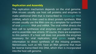
Replication and Assembly
Thereplication mechanism depends on the viral genome.
DNA viruses usually use host cell proteins and enzymes to
make additional DNA that is transcribed to messenger RNA
(mRNA), which is then used to direct protein synthesis. RNA
viruses usually use the RNA core as a template for synthesis
of viral genomic RNA and mRNA. The viral mRNA directs the
host cell to synthesize viral enzymes and capsid proteins,
and to assemble new virions. Of course, there are exceptions
to this pattern. If a host cell does not provide the enzymes
necessary for viral replication, viral genes supply the
information to direct synthesis of the missing proteins.
Retroviruses, such as HIV, have an RNA genome that must
be reverse transcribed into DNA, which then is incorporated
into the host cell genome.
15.
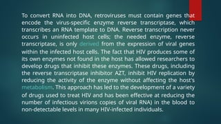
To convert RNAinto DNA, retroviruses must contain genes that
encode the virus-specific enzyme reverse transcriptase, which
transcribes an RNA template to DNA. Reverse transcription never
occurs in uninfected host cells; the needed enzyme, reverse
transcriptase, is only derived from the expression of viral genes
within the infected host cells. The fact that HIV produces some of
its own enzymes not found in the host has allowed researchers to
develop drugs that inhibit these enzymes. These drugs, including
the reverse transcriptase inhibitor AZT, inhibit HIV replication by
reducing the activity of the enzyme without affecting the host's
metabolism. This approach has led to the development of a variety
of drugs used to treat HIV and has been effective at reducing the
number of infectious virions copies of viral RNA) in the blood to
non-detectable levels in many HIV-infected individuals.
16.
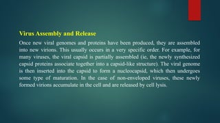
Virus Assembly andRelease
Once new viral genomes and proteins have been produced, they are assembled
into new virions. This usually occurs in a very specific order. For example, for
many viruses, the viral capsid is partially assembled (ie, the newly synthesized
capsid proteins associate together into a capsid-like structure). The viral genome
is then inserted into the capsid to form a nucleocapsid, which then undergoes
some type of maturation. In the case of non-enveloped viruses, these newly
formed virions accumulate in the cell and are released by cell lysis.
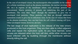
17.
In the caseof enveloped viruses, the nucleocapsids often assemble on the surface
of a cellular membrane (such as the plasma membrane, the nuclear envelope, the
ER, etc.) in regions of the membrane where viral envelope proteins are
concentrated. Matrix proteins, if present, are underlying this part of the
membrane. The virus then "buds" through the membrane to give rise to
enveloped viral particles. These particles can then go through additional
maturation events to give rise to infectious virus. In the case of viruses that form
on the plasma membrane, they can bud from the cell without causing cell lysis.
Other enveloped viruses, however, are lytic
The last stage of viral replication is the release of the new virions
produced in the host organism. They are then able to infect adjacent
cells and repeat the replication cycle. As you have learned, some
viruses are released when the host cell dies, while other viruses can
leave infected cells by budding through the membrane without
directly killing the cell.
18.
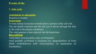
Events of the
1.lytic cycle:
Attachment or adsorption
Requires a receptor
Penetration
T-evens release lysozyme to break down a portion of the cell wall.
The tail sheath contracts and the tail core is driven through the hole
in the wall to the plasma membrane.
The viral genome is then injected into the bacterium.
Biosynthesis
Viral DNA and proteins are synthesized.
Host protein synthesis is stopped by degradation of host
DNA, interference with transcription, or repression of
translation.
19.
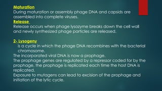
Maturation
During maturation orassembly phage DNA and capsids are
assembled into complete viruses.
Release
Release occurs when phage lysozyme breaks down the cell wall
and newly synthesized phage particles are released.
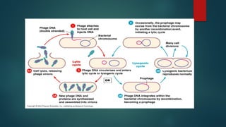
2- Lysogeny
is a cycle in which the phage DNA recombines with the bacterial
chromosome.
The incorporated viral DNA is now a prophage.
The prophage genes are regulated by a repressor coded for by the
prophage, the prophage is replicated each time the host DNA is
replicated.
Exposure to mutagens can lead to excision of the prophage and
initiation of the lytic cycle.
21.
Outcomes of lysogeny
-Bacteriumcan't be reinfected by the same kind of phage.
-Host cell may exhibit new properties due to viral genes carried on the prophage
Specialized transduction - host cell may gain new bacterial genes packaged with
the phage.


![제 23회 보아즈(BOAZ) 빅데이터 컨퍼런스 - [MBOAX] : ABSA를 활용한 소비자 반응 분석 기반 운영 효율화 대시보드 설계](https://cdn.slidesharecdn.com/ss_thumbnails/3-1boaz23rdconferencemboax-260203102709-9d519923-thumbnail.jpg?width=640&height=640&fit=bounds)


