This document describes a study that used computer-aided design and manufacturing (CAD/CAM) to develop an eye prosthesis with osseointegrated retention implants. The researchers used medical imaging and 3D modeling software to design a surgical guide for precisely placing the implants. They then used the guide to plan implant positions and angles to accommodate the prosthesis design. A prototype surgical guide was manufactured and implants were placed using the guide. After osseointegration, a custom prosthesis was made to attach to the implants. The researchers concluded that CAD/CAM methods improved accuracy, reduced errors and appointments, and represented a promising technique for facial prosthesis development.
The Ferrara Intrastromal Corneal Ring is a minimally invasive surgical option to treat ectatic corneal disorders like keratoconus. It improves the shape of the cornea, decreases astigmatism, and improves vision. The PMMA ring segments are inserted into the peripheral stroma, preserving the anterior corneal shape. A review of 340 patients found the procedure improved uncorrected and best corrected visual acuity for keratoconus grades 1 through 3. By 3 months post-op, patients achieved visual rehabilitation and refractive stability. Complications were low at 3-5% but higher for more advanced keratoconus.
The Indian Dental Academy is the Leader in continuing dental education , training dentists in all aspects of dentistry and
offering a wide range of dental certified courses in different formats.for more details please visit
www.indiandentalacademy.com
PURPOSE: To report the clinical outcomes of implantation of a new Ferrara intrastromal corneal ring segment (ICRS) with a 210-degree arc length in eyes with keratoconus.
This document discusses principles and techniques for strabismus surgery. It covers goals of strabismus surgery such as sensory fusion and cosmesis. Factors affecting surgical success like timing and tables are presented. Techniques for weakening and strengthening muscles like recession, resection, and plication are described. Measurement, anchoring, and closure techniques are illustrated. References on strabismus surgery techniques are provided.
This document contains 44 images from www.indiandentalacademy.com depicting the use of digital technology in orthodontics and dentistry. The images show a digital cephalometric tracing superimposed on a patient video image, digital patient records and images, a DigiGraph head holder and model board, a DigiGraph monitor and keyboard, a video printer, a digitizing handpiece, and various orthodontic model trimming and analysis techniques using digital tools.
This document describes yellow Ferrara rings, which are plastic intracorneal rings used to treat keratoconus. The rings contain a yellow filter and prismatic effect to reduce light scattering. Studies using C-Quant and point spread function measurements found the rings decreased straylight parameters, indicating less retinal light scattering. Clinical evidence showed the rings resolved patients' complaints of halos after exchanging a clear ring for a yellow one. The pigmented ring is better because it absorbs wavelengths that cause more light scattering.
This document discusses the osteo-odonto-keratoprosthesis (OOKP), a surgical procedure used to treat corneal blindness. It involves implanting an artificial cornea attached to a patient's tooth and bone. The OOKP provides the best option for restoring vision in severe corneal disease. The procedure is complex, involving both ophthalmological and oral surgeons. It has high success rates but also risks like infection. Lifelong follow up is required as it is a two-stage surgery. Overall, the OOKP offers a successful method of visual rehabilitation for corneal blindness.
A three dimensional finite element analysis of tilted or parallel implant.ppt...enochrao
In the Nobel biocare's model , the so-called All-on-Four protocol, four dental implants are placed in the interforaminal region of the edentulous mandible to support full-arch fixed prostheses.In Nobel biocare’s design, the posterior implants are tilted distally to a maximum of 45 degrees. The soul purpose of this finite element study was to determine and evaluate the stress concentration in peri-implant bone during two different loading conditions and the values obtained , will be used to compare with another design in which the four implants are placed parallel to each other and perpendicular to the occlusal plane. Three-dimensional finite element models consisted of mandibular bone, four dental implants inserted in two different configurations—with the distal implants tilted (model A) or four parallel implants (model S)—and hybrid superstructures. Two loading conditions (178 N/central incisors or 300 N/left first molar) were considered, and von Mises stress values were determined. During anterior loading, higher stress concentrations were detected in the peri-implant bone of all four implants in model A. During posterior loading, lower stress concentrations were observed around the anterior implants of model A; however, the tilted posterior implants were subjected to higher stresses in every condition. Application of either of these designs was successful in reducing peri-implant stress in one loading condition. However, neither design demonstrated better performance in both loading conditions; therefore, within the limitations of this study, neither design demonstrated clearly superior performance.
CBCT has many uses in orthodontics including for impacted teeth, root resorption, and boundary conditions. It provides more accurate 3D information than 2D imaging. CBCT is particularly useful for impacted or transposed teeth to localize them, for root resorption diagnosis, and to understand boundary conditions that may impact treatment planning. CBCT also has applications for craniofacial anomalies, TMJ assessment, and orthognathic surgery planning by providing detailed 3D visualization of structures.
This document summarizes various approaches for restoring functional vision at all distances, like a young adult, after cataract or presbyopia surgery. It focuses on accommodative intraocular lenses (AIOLs), which aim to provide accommodation through optical or positional changes. However, past AIOL studies showed limitations in near vision outcomes due to poor methodology and commercial bias. Future AIOLs would need to be independent of the capsular bag, with outcomes proven in large, long-term studies using standardized tests to measure accommodation and distinguish it from pseudo accommodation. While multifocal IOLs provide functional vision at all distances, they require neuroadaptation and always disperse some light between foci, rather than using it all efficiently.
finite element analysis of dental implantshldtpaul2
This document summarizes a student's finite element analysis project on dental implants using Abaqus software. The student created 2D models of a dental implant, abutment, screw, and jawbone. Materials properties were defined and meshes with varying sizes were tested. The analysis found maximum principal and von Mises stresses occurred at the lower lateral corner of the screw. While the stresses were higher than reported in literature, finite element analysis has advantages over experiments by avoiding risks to patients or animals. Simplifications mean results must be interpreted carefully compared to clinical studies.
Every patient has special needs and requirements, which have to be considered when determining the appropriate treatment.
Younger patients want esthetic closure of gaps to retain their young look as long as possible.
Older patients who are facing edentulousness still want to enjoy life and therefore prefer immediate fixed restorations, allowing them to continue participating in their social life and enjoy eating.
The quality of life of edentulous patients could be considerably increased with the help of a firmly fixed prosthesis, especially regarding the quality of their nutrition and the ability to make an untroubled appearance in public.
For us, implant prosthetics starts with the preservation of the hard and soft tissue.
By effectively fighting bacteria with the HELBO therapy, periodontally damaged teeth can frequently be preserved, and, if this is not possible, the foundation can be laid for
reliable early implant placement.
Endoscopic repair of orbital floor fractures /certified fixed orthodontic cou...Indian dental academy
The Indian Dental Academy is the Leader in continuing dental education , training dentists in all aspects of dentistry and offering a wide range of dental certified courses in different formats.
This study evaluated the outcomes of MyoRing intracorneal implantation in 15 eyes of 15 patients with pellucid marginal degeneration (PMD). Following MyoRing implantation, uncorrected and best corrected visual acuity improved significantly, refractive errors were reduced, and keratometry values decreased. Patients reported low rates of visual symptoms and high satisfaction scores. The study demonstrated that MyoRing implantation via microkeratome is a safe and effective procedure for treating PMD.
Keraring intracorneal ring segments are designed to treat keratoconus and other corneal ectatic disorders. They work by remodeling the cornea to regularize its surface and reduce refractive errors through addition techniques. Keraring comes in different sizes and thicknesses to customize treatment and provides long-term stabilization and improvement in vision. Extensive clinical use has demonstrated its safety and effectiveness for correcting refractive errors.
BASING AND TRIMMING OF ORTHODONTIC MODELSDr Susna Paul
This document discusses study model construction and trimming for orthodontic diagnosis. It describes making impressions, casts, and bases for maxillary and mandibular study models. Proper trimming involves using templates, squares, and guides to ensure models are symmetrical and meet standardized measurements for anatomical and artistic portions. Well-trimmed models accurately reproduce teeth and soft tissues for evaluating malocclusions and treatment planning.
Premier IOL choices Technique & Decision Making do we really need femtosecond...presmedaustralia
This document discusses techniques for cataract surgery and considerations for improving patient outcomes and satisfaction. It recommends performing cataract surgery earlier based on symptoms to improve vision and quality of life. Achieving accurate refractive outcomes is emphasized through repeatable capsulorhexis, minimizing induced astigmatism, and using toric and multifocal IOLs. Sources of refractive surprise are examined. Markings and alignment of toric IOLs are covered. Stable lens position is important for predictable results. Multifocal IOLs are presented as an option for reducing glasses dependence.
Doktor Vedat Kaya, Canan Aslı Utine, Sezen Harmancı Karakuş, Işılay Kavadarlı ve Ömer Faruk Yılmaz tarafından hazırlanmış olan bu makaleyi ilginize sunarız.
This document summarizes the current treatment options, outcomes, and controversies regarding refractive surgery in children. It reviews literature on various refractive surgery techniques like LASIK and PRK. While small studies show promise, there is no long-term data from large controlled trials. Key issues include risks of regression, flap complications, the impact of amblyopia on vision, and unknowns around healing in children. More research is still needed before refractive surgery can be considered routine for pediatric patients.
Dr. Francisco Sanchez presentation at the Mediphacos User Meeting 2013 - KERA...Mediphacos
This document discusses a new 355 degree Keraring for keratoconus correction. It presents the design of the Keraring, which has a larger arc length than previous models to improve central corneal flattening for advanced nipple cone cases. Surgical techniques for implanting the Keraring using either femtosecond laser ring dissection or pocket creation are shown. Preliminary results on 7 eyes found improvement in visual acuity and refractive error, with 100% of eyes within 1 diopter of the target refraction at 6 months. The conclusion is that the new 355 degree Keraring provides reasonable outcomes, indicating it is an option for nipple cone keratoconus correction.
Imaging in implantology /certified fixed orthodontic courses by Indian denta...Indian dental academy
The Indian Dental Academy is the Leader in continuing dental education , training dentists in all aspects of dentistry and offering a wide range of dental certified courses in different formats.
Indian dental academy provides dental crown & Bridge,rotary endodontics,fixed orthodontics,
Dental implants courses.for details pls visit www.indiandentalacademy.com ,or call
00919248678078
Technological advances in dental implant surgeryPeriowiki.com
This document discusses recent technological advances in dental implant surgery, including computer-aided design/computer-aided manufacturing (CAD/CAM) technology and computer-guided implant surgery techniques. It describes computerized tomography (CT) imaging and how CT data can be used for virtual surgical planning and fabrication of surgical guides. The document compares computer-guided implant surgery (CGIS), which uses static surgical guides, to computer-navigated implant surgery (CNIS), which allows for intraoperative modification of the surgical plan. Both techniques aim to increase the accuracy and predictability of dental implant placement.
The document discusses the "gold standard" of secondary intraocular lens (IOL) implantation in the era of minimally invasive vitreoretinal surgery (MIVS). It describes scleral fixation and iris-claw IOLs as options for secondary IOL implantation when capsular support is absent. The iris-claw IOL implanted in the posterior chamber is presented as the preferred option, allowing preservation of anterior segment anatomy and low risk of spontaneous dislocation. The trans-conjunctival, mini-invasive approach is also described as beneficial for the treatment of cataract surgery complications during the same setting to minimize trauma.
recent advances in prosthodontics/dental lab technology courses by Indian den...Indian dental academy
This document summarizes several new developments in dental ceramics. It discusses materials like In-Ceram, Procera, and IPS Empress 2 which can be used for posterior dental restorations due to improvements in strength and toughness compared to conventional ceramics. In-Ceram cores are crystalline rather than glass-based, increasing strength. Procera uses a titanium substructure for strength while Empress 2 uses lithium disilicate glass ceramics with 60% crystal content for high strength without compromising translucency. These new materials allow for all-ceramic restorations in areas requiring more load bearing than previous ceramics.
Recent advances in prosthodontics / crown & bridge courses by indian dental a...Indian dental academy
The Indian Dental Academy is the Leader in continuing dental education , training dentists in all aspects of dentistry and offering a wide range of dental certified courses in different formats.
Indian dental academy provides dental crown & Bridge,rotary endodontics,fixed orthodontics,
Dental implants courses.for details pls visit www.indiandentalacademy.com ,or call
0091-9248678078
1) The document describes an innovative technique called the "anteriorly pedicled retroauricular flap" for reconstructing auricular defects.
2) This technique uses a skin flap from behind the ear that is initially pedicled in an anterior direction, allowing for easy wound access and dressing.
3) In three cases, the technique successfully reconstructed large ear defects, with good aesthetic outcomes and no complications.
Title: Otoplasty: New Modification of the Mustardé technique
Author: Mohamed A.S.M. El-Rouby, MD,
Assistant Professor of Plastic surgery, Ain Shams University, Cairo, Egypt.
Abstract
Background: one of the most established techniques for management of protruding ears is the Mustardé technique (1). Many modifications had been published for this technique; however, all these modifications started by retro-auricular incision. We modify the Mustardé technique using three retroauricular microincisions to correct several deformities of the auricular cartilage in protruding ears.
Patients and Methods: 46 patients (7unilateral, 39 bilateral) (85 ears) who were candidates for this technique, their age (25 ± 2.8 years), 38 males, 8 females. The operation time, steps, follow up sessions (2 weeks, 3, 6 and 18 months) data was recorded. Preoperative and postoperative (1,18 months) photos were compared and analyzed by custom made computer program the evaluated the results.
Results: 42 patients achieve a natural appearance. extrusion of threads occurred in 8 ears. Asymmetrical ears were noticed in 4 patients and recurrence in 11 patients. These patients were revised by Mustardé technique with retro-auricular incisions. None of the patients developed retro-auricular scars.
Conclusion: this versatile modification allows for better asthenic results of otoplasty and minimizes complications of skin incision unless cartilage and/or skin resection is needed.
Th e use of premium IOLs requires more specifically than standard monofocal IOLs a thorough clinical and para clinical examination using modern equipments.
We will only mention micro-incision premium IOLs that are used
in our daily practice. All information regarding the characteristics of all available and especially multifocal IOLs are available in the SFO 2012 Report on presbyopia
This case report describes a 21-year-old male patient who presented with a naso-orbito-ethmoid fracture following a motor vehicle accident. Imaging revealed a comminuted NOE fracture. The patient underwent open reduction and internal fixation through a bicoronal approach to repair the fracture. Medial canthopexy was performed via transnasal wiring. Post-operatively, the patient's telecanthus was corrected and intercanthal distance was restored. Follow-up imaging showed good repair of the fracture. NOE fractures can be complex to diagnose and treat, and open reduction may be necessary for comminuted fractures to properly restore facial contours and nasal projection.
The Journal of Implant & Advanced Clinical Dentistry - Oct. 2015Jay True
Amr Hosny Elkhadem, DDS, MSc
Lecturer, Prosthodontics, Faculty of Oral & Dental medicine, Cairo University
The keyless partial guidance using the simple guide kit and c-shaped sleeves is a promising economic alternative to conventional guided approach. Further investigations are required to evaluate
its accuracy and long term success rates.
www.SimpleGuideSystem.com
ABSTRACT- The purpose of this study was to access the outcome of modified manual small incision cataract surgery (M-MSICS) in terms of postoperative visual recovery (Best Corrected Visual Acuity). In this prospective study, the patients having cataracts with nuclear sclerosis not more than early grade 3 were randomly assigned in 2-groups with 50- patients in each group [Group A (C-MSICS), Group B (M-MSICS)]. Both techniques were compared for each stage in terms of postoperative visual recovery (Best Corrected Visual Acuity). Follow ups in postoperative period were carried out on 1st and 3rd postoperative days, 2 weeks, 4 weeks and 6 weeks. Significant early postoperative visual recovery was observed in Modified manual small incision cataract surgery (M-MSICS) as compare to conventional technique. Postoperative surgical induced astigmatism at 6 weeks was significantly less in M-MSICS group (p<0.05%). So it can be concluded that M-MSICS is better technique than C-MSICS in terms of early postoperative visual recovery & less postoperative surgical induced astigmatism.
Key-words- Conventional manual small incision cataract surgery (C-MSICS), Modified manual small incision cataract surgery (M-MSICS), Postoperative visual outcome
This study examined the effect of the sequence of lateral osteotomy and hump removal on the aesthetic outcome in rhinoplasty. Lateral osteotomies were performed on 36 cadaver heads, with the osteotomies either done before or after hump removal. Analysis found that performing osteotomies before removing larger humps resulted in more regular bone fragment patterns compared to performing osteotomies after hump removal. For noses with small humps, there was no difference in bone fragment patterns between the osteotomy sequences. The study concludes that performing osteotomies before hump removal helps prevent irregular bone fragments when reducing larger humps.
Implant supported maxillofacial prosthesis/cosmetic dentistry coursesIndian dental academy
This document discusses maxillofacial prosthodontics and the use of osseointegrated implants to support facial prostheses. It covers the history and development of maxillofacial osseointegration, differences from oral osseointegration, advantages over adhesives, criteria for success, and treatment planning considerations. Key aspects include improved retention and stability of prostheses supported by implants compared to adhesives, as well as increased longevity, comfort, and hygiene. Success rates are generally high but lower for irradiated patients. Careful patient selection and consideration of medical conditions is important.
International Journal of Engineering and Science Invention (IJESI) is an international journal intended for professionals and researchers in all fields of computer science and electronics. IJESI publishes research articles and reviews within the whole field Engineering Science and Technology, new teaching methods, assessment, validation and the impact of new technologies and it will continue to provide information on the latest trends and developments in this ever-expanding subject. The publications of papers are selected through double peer reviewed to ensure originality, relevance, and readability. The articles published in our journal can be accessed online.
Orthognathic surgery involves correcting jaw deformities through combining orthodontics and maxillofacial surgery. It is primarily used in adults once growth has ceased to treat conditions too severe for orthodontics alone, such as malocclusions, airway issues, pain, or poor aesthetics. Planning involves clinical exams, models, virtual surgical planning, and splint fabrication to simulate and guide the surgery, which may include Le Fort osteotomies of the maxilla, sagittal split or vertical ramus osteotomies of the mandible, and genioplasty. Risks include nerve damage, bleeding, infection, and relapse, so patients must be informed and compliant.
Goals of anophthalmic socket surgery are-
-Maximizing orbital implant volume with good centration within the orbit
- Achieving optimal eyelid contour, volume, and tone
- Establishing a socket lining with deep fornices to retain the prosthesis
- Transmitting motility from the implant to the overlying prosthesis
- Achieving comfort and symmetry
Implant supported maxillofacial prosthesis./ lingual orthodontics coursesIndian dental academy
Indian Dental Academy: will be one of the most relevant and exciting training center with best faculty and flexible training programs for dental professionals who wish to advance in their dental practice,Offers certified courses in Dental implants,Orthodontics,Endodontics,Cosmetic Dentistry, Prosthetic Dentistry, Periodontics and General Dentistry.
Dr. Guilherme Rocha, Dr. Paulo Ferrara, Dr. Leonardo Torquetti, Dra. Luciene Barbosa analisam os resultados dos implantes de Anel de Ferrara de Arco longo no pós operatório de 6 meses
The Indian Dental Academy is the Leader in continuing dental education , training dentists in all aspects of dentistry and offering a wide range of dental certified courses in different formats.
Indian dental academy provides dental crown & Bridge,rotary endodontics,fixed orthodontics,
Dental implants courses.for details pls visit www.indiandentalacademy.com ,or call
00919248678078
Indian Dental Academy: will be one of the most relevant and exciting training center with best faculty and flexible training programs for dental professionals who wish to advance in their dental practice,Offers certified courses in Dental implants,Orthodontics,Endodontics,Cosmetic Dentistry, Prosthetic Dentistry, Periodontics and General Dentistry.
2012-06-21-inauguration-labo-additive-manufacturingadvanced-engineering-innov...Carsten Engel
This document discusses Advanced Electrical Tools' use of additive manufacturing to produce titanium tools for the aerospace industry. It summarizes the company's history and products, the market requirements for lightweight, high-quality tools, and why the company chose additive manufacturing over traditional machining. Specifically, it discusses how additive manufacturing allows for optimized designs with integrated functions at lower costs than conventional titanium machining. The perspectives section outlines plans to increase production rates and design new products using additive manufacturing.
2012-11-15-high-vlaue-manufacturing-formulagroupt-121204101004-phpapp02Carsten Engel
- Formula Group T is a team of 15 master's students from KU Leuven who design and build a formula-style race car called Areion to compete in Formula Student competitions.
- Areion is an electric vehicle with a steel space frame, double wishbone suspension, axial flux motor, and 8.5 kWh lithium polymer battery pack. It can accelerate from 0-100 km/h in 3.5 seconds.
- Formula Student competitions involve static design events and dynamic driving events that are judged by over 50 automotive industry specialists. In 2012, Formula Group T won several awards including the Airbus Teamwork Award.
3D Printing - A revolution capable of changing the supply chain of the medica...Carsten Engel
This document discusses the potential impact of 3D printing on supply chains and logistics. It suggests that 3D printing could accelerate a shift from "push supply chains" driven by mass production to "pull supply chains" driven by customer demand. This would enable more agile manufacturing and mass customization. Products could be made and distributed closer to consumers with lower transport costs. However, the impact will depend on the specific industry and product. 3D printing is best suited to small batch and customized production rather than mass manufacturing standard products. The technology's disruptive effects may be less transformative than some predictions.
Selective Laser Melting versus Electron Beam MeltingCarsten Engel
This document summarizes research on additive manufacturing technologies for metal applications. It discusses Sirris, an organization that provides technology services to industry, and their expertise in additive manufacturing. Two key additive manufacturing technologies for metals are described - Electron Beam Melting (EBM) and Laser Beam Melting (LBM). EBM uses an electron beam to sinter metal powder in a vacuum environment, while LBM uses a laser beam under argon gas. Their differences in terms of process parameters, material properties, and advantages/disadvantages are summarized. Metallurgical analysis shows EBM produces a uniform fine-grained microstructure while LBM microstructure depends on build orientation. Mechanical properties are also compared between the two technologies.
3D printing has many medical applications including printing 96% of hearing aids, reducing surgery time from 97 to 23 hours thanks to 3D printed models, and being the only solution for 3D printed bone in some cases. It has also saved babies through techniques like 3D printing. The technology continues to advance and its potential applications in medicine seem limitless.
1sirrisstateoftheartengel-140519033927-phpapp01Carsten Engel
The document discusses metal additive manufacturing (AM) technologies. It provides an overview of common metal AM processes like laser beam melting and electron beam melting, which work by melting metal powder layer by layer. Metal AM allows for complex geometries, nearly unlimited design freedom, and production without tools. While initially used for prototypes, metal AM is gaining acceptance for production applications in industries like aerospace, medical, and dental. For metal AM to see wider adoption, issues like standardization, reproducibility, costs, and material variety must still be addressed.
This document discusses 3D bioprinting and its potential applications. It begins with definitions of bioprinting and discusses its goals in tissue engineering. Current achievements are summarized, including the first 3D printed bladder in 2006 and liver in 2009. Requirements for organ bioprinting are outlined, including cell sources, scaffold materials, and bioprinting technologies. The document concludes that bioprinting has potential to help address the shortage of organs for transplantation.
Sirris is a collective center for the Belgian technology industry located in Belgium. It has extensive additive manufacturing capabilities and competencies, including 15 engineers and technicians and machines in Liège and Charleroi. Some of its key additive manufacturing technologies are stereolithography, laser sintering of metals, electron beam melting, and its own Optoform process for producing custom implants and prosthetics in ceramics and metals for the medical industry. Sirris has applied these technologies to produce porous bone scaffolds and over 10,000 spinal implants.
Medical applications of Additive Manufacturing Technologies in generalCarsten Engel
The document summarizes Sirris, the collective centre of the Belgian technology industry. Sirris is a non-profit organization that works to increase the competitiveness of technology companies in Belgium. The document outlines Sirris' local presence across Belgium and expertise in additive manufacturing technologies. It discusses applications in medical fields like customizing implants using 3D printing based on patient CT scans.
Medical applications of Additive Manufacturing Technologies in general
6036-10094-1-PB
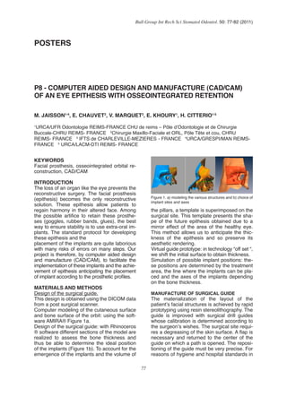
1. 77
Bull Group Int Rech Sci Stomatol Odontol. 50: 77-82 (2011)
POSTERS
1
URCA/UFR Odontologie REIMS-FRANCE CHU de reims – Pôle d’Odontologie et de Chirurgie
Buccale-CHRU REIMS- FRANCE 2
Chirurgie Maxillo-Faciale et ORL, Pôle Tête et cou, CHRU
REIMS- FRANCE 3
IFTS de CHARLEVILLE-MEZIERES - FRANCE 4
URCA/GRESPI/MAN REIMS-
FRANCE 5
URCA/LACM-DTI REIMS- FRANCE
M. JAISSON1-4
, E. CHAUVET2
, V. MARQUET3
, E. KHOURY1
, H. CITTERIO1-5
P8 - COMPUTER AIDED DESIGN AND MANUFACTURE (CAD/CAM)
OF AN EYE EPITHESIS WITH OSSEOINTEGRATED RETENTION
KEYWORDS
Facial prosthesis, osseointegrated orbital re-
construction, CAD/CAM
INTRODUCTION
The loss of an organ like the eye prevents the
reconstructive surgery. The facial prosthesis
(epithesis) becomes the only reconstructive
solution. These epithesis allow patients to
regain harmony in their altered face. Among
the possible artifice to retain these prosthe-
ses (goggles, rubber bands, glues), the best
way to ensure stability is to use extra-oral im-
plants. The standard protocol for developing
these epithesis and the
placement of the implants are quite laborious
with many risks of errors on many steps. Our
project is therefore, by computer aided design
and manufacture (CAD/CAM), to facilitate the
implementation of these implants and the achie-
vement of epithesis anticipating the placement
of implant according to the prosthetic profiles.
MATERIALS AND METHODS
Design of the surgical guide.
This design is obtained using the DICOM data
from a post surgical scanner.
Computer modeling of the cutaneous surface
and bone surface of the orbit: using the soft-
ware AMIRA® Figure 1a.
Design of the surgical guide: with Rhinoceros
® software different sections of the model are
realized to assess the bone thickness and
thus be able to determine the ideal position
of the implants (Figure 1b). To account for the
emergence of the implants and the volume of
the pillars, a template is superimposed on the
surgical site. This template presents the sha-
pe of the future epithesis obtained due to a
mirror effect of the area of the healthy eye.
This method allows us to anticipate the thic-
kness of the epithesis and so preserve its
aesthetic rendering.
Virtual guide prototype: in technology “off set “,
we shift the initial surface to obtain thickness.
Simulation of possible implant positions: the-
se positions are determined by the treatment
area, the line where the implants can be pla-
ced and the axes of the implants depending
on the bone thickness.
MANUFACTURE OF SURGICAL GUIDE
The materialization of the layout of the
patient’s facial structures is achieved by rapid
prototyping using resin stereolithography. The
guide is improved with surgical drill guides
whose calibration is determined according to
the surgeon’s wishes. The surgical site requi-
res a degreasing of the skin surface. A flap is
necessary and returned to the center of the
guide on which a path is opened. The reposi-
tioning of the guide must be very precise. For
reasons of hygiene and hospital standards in
Figure 1. a) modeling the various structures and b) choice of
implant sites and axes
2. 78
Bull Group Int Rech Sci Stomatol Odontol. 50: 77-82 (2011)
terms of sterilization, the prototype was made
of titanium sintered by SIRRIS (Charleroi) (Fi-
gure 2).
DESIGN AND MANUFACTURE OF EPITHESIS
The underside of the epithesis will receive the
retention elements (magnets). We must use a
rigid material for this part. It is computer de-
signed by reduction of the skin surface repo-
sitioned by mirroring the unaffected side. The
volumes of the substructure pillars are also
provided.
We manufactured by machining a block of
dental resin. The lost wax process is used to
construct the superficial part. The mold is in-
vested by the silicone-dyed. The make-up of
the surface is done in a conventional manner.
SURGICAL PHASES
The surgeon begins by positioning the guide
and by marking the skin at the future implant
sites. Then he realizes the flap design, incises
and displaces the tissue to the orbital cavity. It
replaces the guide and directed drilling. Four
Vistafix ™ implants are placed and protected
by screws.
The flap is repositioned and sutured. Given
the fragility of the surgical site, a phase of os-
seointegration under skin coverage is neces-
sary before the final discovery of the implants.
The second surgical stage is to find the fix-
tures, redesign peri-implant sites by refining
the skin to reduce the risk of infection in these
areas of transcutaneous permanent commu-
nication. The healing abutments are in place
for a few weeks. The healing abutments are
replaced with magnetic pillars.
RESULTS
After this osseointegration phase of a few
months, we can complete the cosmetic part of
the epithesis and add retainers initially plan-
ned (magnets). All implants were osseointe-
grated and the skin around the pillars
appears very healthy. The rules of hygiene
must be fully explained to the patient to avoid
any risk of infection.
DISCUSSION
Retention achieved with implant systems (ie
magnets) is very safe and comfortable for
the patient. Oncologic surveillance or a futu-
re reconstruction surgery is not disturbed by
the implants. Similarly, the removable epithe-
sis authorizes a healthy site. The precision
of the surgery is improved: the sites can be
selected according to their higher density and
bone implants provide an ideal orientation to
the contours and volumes of future epithe-
sis. The second stage procedure is simplified
with direct location of the implants through
the guide and by discovering the fixtures by
coring percutaneous technique. CAD/CAM
eliminates the facial impression often unplea-
sant for the patient as well as errors related
to this impression. It happens to reduce the
number of appointments needed to develop
such prosthesis. Our approach should lead
to achieving almost total CAD/CAM process.
We are working on the manufacturing phase
of a wax model of the outer part, which will be
replaced by tinted silicone and makeup. The
choice of a digital scanning process authori-
zing open eye is essential. CAD will anticipa-
te the location of the artificial iris. This model
will be a mirrored replica of the contralateral
healthy side. No CAM process uses this ma-
terial. The positioning of the iris and the
silicone work will be the only manual steps for
the epithesist.
CONCLUSION
Computer aided design and manufacture
(cad-cam) can reduce the overall processing
time, increase the accuracy of surgery and
the quality of epithesis. They therefore repre-
sent a promising technique for all these peo-
ple maimed by life, that reconstructive surgery
couldn’t help. We propose to finalize the deve-
lopment of the wax pattern to be replaced by
tinted silicone (the simulated skin) through the
use of an optical or holographic sensor.
ACKNOWLEDGEMENTS
AssociationduLions’ club de Charleville
Associationdes Gueules Cassé es PARIS FR
ANCE
Figure 2. The surgical guide
3. 79
Bull Group Int Rech Sci Stomatol Odontol. 50: 77-82 (2011)
PrPierre Millet. URCA/UFRO dontologie REI
MS - FRANCECHU de Reims – Pôl e d ’ O d
ontologie et de Chirurgie
Buccale - CHRU REIMS- FRANCE. LACM-
DTI REIMS
R E F E R E N C E S
1. BONNEFOYH, GARDANY (2003) IntJof
D e s i g n a n d I n n o v a t i o n R e s e a
r c h 3 ( 3 ) , 3 1 - 4 8
2. K H O U R Y E . D i p l ô m e d ’ é t a t d e
d o c t e u r e n c h i r u r g i e d e n t a i r
e , 2 0 1 1 , R e i m s
3. V I G A R I O S E . e t a l . ( 2 0 0 5 ) L e
s c a h i e r s d e p r o t h è s e , 1 3 0 , 6
7 - 7 3 .
4. U D O F . e t a l . ( 2 0 0 2 ) R e v M e n s
S u i s s e O d o n t o s t o m a t o l , 1 1 2
, 3 5 1 - 3 5 4 . T A Y L O R T D C l i n i c a
l m a x i l l o f a c i a l p r o s t h e t i c s , Q
u i n t e s s e n c e 2 0 0 0
4. 80
Bull Group Int Rech Sci Stomatol Odontol. 50: 77-82 (2011)
POSTERS
1
URCA/UFR Odontologie REIMS-FRANCE CHU de reims – Pôle d’Odontologie et de Chirurgie
Buccale-CHRU REIMS- FRANCE 2
Chirurgie Maxillo-Faciale et ORL, Pôle Tête et cou, CHRU
REIMS- FRANCE 3
IFTS de CHARLEVILLE-MEZIERES - FRANCE 4
URCA/GRESPI/MAN REIMS-
FRANCE 5
URCA/LACM-DTI REIMS- FRANCE
M. JAISSON1-4
, E. CHAUVET2
, V. MARQUET3
, E. KHOURY1
, H. CITTERIO1-5
P8 - CONCEPTION ET FABRICATION ASSISTÉES PAR ORDINATEUR
(CFAO) D’UNE ÉPITHÈSE OCULO-PALPÉBRALE IMPLANTO-PORTÉE
MOTS CLÉS
Prothèse maxillo-faciale, implantologie extra-
orale (mot libre), CFAO_Systeme de
INTRODUCTION
La perte d’un organe tel que l’œil est encore
incompatible avec la chirurgie réparatrice et
reconstructrice.
La prothèse faciale (épithèse) devient la seule
solution de reconstruction. Ces épithèses per-
mettent aux patients de retrouver l’harmonie
de leur visage altéré.
Parmi les artifices possibles pour retenir ces
prothèses (lunettes, élastiques, colles), le
meilleur moyen de garantir une bonne stabi-
lité est de recourir aux implants extra-oraux.
Le protocole classique d’élaboration de ces
épithèses et la mise en place des implants sont
assez laborieux avec des risques d’erreurs
sur beaucoup d’étapes. Notre projet consiste
donc, grâce à la conception et la fabrication
assistées par ordinateur (CFAO), à faciliter la
mise en place de ces implants et la réalisation
de l’épithèse en anticipant le positionnement
des implants selon les profils prothétiques.
MATÉRIEL ET MÉTHODE
Conception du guide chirurgical.
Cetteconceptionsefaitàpartirdel’exploitation
du scanner post chirurgical du patient.
Modélisation sur ordinateur de la surface cu-
tanée et osseuse de la cavité orbitaire : grâce
au logiciel AMIRA® Figure 1a.
Conception du guide chirurgical : avec le lo-
giciel RHINOCEROS® on réalise différentes
sections du modèle pour évaluer l’épaisseur
osseuse et ainsi pouvoir déterminer la posi-
tion idéale des implants (figure 1b). Pour tenir
compte de l’émergence des implants et du
volume des piliers on superpose sur le site à
appareiller un gabarit de la surface de la future
épithèse obtenu un effet miroir de la zone de
l’œil restant. Ce procédé permet d’anticiper
l’épaisseur de l’épithèse et ainsi de préser-
ver le rendu esthétique de la future épithèse,
Réalisation du prototype virtuel du guide : par
la technique « off set » on prend la surface, et
on la décale pour avoir de l’épaisseur.
Simulation des positions implantaires possi-
bles : celles-ci sont déterminées par la zone
à traiter, la ligne sur laquelle les implants peu-
vent êtres placés, ainsi que des axes des im-
plants en fonction de l’épaisseur osseuse.
FABRICATION DU GUIDE CHIRURGICAL
La matérialisation de la maquette des struc-
tures faciales du patient est réalisée par pro-
totypage rapide par stéréolithographie de ré-
sine.
Le guide chirugical est aménagé avec des
guides de forage dont le calibrage est établi
selon les souhaits du chirurgien. Le site opé-
ratoire nécessite un dégraissage de la surface
Figure 1. a)modélisation des différentes structures et b)
choix des sites etaxes implantaires
5. 81
Bull Group Int Rech Sci Stomatol Odontol. 50: 77-82 (2011)
cutanée. Un lambeau est nécessaire et rame-
né vers le centre du guide sur lequel passage
est aménagé. Le repositionnement du guide
doit rester très précis.
Pour des raisons d’hygiène et de normes hos-
pitalière en terme de stérilisation le prototype
a été réalisé en titane fritté par la société Sirris
de Charleroi (figure 2).
DESIGN AND MANUFACTURE OF EPITHE-
SIS
The underside of the epithesis will receive the
retention elements (magnets). We must use a
rigid material for this part. It is computer de-
signed by reduction of the skin surface repo-
sitioned by mirroring the unaffected side. The
volumes of the substructure pillars are also
provided.
We manufactured by machining a block of
dental resin. The lost wax process is used to
construct the superficial part. The mold is in-
vested by the silicone-dyed. The make-up of
the surface is done in a conventional manner.
SURGICAL PHASES
The surgeon begins by positioning the guide
and by marking the skin at the future implant
sites. Then he realizes the flap design, incises
and displaces the tissue to the orbital cavity. It
replaces the guide and directed drilling. Four
Vistafix ™ implants are placed and protected
by screws.
The flap is repositioned and sutured. Given
the fragility of the surgical site, a phase of os-
seointegration under skin coverage is neces-
sary before the final discovery of the implants.
The second surgical stage is to find the fix-
tures, redesign peri-implant sites by refining
the skin to reduce the risk of infection in these
areas of transcutaneous permanent commu-
nication. The healing abutments are in place
for a few weeks. The healing abutments are
replaced with magnetic pillars.
RESULTS
After this osseointegration phase of a few
months, we can complete the cosmetic part of
the epithesis and add retainers initially plan-
ned (magnets). All implants were osseointe-
grated and the skin around the pillars
appears very healthy. The rules of hygiene
must be fully explained to the patient to avoid
any risk of infection.
DISCUSSION
Retention achieved with implant systems (ie
magnets) is very safe and comfortable for
the patient. Oncologic surveillance or a futu-
re reconstruction surgery is not disturbed by
the implants. Similarly, the removable epithe-
sis authorizes a healthy site. The precision
of the surgery is improved: the sites can be
selected according to their higher density and
bone implants provide an ideal orientation to
the contours and volumes of future epithe-
sis. The second stage procedure is simplified
with direct location of the implants through
the guide and by discovering the fixtures by
coring percutaneous technique. CAD/CAM
eliminates the facial impression often unplea-
sant for the patient as well as errors related
to this impression. It happens to reduce the
number of appointments needed to develop
such prosthesis. Our approach should lead
to achieving almost total CAD/CAM process.
We are working on the manufacturing phase
of a wax model of the outer part, which will be
replaced by tinted silicone and makeup. The
choice of a digital scanning process authori-
zing open eye is essential. CAD will anticipa-
te the location of the artificial iris. This model
will be a mirrored replica of the contralateral
healthy side. No CAM process uses this ma-
terial. The positioning of the iris and the silico-
ne work will be the only manual steps for the
epithesist.
CONCLUSION
Computer aided design and manufacture
(cad-cam) can reduce the overall processing
time, increase the accuracy of surgery and
the quality of epithesis. They therefore repre-
sent a promising technique for all these peo-
ple maimed by life, that reconstructive surgery
couldn’t help. We propose to finalize the deve-
lopment of the wax pattern to be replaced by
tinted silicone (the simulated skin) through the
use of an optical or holographic sensor.
Figure 2. The surgical guide
6. 82
Bull Group Int Rech Sci Stomatol Odontol. 50: 77-82 (2011)
ACKNOWLEDGEMENTS
A s s o c i a t i o n d u L i o n s ’ c l u b d e C h
a r l e v i l l e
A s s o c i a t i o n d e s G u e u l e s C a s s é
e s P A R I S F R A N C E
P r P i e r r e M i l l e t . U R C A / U F R O d o
n t o l o g i e R E I M S - F R A N C E C H U d
e R e i m s – P ô l e d ’ O d o n t o l o g i e e t
d e C h i r u r g i e
B u c c a l e - CHRU REIMS- FRANCE. LACM-
DTI REIMS
R E F E R E N C E S
1. B O N N E F O Y H , G A R D A N Y . ( 2 0
0 3 ) I n t J o f D e s i g n a n d I n n o v a t
i o n R e s e a r c h 3 ( 3 ) , 3 1 - 4 8
2. K H O U R Y E . D i p l ô m e d ’ é t a t d e
d o c t e u r e n c h i r u r g i e d e n t a i r
e , 2 0 1 1 , R e i m s
3. V I G A R I O S E . e t a l . ( 2 0 0 5 ) L e
s c a h i e r s d e p r o t h è s e , 1 3 0 , 6
7 - 7 3 .
4. U D O F . e t a l . ( 2 0 0 2 ) R e v M e n s
S u i s s e O d o n t o s t o m a t o l , 1 1 2
, 3 5 1 - 3 5 4 . T A Y L O R T D C l i n i c a
l m a x i l l o f a c i a l p r o s t h e t i c s , Q
u i n t e s s e n c e 2 0 0 0