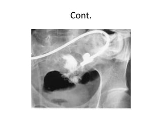
2. Introduction
• A fistula is defined as an abnormal
communication between two epithelialized
surfaces
• 2 types
– Internal fistula: the communication occurs between
two parts of the GIT or adjacent organs
• e.g., enterocolonic fistula or colovesicular fistula
– External fistula: involves the skin or another external
surface epithelium
• e.g., enterocutaneous fistula or rectovaginal fistula
3. Cont.
• Types of enterocutaneous fistulas
– Low-output
• Enterocutaneous fistulas that drain less than 200 mL of
fluid per day
– High-output fistulas
• Enterocutaneous fistulas that drain more than 500 mL
of fluid per day
4. Cont.
• More than 80% of enterocutaneous fistulas
represent iatrogenic complications that occur
as the result of enterotomies or intestinal
anastomotic dehiscences
• Fistulas that arise spontaneously without
antecedent iatrogenic injury are usually
manifestations of progression of underlying
Crohn's disease or cancer
5. Clinical Presentation
• Iatrogenic enterocutaneous fistulas usually become
clinically evident between the 5th and 10th postop
days
• Fever, prolonged ileus, abdominal tenderness,
wound infection, and leukocytosis are the initial
signs
• The diagnosis becomes obvious when drainage of
enteric material through the abdominal wound or
through existing drains occurs
• These fistulas are often associated with intra-
abdominal abscesses
6. Cont.
• Fistulas have the potential to close spontaneously
• Factors inhibiting spontaneous closure
– Fistula related
• High output fistulas
• Epithelialization of the fistula tract
– Systemic factors
• Malnutrition
• Sepsis
– Bowel related
• Inflammatory bowel disease
• Cancer
• Radiation
• Obstruction of the intestine distal to the origin of the fistula
• Foreign bodies
7. Complications
• Dehydration
• Electrolyte abnormalities
• Malnutrition
• Skin excoriation
• Reccurrent UTIs
(Enterovesicular fistulas)
The loss of enteric luminal
contents, particularly from
high-output fistulas
originating from the
proximal small intestine
8. Diagnosis
• CT scan (with enteral contrast) is the most useful initial
test
– Leakage of contrast material from the intestinal lumen can
be observed
– Intra-abdominal abscesses can also be seen and drained
percutaneously
• Small-bowel series or enteroclysis
– If the anatomy of the fistula is not clear on CT scanning
• Fistulography
– A contrast is injected under pressure through a catheter
placed percutaneously into the fistula tract
– It may offer greater sensitivity in localizing the fistula origin
10. Treatment
• The treatment of enterocutaneous fistulas
should proceed through an orderly sequence
of steps
11. Cont.
• Step 1: Stabilization
– Fluid and electrolyte resuscitation
– Nutritional rehabilitation
• Usually through the parenteral route initially
– Control of epsis
• Antibiotics and drainage of abscesses
– Protection of skin from the fistula effluent
• Ostomy appliances or fistula drains
12. Cont.
• Step 2: Investigation
– The anatomy of the fistula is defined using the
studies
13. • Step 3: Rehabilitation
– Probability of spontaneous closure is maximized
– Nutrition and time are the key components of this
phase
• Most patients will require TPN; however, a trial of oral or
enteral nutrition should be attempted in patients with low-
output fistulas originating from the distal intestine
• 2 to 3 months are allowed for spontaneous closure
– Fistulas that do not close during this period are unlikely to do so
– ?The somatostatin analogue octreotide
14. Cont.
• Surgery
– Procedure
• Resection of the fistula tract, together with the segment of
intestine from which it originates
• Simple closure of the opening in the intestine from which
the fistula originates is associated with high recurrence
rates
– Patients with intestinal fistulas typically have
extensive and dense intra-abdominal adhesions
• As a result, operations performed for nonhealing fistulas
can present formidable challenges
15. Prognosis
• Enterocutaneous fistulas are associated with a
10 to 15% mortality rate, mostly related to
sepsis or underlying disease
• Overall, 50% of intestinal fistulas close
spontaneously
A useful mnemonic designates factors that inhibit spontaneous closure of intestinal fistulas: FRIEND (foreign body within the fistula tract; radiation enteritis; infection/inflammation at the fistula origin; epithelialization of the fistula tract; neoplasm at the fistula origin; distal obstruction of the intestine)