Download as PDF, PPTX


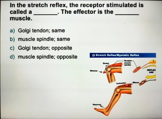


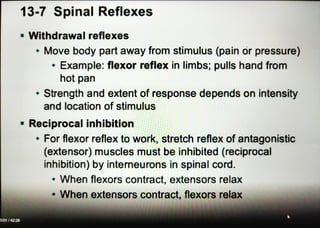


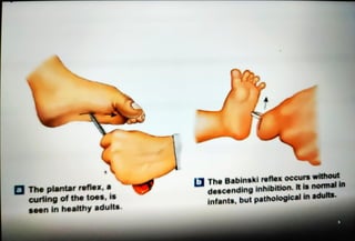

The document discusses spinal reflexes including stretch reflexes, polysynaptic reflexes, and crossed extensor reflexes. It describes the receptors, reflex arcs, and spinal cord pathways involved in these reflexes. It also mentions how the brain can alter spinal reflexes, giving examples of the plantar reflex and Babinski reflex.