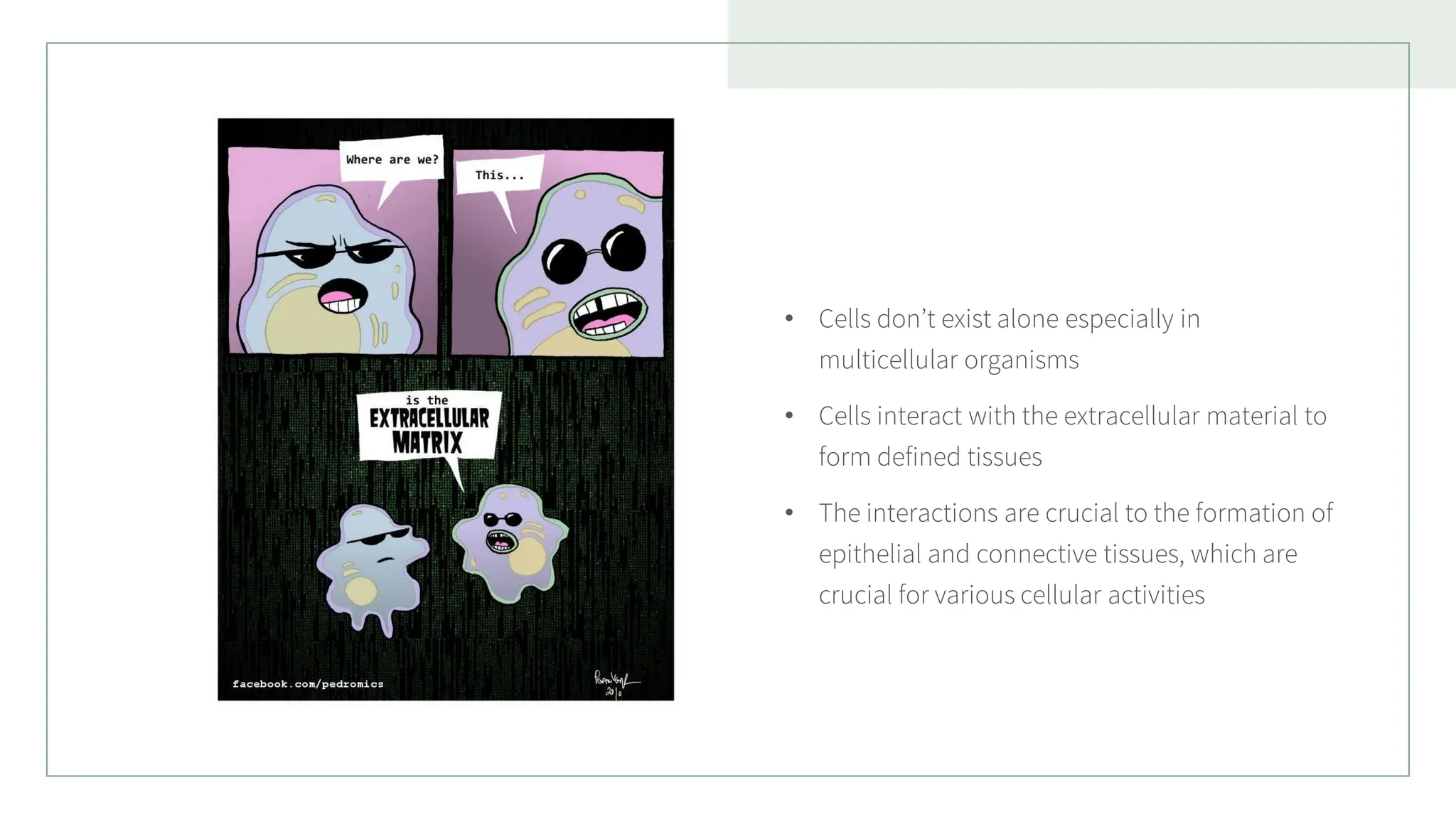
• Cells don’texist alone especially in
multicellular organisms
• Cells interact with the extracellular material to
form defined tissues
• The interactions are crucial to the formation of
epithelial and connective tissues, which are
crucial for various cellular activities
3.
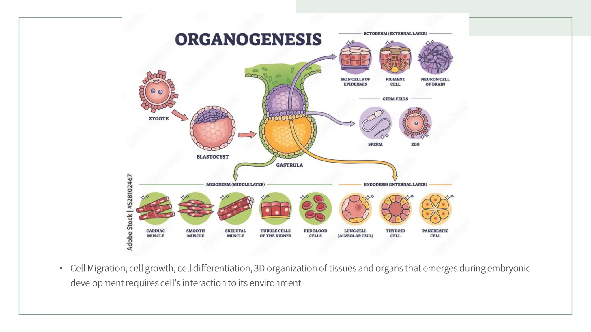
• Cell Migration,cell growth, cell differentiation, 3D organization of tissues and organs that emerges during embryonic
development requires cell’s interaction to its environment
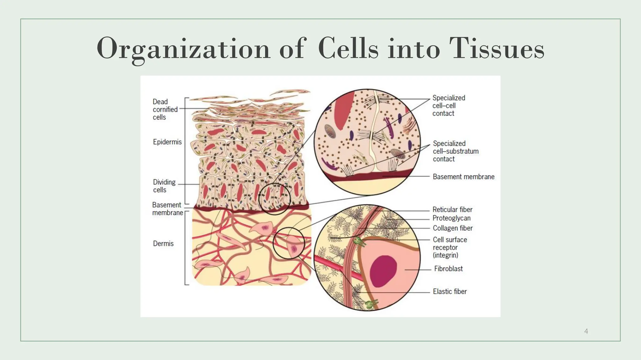
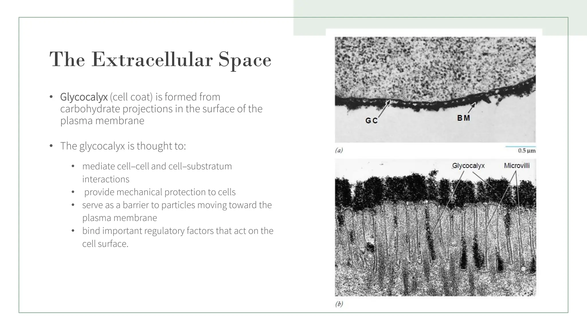
The Extracellular Space
•Glycocalyx (cell coat) is formed from
carbohydrate projections in the surface of the
plasma membrane
• The glycocalyx is thought to:
• mediate cell–cell and cell–substratum
interactions
• provide mechanical protection to cells
• serve as a barrier to particles moving toward the
plasma membrane
• bind important regulatory factors that act on the
cell surface.
5
6.
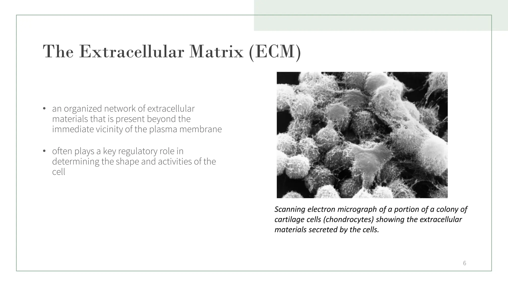
The Extracellular Matrix(ECM)
• an organized network of extracellular
materials that is present beyond the
immediate vicinity of the plasma membrane
• often plays a key regulatory role in
determining the shape and activities of the
cell
6
Scanning electron micrograph of a portion of a colony of
cartilage cells (chondrocytes) showing the extracellular
materials secreted by the cells.
7.
7
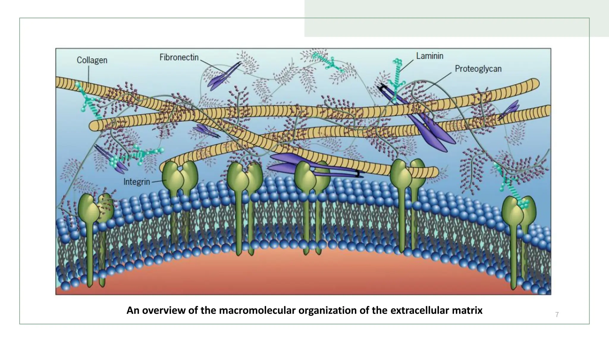
An overview ofthe macromolecular organization of the extracellular matrix
8.
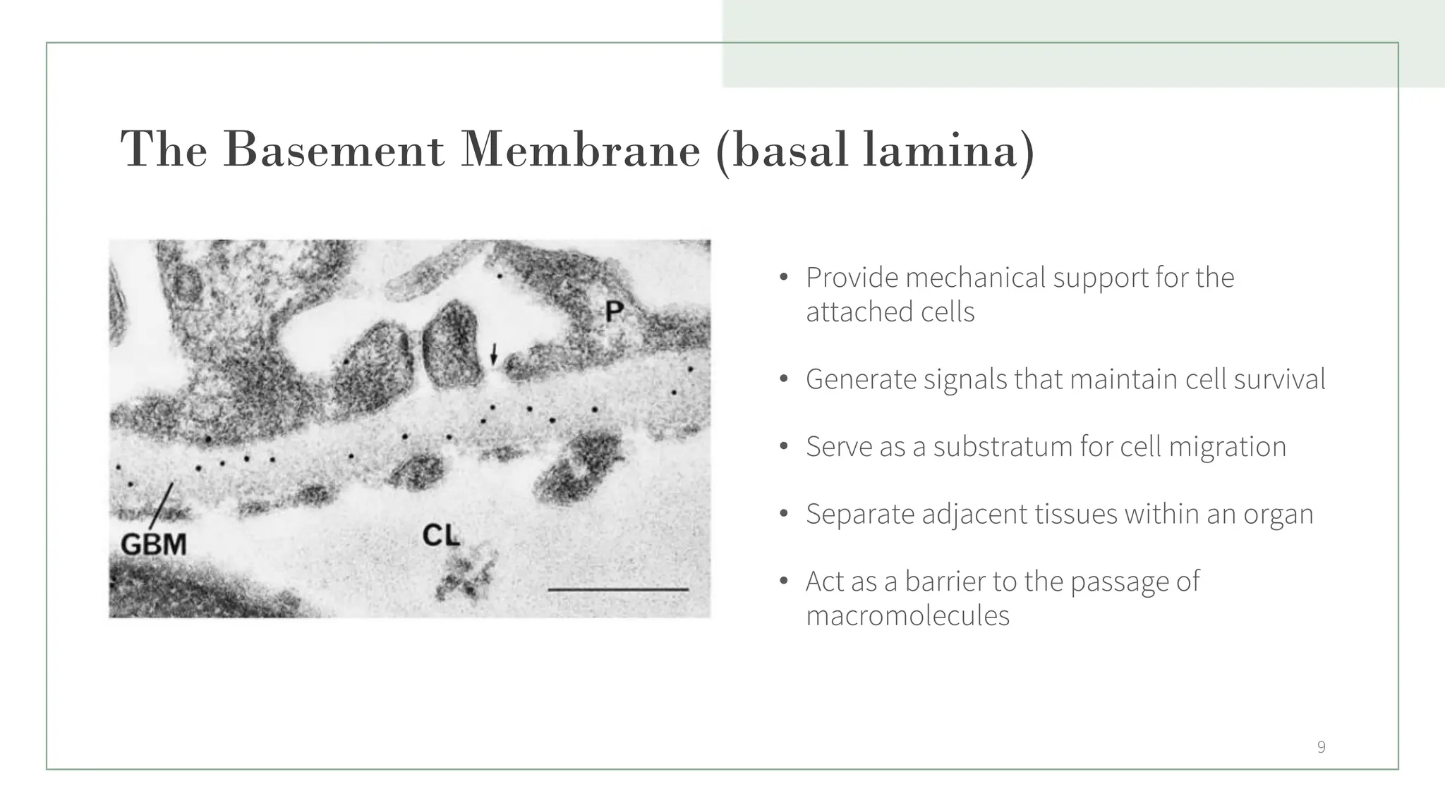
The Basement Membrane(basal lamina)
• One of the best defined extracellular
matrices
1. surrounds nerve fibers, muscles, and fat
cells
2. underlies the basal surface of epithelial
tissues, such as the epidermis of the
skin or the lining of the digestive and
respiratory tracts, and
3. underlies the inner endothelial lining of
blood vessels.
8
9.
The Basement Membrane(basal lamina)
• Provide mechanical support for the
attached cells
• Generate signals that maintain cell survival
• Serve as a substratum for cell migration
• Separate adjacent tissues within an organ
• Act as a barrier to the passage of
macromolecules
9
10.
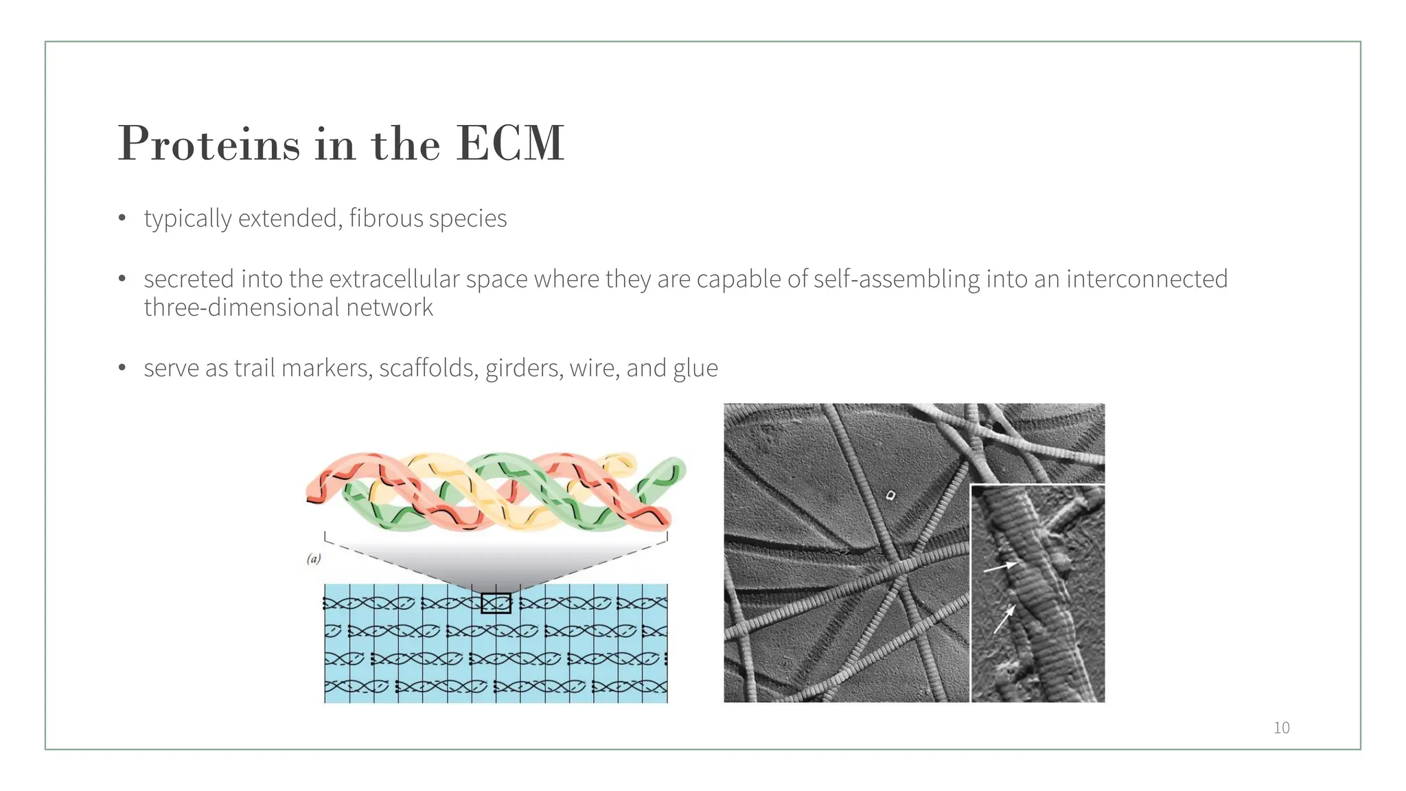
Proteins in theECM
• typically extended, fibrous species
• secreted into the extracellular space where they are capable of self-assembling into an interconnected
three-dimensional network
• serve as trail markers, scaffolds, girders, wire, and glue
10
11.
Collagen
• comprise afamily of fibrous
glycoproteins that are present only in
extracellular matrices
• noted for their high tensile strength
• single most abundant protein in the
human body
• produced primarily by fibroblasts
Collagen’s specific roles include:
• Helping fibroblasts form in your dermis (middle skin layer),
which helps new cells grow.
• Playing a role in replacing dead skin cells.
• Providing a protective covering for organs.
• Giving structure, strength and elasticity to your skin.
• Helping your blood to clot.
11
12.
Types of Collagen
•Type I. This type makes up 90% of your body’s collagen. Type I is densely packed and used to provide
structure to your skin, bones, tendons and ligaments.
• Type II. This type is found in elastic cartilage, which provides joint support.
• Type III. This type is found in muscles, arteries and organs.
• Type IV. This type is found in the layers of your skin.
• Type V. This type is found in the cornea of your eyes, some layers of skin, hair and tissue of the placenta.
12
13.
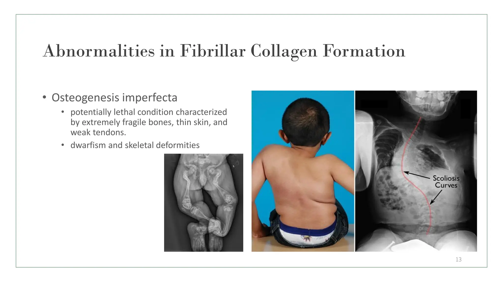
Abnormalities in FibrillarCollagen Formation
13
• Osteogenesis imperfecta
• potentially lethal condition characterized
by extremely fragile bones, thin skin, and
weak tendons.
• dwarfism and skeletal deformities
14.
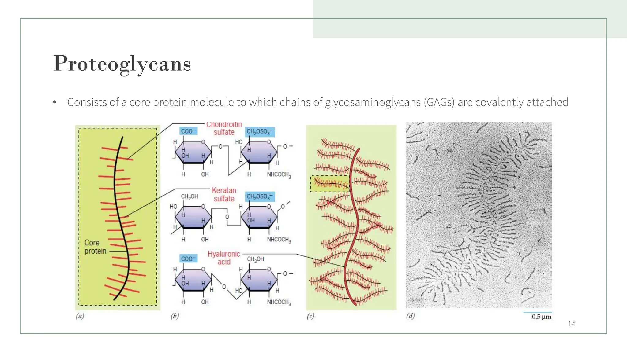
Proteoglycans
• Consists ofa core protein molecule to which chains of glycosaminoglycans (GAGs) are covalently attached
14
15.
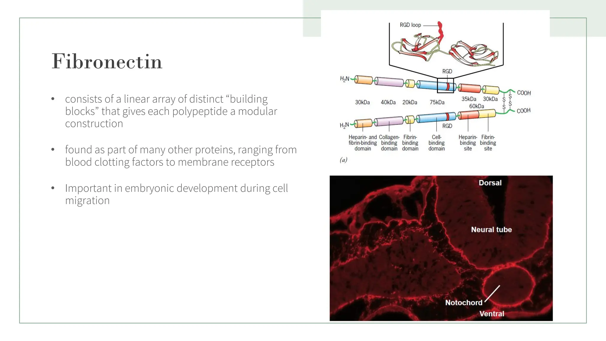
Fibronectin
• consists ofa linear array of distinct “building
blocks” that gives each polypeptide a modular
construction
• found as part of many other proteins, ranging from
blood clotting factors to membrane receptors
• Important in embryonic development during cell
migration
15
16.
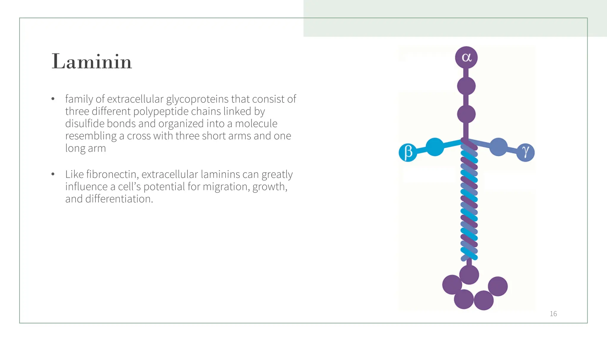
Laminin
• family ofextracellular glycoproteins that consist of
three different polypeptide chains linked by
disulfide bonds and organized into a molecule
resembling a cross with three short arms and one
long arm
• Like fibronectin, extracellular laminins can greatly
influence a cell’s potential for migration, growth,
and differentiation.
16
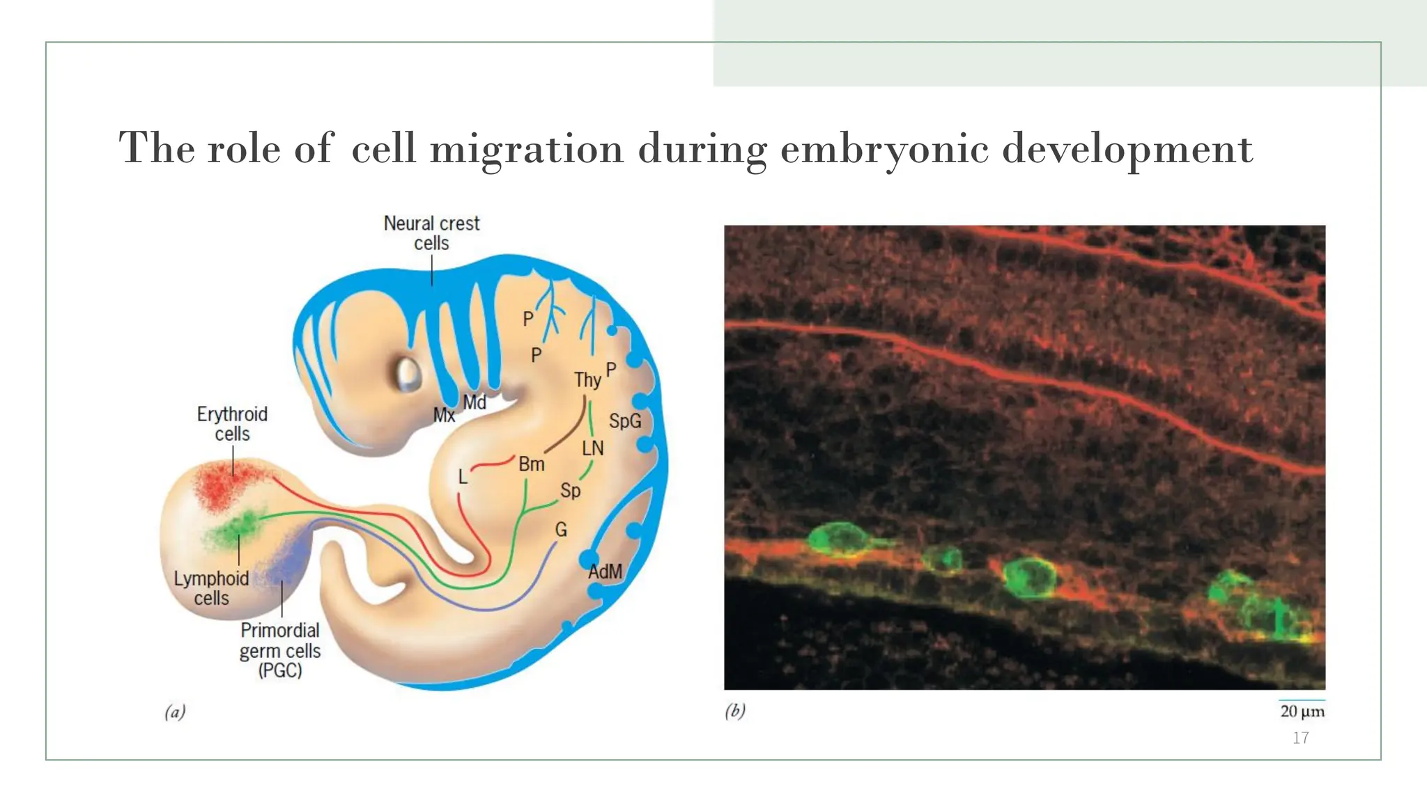
17.
The role ofcell migration during embryonic development
17
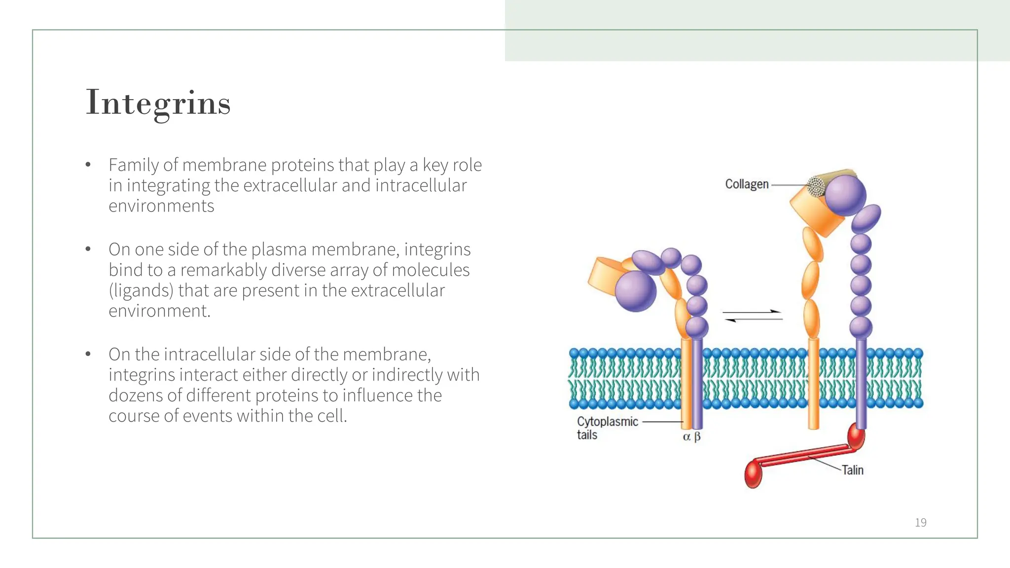
Integrins
• Family ofmembrane proteins that play a key role
in integrating the extracellular and intracellular
environments
• On one side of the plasma membrane, integrins
bind to a remarkably diverse array of molecules
(ligands) that are present in the extracellular
environment.
• On the intracellular side of the membrane,
integrins interact either directly or indirectly with
dozens of different proteins to influence the
course of events within the cell.
19
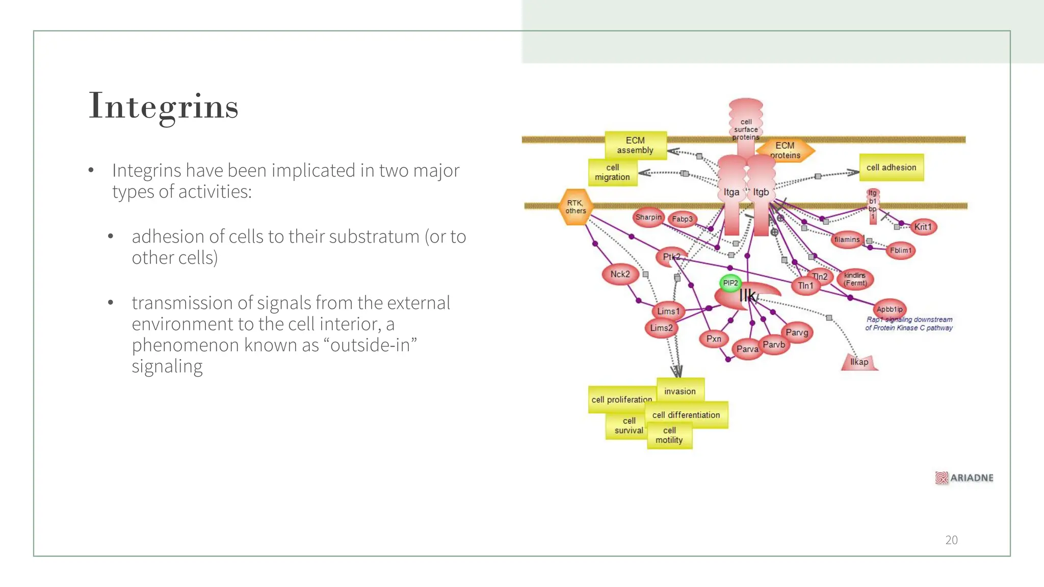
20.
Integrins
• Integrins havebeen implicated in two major
types of activities:
• adhesion of cells to their substratum (or to
other cells)
• transmission of signals from the external
environment to the cell interior, a
phenomenon known as “outside-in”
signaling
20
21.
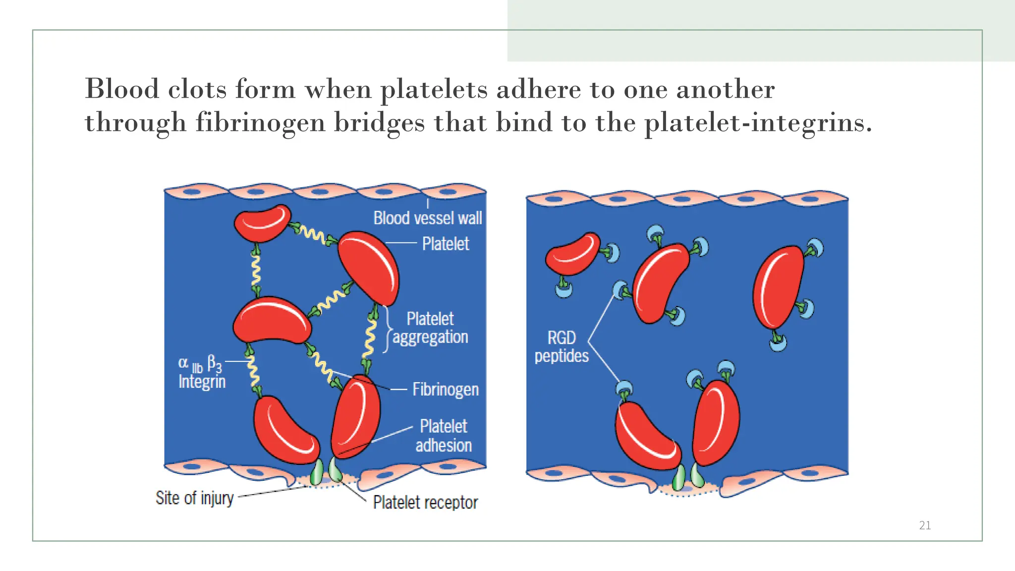
Blood clots formwhen platelets adhere to one another
through fibrinogen bridges that bind to the platelet-integrins.
21
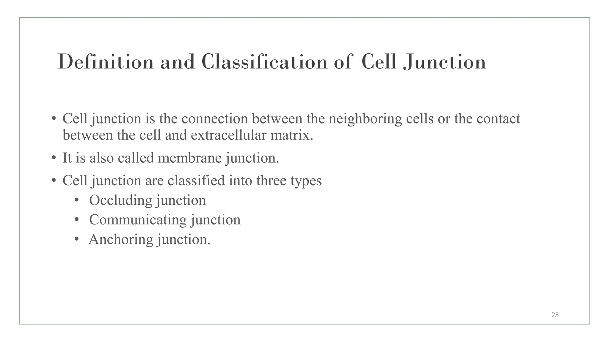
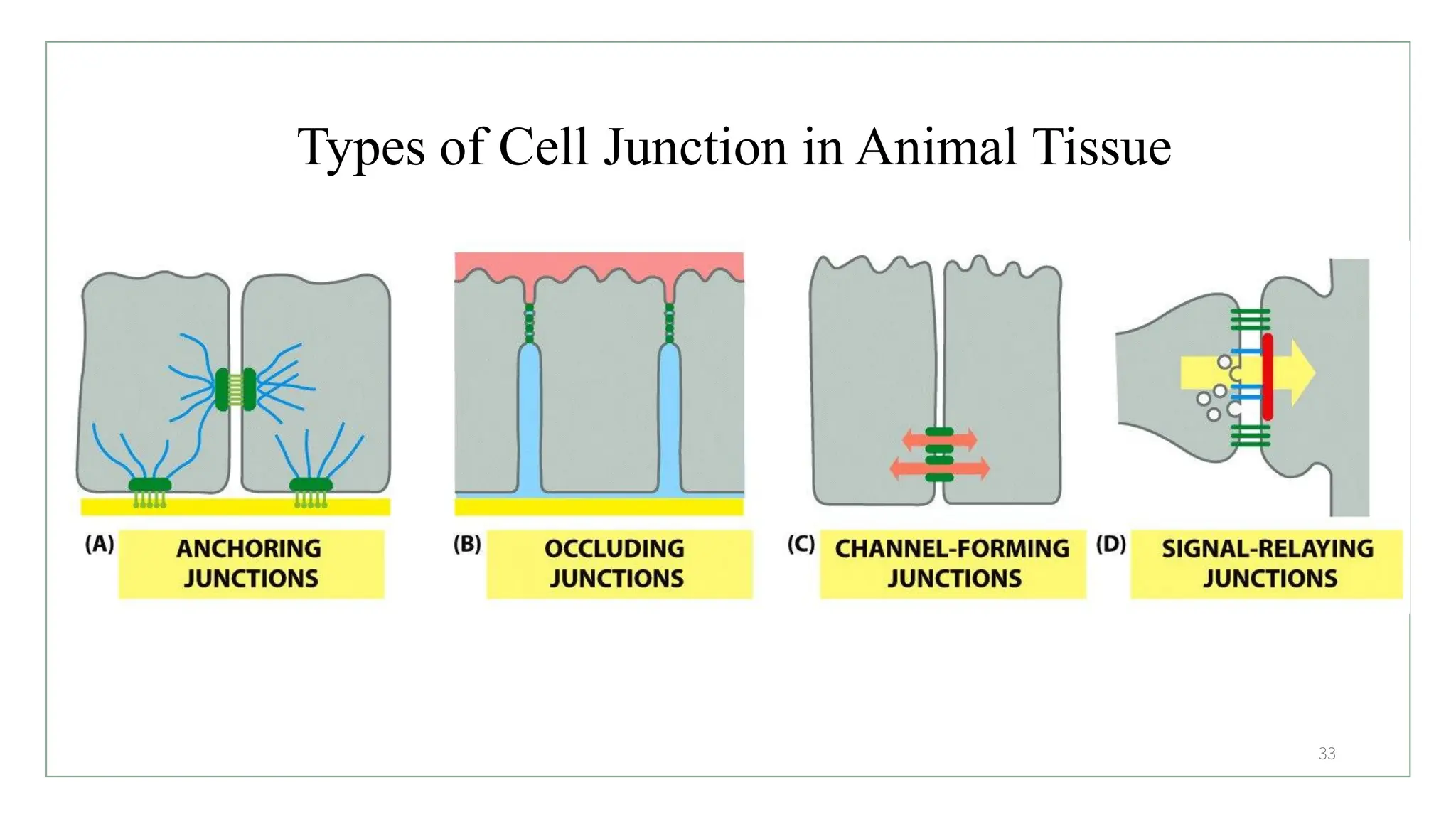
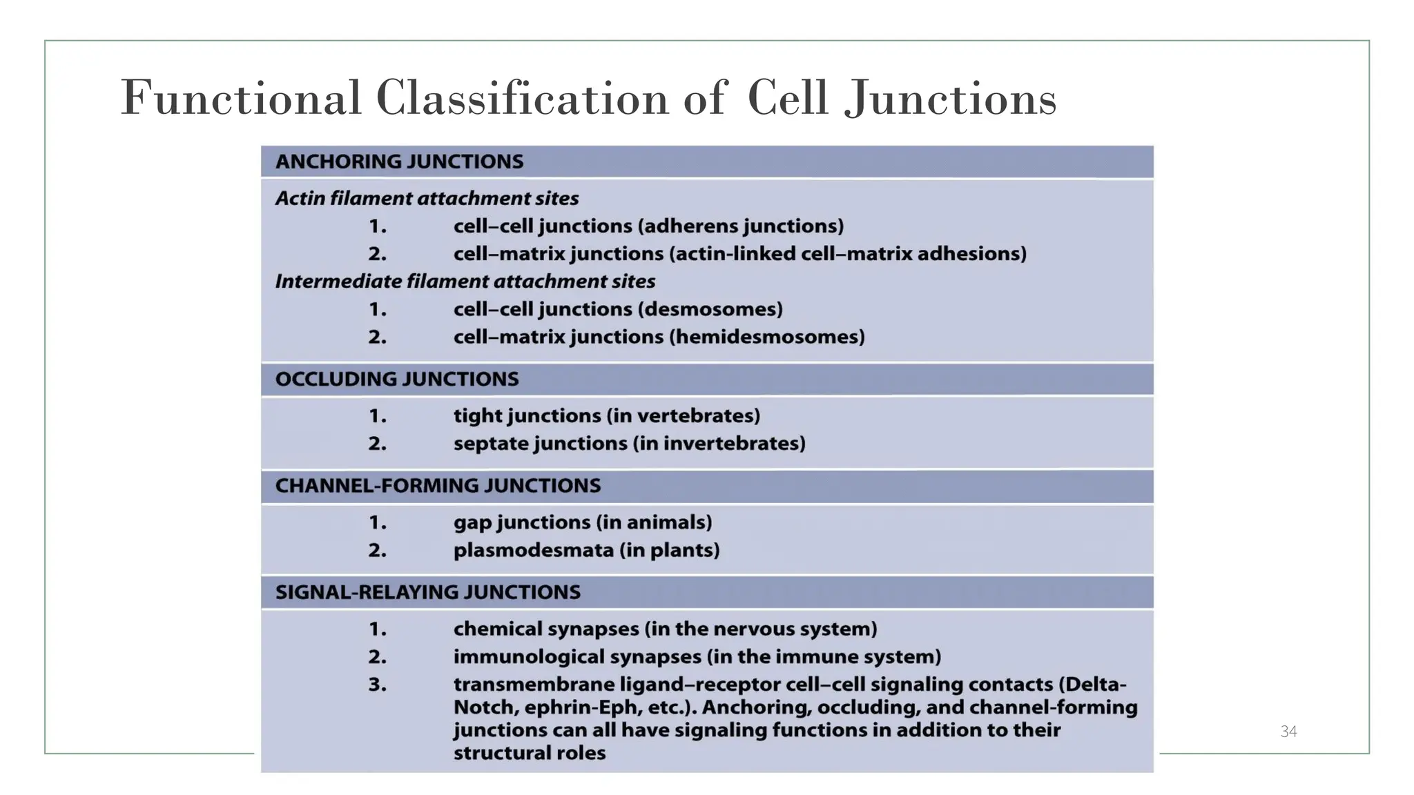
Definition and Classificationof Cell Junction
• Cell junction is the connection between the neighboring cells or the contact
between the cell and extracellular matrix.
• It is also called membrane junction.
• Cell junction are classified into three types
• Occluding junction
• Communicating junction
• Anchoring junction.
23
24.
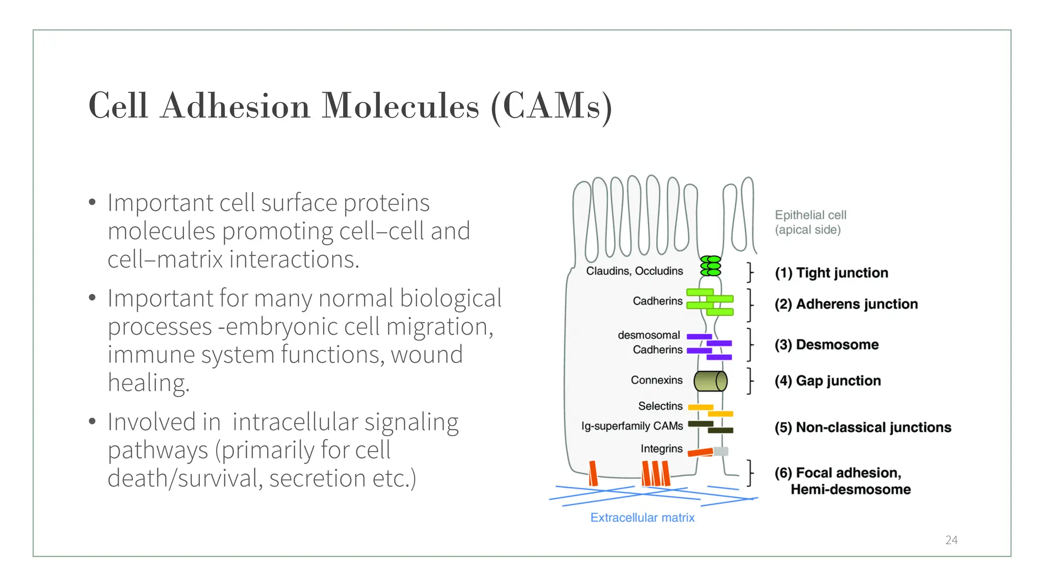
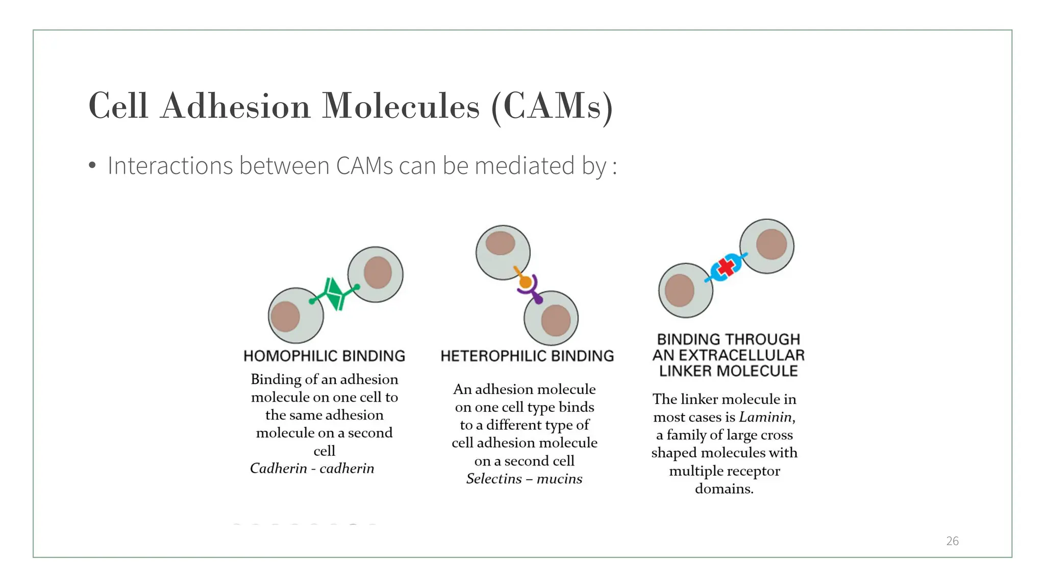
Cell Adhesion Molecules(CAMs)
• Important cell surface proteins
molecules promoting cell–cell and
cell–matrix interactions.
• Important for many normal biological
processes -embryonic cell migration,
immune system functions, wound
healing.
• Involved in intracellular signaling
pathways (primarily for cell
death/survival, secretion etc.)
24
25.
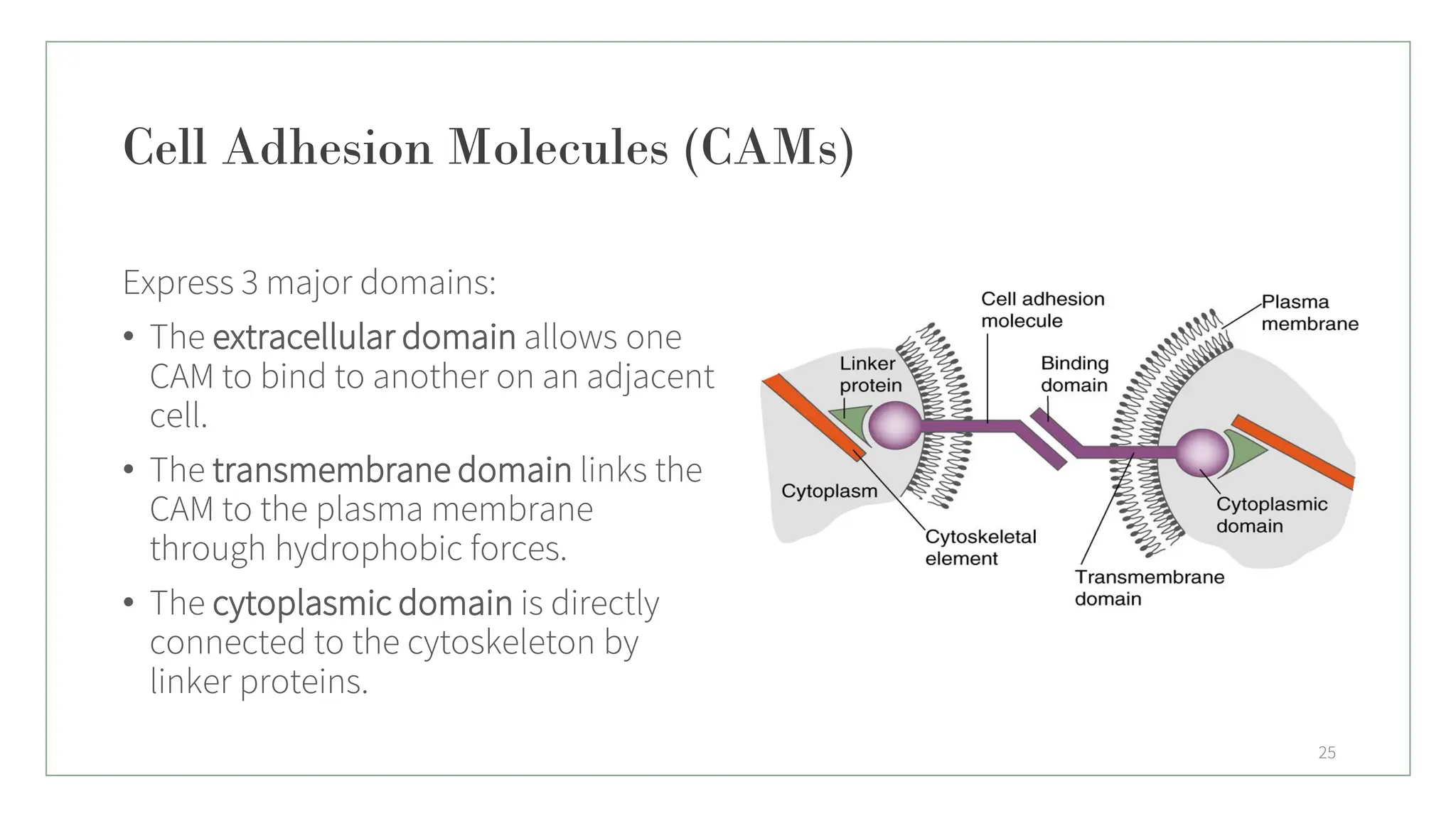
Cell Adhesion Molecules(CAMs)
Express 3 major domains:
• The extracellular domain allows one
CAM to bind to another on an adjacent
cell.
• The transmembrane domain links the
CAM to the plasma membrane
through hydrophobic forces.
• The cytoplasmic domain is directly
connected to the cytoskeleton by
linker proteins.
25
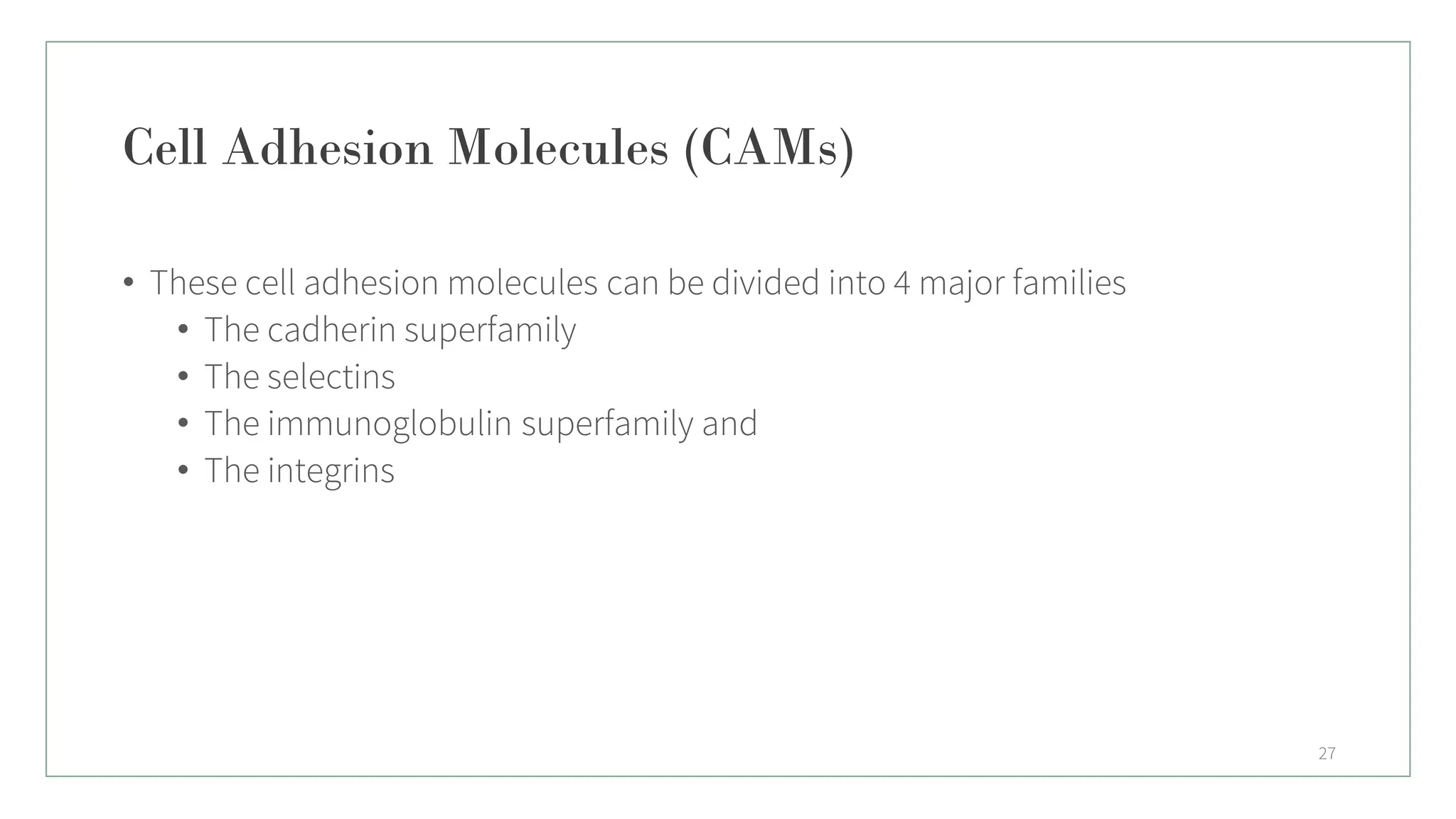
Cell Adhesion Molecules(CAMs)
• These cell adhesion molecules can be divided into 4 major families
• The cadherin superfamily
• The selectins
• The immunoglobulin superfamily and
• The integrins
27
28.
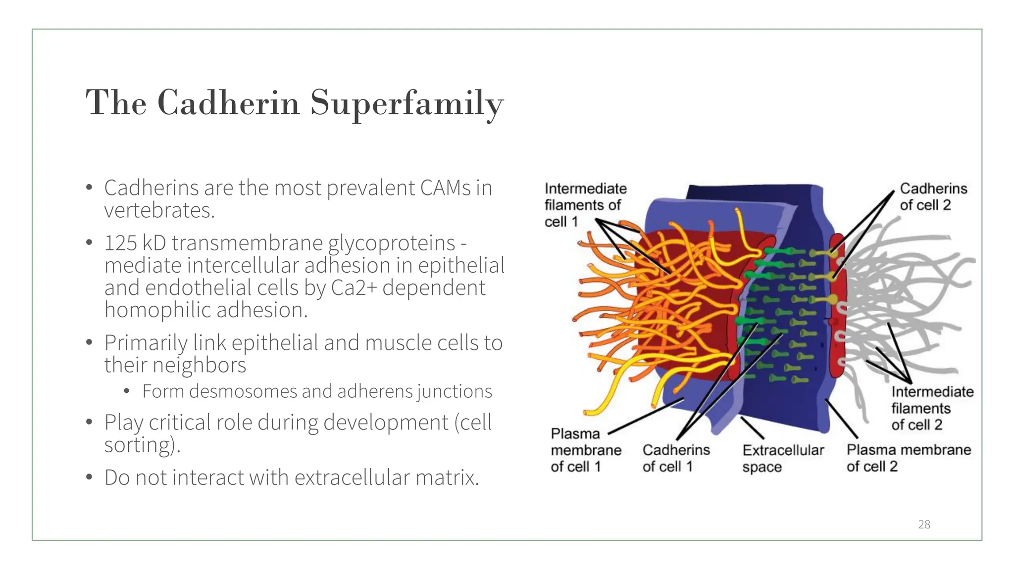
The Cadherin Superfamily
•Cadherins are the most prevalent CAMs in
vertebrates.
• 125 kD transmembrane glycoproteins -
mediate intercellular adhesion in epithelial
and endothelial cells by Ca2+ dependent
homophilic adhesion.
• Primarily link epithelial and muscle cells to
their neighbors
• Form desmosomes and adherens junctions
• Play critical role during development (cell
sorting).
• Do not interact with extracellular matrix.
28
29.
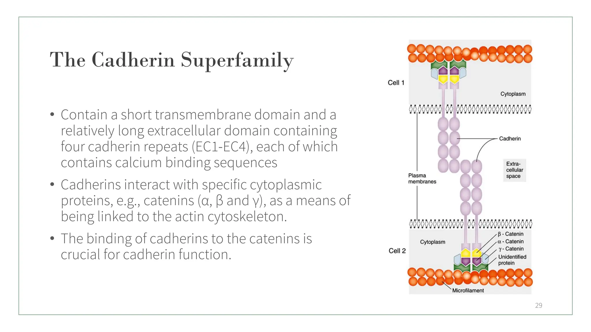
The Cadherin Superfamily
•Contain a short transmembrane domain and a
relatively long extracellular domain containing
four cadherin repeats (EC1-EC4), each of which
contains calcium binding sequences
• Cadherins interact with specific cytoplasmic
proteins, e.g., catenins (α, β and γ), as a means of
being linked to the actin cytoskeleton.
• The binding of cadherins to the catenins is
crucial for cadherin function.
29
30.
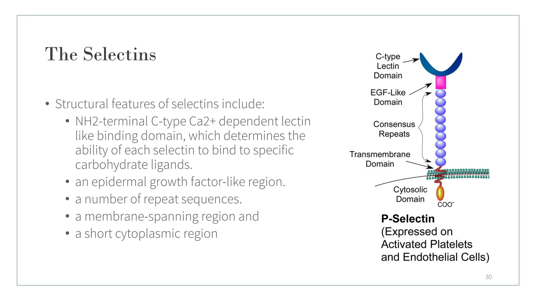
The Selectins
• Structuralfeatures of selectins include:
• NH2-terminal C-type Ca2+ dependent lectin
like binding domain, which determines the
ability of each selectin to bind to specific
carbohydrate ligands.
• an epidermal growth factor-like region.
• a number of repeat sequences.
• a membrane-spanning region and
• a short cytoplasmic region
30
31.
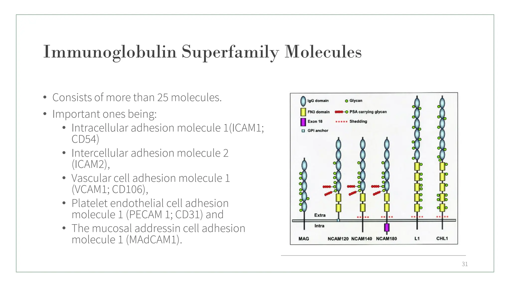
Immunoglobulin Superfamily Molecules
•Consists of more than 25 molecules.
• Important ones being:
• Intracellular adhesion molecule 1(ICAM1;
CD54)
• Intercellular adhesion molecule 2
(ICAM2),
• Vascular cell adhesion molecule 1
(VCAM1; CD106),
• Platelet endothelial cell adhesion
molecule 1 (PECAM 1; CD31) and
• The mucosal addressin cell adhesion
molecule 1 (MAdCAM1).
31
32.
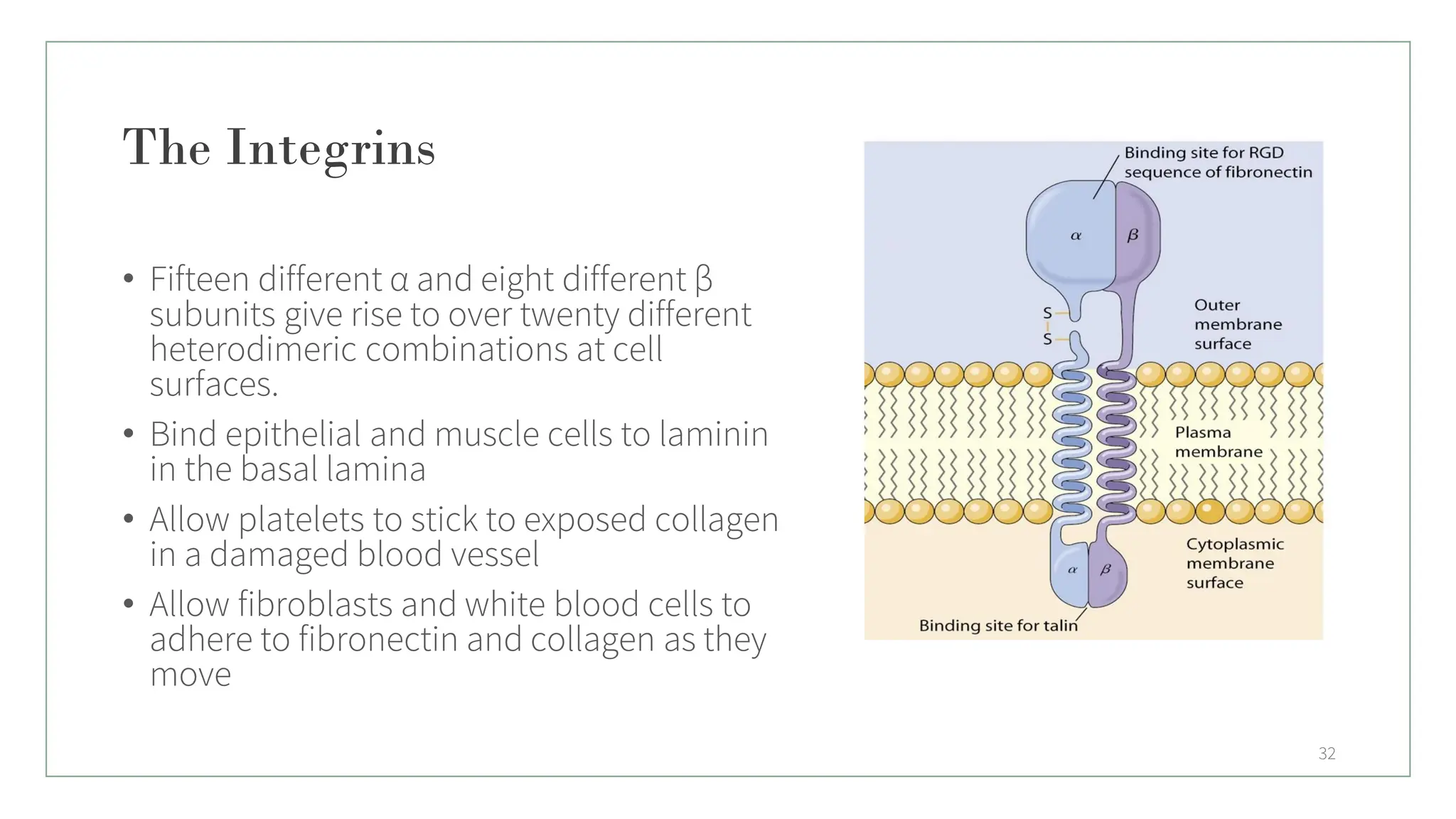
The Integrins
• Fifteendifferent α and eight different β
subunits give rise to over twenty different
heterodimeric combinations at cell
surfaces.
• Bind epithelial and muscle cells to laminin
in the basal lamina
• Allow platelets to stick to exposed collagen
in a damaged blood vessel
• Allow fibroblasts and white blood cells to
adhere to fibronectin and collagen as they
move
32
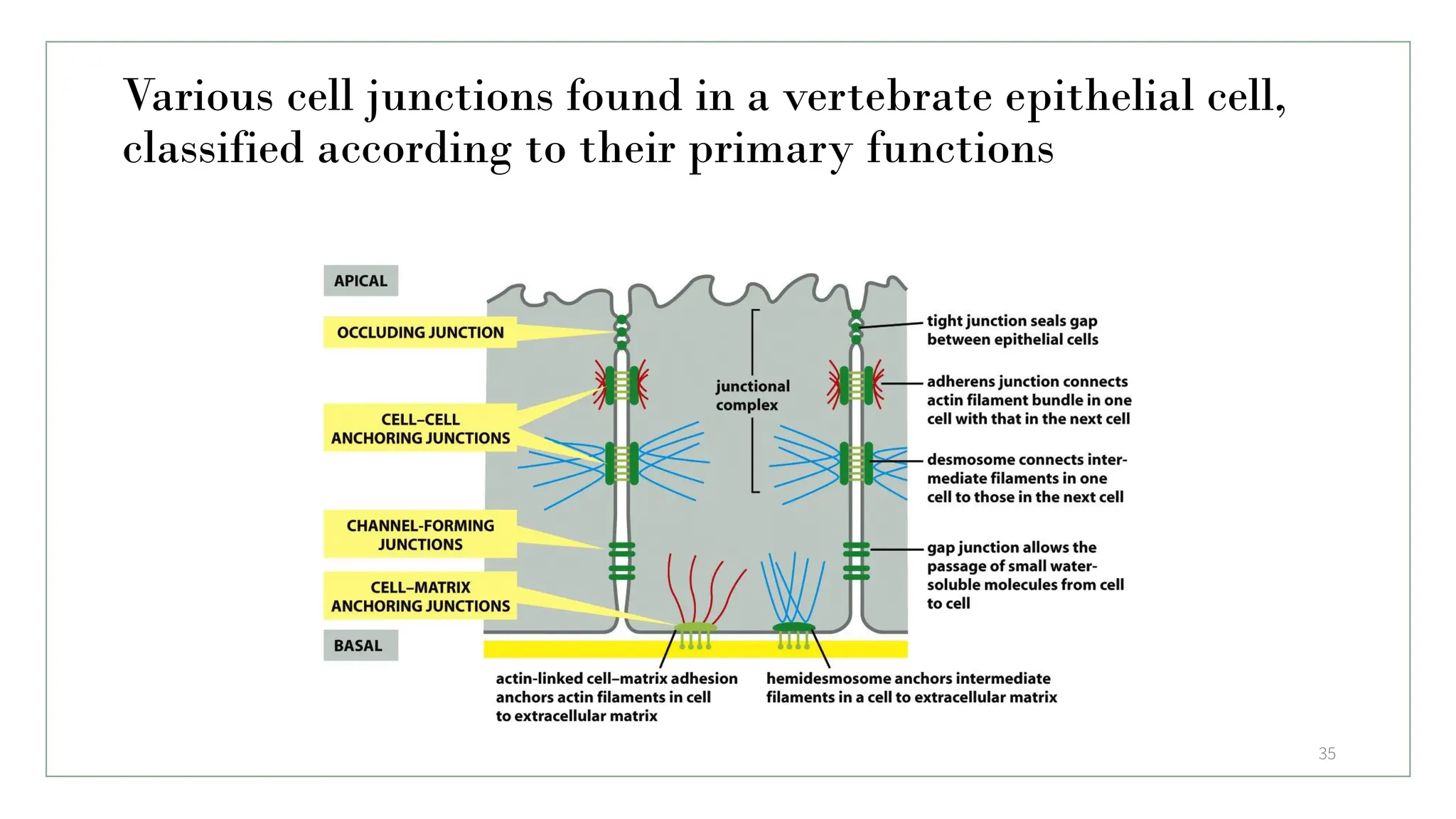
Various cell junctionsfound in a vertebrate epithelial cell,
classified according to their primary functions
35
36.
Occluding Junction
• Acell-cell junction that seals cells together in an epithelium in a way that prevents even
small molecules from leaking from one side of the sheet to the other.
Tight Junction
• Tight Junction- (occluding junctions / zonulae occludens - zonula occludens), are the
closely associated areas of two cells whose membranes join together forming a virtually
impermeable barrier to fluid.
• A type of junctional complex present only in vertebrates.
• Consist of linear array of several integral proteins.
• Junctional proteins occludins and claudins & members of IG superfamily are
transmembrane proteins.
36
37.
Function of TightJunction
• Strength and stability
• Selective permeable for ions.
• Fencing function
• Maintenance of cell polarity
• Blood-brain barrier
• Cludin -16 in Thick Junctions of Ascending Loop of Henle.
• Cludin- 15 Permability of cations / anions.
37
38.
Adhering Junctions
• Desmosome-connects intermediate filament of one cell with other cells.
• Claudin
• Hemidesmosome
• Desmoplakin is essential for normal desmosomal adhesion.
38
39.
Communicating Junction
• Celljunction which permit the intercellular exchange of substance are called
communicating junction, these junction permit the movement of ions and
molecules from one cell to another cell.
• Gap junction
• Chemical synapse
39
40.
Function of GapJunction
• Channel passage the substance have molecular weight less than 1000 amu.
• Exchange of chemical messenger between cells
• Rapid propagation of action potential from one cell to another cell.
40
41.
Desmosomes
• Also knownas macula adherens; a cell structure specialized for cell-to-cell
adhesion.
• Are molecular complexes of cell adhesion proteins and linking proteins that
attach the cell surface adhesion proteins to intracellular keratin cytoskeletal
filaments.
• The cell adhesion proteins of the desmosome, desmoglein and desmocollin, are
members of the cadherin family.
41
42.
Hemidesmosomes
• Hemidesmosomes looklike half-desmosomes that attach cells to the underlying
basal lamina.
• Rather than using desmogleins, hemidesmosomes use desmopenetrin cell
adhesion proteins, which are members of Integrin family.
• The integrin molecule attach to one of many multi-adhesive proteins such as
laminin, resident within the extracellular matrix, thereby forming one of many
potential adhesions between cell and matrix.
42
43.
Anchoring Junction
• Anchoringjunction are the junction, which provides strength to the cell by acting
like mechanical attachment.
• These junction provide firm structural attachment between two cells or between
a cell and extracellular matrix
• Anchoring junction are responsible for structural integrity of the tissue.
43