Download to read offline
![Prathigudupu RS et al. Facial Asymmetry Post TMJ Ankylosis.
41
Journal of Advanced Medical and Dental Sciences Research |Vol. 6|Issue 7| July 2018
Journal of Advanced Medical and Dental Sciences Research
@Society of Scientific Research and Studies
Journal home page: www.jamdsr.com doi: 10.21276/jamdsr UGC approved journal no. 63854
Case Report
Orthomorphic and Orthognathic treatment for Aesthetics and Function in Facial
Asymmetry Post TMJ Ankylosis - A Case Report
1
Raja Satish Prathigudupu, 2
Ranjith Kumar P, 3
Phani Kumar K, 4
Surekha Kanala, 5
Rahul VC Tiwari, 6
Latheef Saheb S.
1
Senior Registrar, Ministry of Health, Amiri Dental Casuality, Kuwait,
2
Professor & Consultant, Confydenz Dental Clinic, Guntur, Andhra Pradesh, India,
3
Professor, OMFS, Sibar Institute of Dental Sciences, Takkellapadu, Guntur, AP, India,
4
Professor & HOD, Government Dental College, Vijayawada, Andhra Pradesh, India,
5
FOGS, MDS, OMFS & Dentistry, JMMCH & RI, Thrissur, Kerala, India,
6
Consultant, Partha Dental Clinic, Hyderabad, Telangana, India
ABSTRACT:
Introduction: Orthomorphic surgery is performed when alone orthognathic is unable to correct the condition. It refers to correction
offacial asymmetry or jaw deviation without changing the occlusion. It is the best treatment option available so far for such deformities.
Case Report: A 23-year-old male patient presented to us with the complaint of flattening of the right side of face. Patient gave a history
of childhood trauma which lead to temporomandibular joint ankylosis and was operated for the same at the age of 10 years. He reported
for unaesthetic appearance and was diagnosed as facial asymmetry with occlusal cant. Treatment was planned in two stages by giving
priority to correctfunction first and then proceeding for the aesthetics 6 months later. Conclusion: An optimal function and aesthetic was
achieved post-operatively. The described technique has the quality to alter the dentofacial deformity in any magnitude and directionin
accurate dimension.
Key words: Orthognathic surgery, orthomorphic surgery, facial asymmetry, two stage procedure.
Received: 2 May 2018 Revised: 16 May 2018 Accepted: 17 May 2018
Corresponding Author: Dr. Raja Satish Prathigudupu, Senior Registrar, Ministry of Health, Amiri Dental Casuality,
Kuwait.
This article may be cited as: Prathigudupu RS, P Kumar R, K Kumar P, Kanala S, Tiwari RV, S Saheb L. Orthomorphic
and Orthognathic treatment for Aesthetics and Function in Facial Asymmetry Post TMJ Ankylosis - A Case Report. J Adv
Med Dent Scie Res 2018;6(7):41-45.
INTRODUCTION:
All normal human faces have some degree of asymmetry.
The ancient Greeks were probably the first to notice these
variations between the two sides of the face, as discovered
much later by analysis of their statuary, which included
mild to moderate facial asymmetries. Normal asymmetries
such as these often go unnoticed by the general public.
Esthetically pleasing and apparently symmetrical faces do
indeed exhibit skeletal asymmetries and one side of the face
can be rather different from the other and still be considered
completely normal. The level at which asymmetry becomes
unacceptable to a patient is variable and depends on many
factors, most of which are psychological.[1] Facial
asymmetry, when obvious, has enormous
sociopsychological impact on the affected individuals. It
can occur as a consequence of developmental anomalies or
disease or after trauma or surgery. Surgical reconstruction is
usually indicated in most instances involving a noticeable
facial asymmetry. This is usually accomplished by
reconstructing the deformed portion with its normal
counterpart working as a reference.[2] Patients who present
with significant facial asymmetry are not only concerned
with restoring functional occlusion but also with improving
esthetics and beauty. This has often been a source of social
scorn for many of these patients. Beauty and symmetry have
often been thought of synonymously; hence, the belief that
(e) ISSN Online: 2321-9599; (p) ISSN Print: 2348-6805 SJIF (Impact factor) 2017= 6.261; Index Copernicus value 2016 = 76.77](https://image.slidesharecdn.com/42ndpublication-jamdsr-5thname-200113073924/85/42nd-publication-jamdsr-5th-name-1-320.jpg)
![Prathigudupu RS et al. Facial Asymmetry Post TMJ Ankylosis.
41
Journal of Advanced Medical and Dental Sciences Research |Vol. 6|Issue 7| July 2018
Journal of Advanced Medical and Dental Sciences Research
@Society of Scientific Research and Studies
Journal home page: www.jamdsr.com doi: 10.21276/jamdsr UGC approved journal no. 63854
Case Report
Orthomorphic and Orthognathic treatment for Aesthetics and Function in Facial
Asymmetry Post TMJ Ankylosis - A Case Report
1
Raja Satish Prathigudupu, 2
Ranjith Kumar P, 3
Phani Kumar K, 4
Surekha Kanala, 5
Rahul VC Tiwari, 6
Latheef Saheb S.
1
Senior Registrar, Ministry of Health, Amiri Dental Casuality, Kuwait,
2
Professor & Consultant, Confydenz Dental Clinic, Guntur, Andhra Pradesh, India,
3
Professor, OMFS, Sibar Institute of Dental Sciences, Takkellapadu, Guntur, AP, India,
4
Professor & HOD, Government Dental College, Vijayawada, Andhra Pradesh, India,
5
FOGS, MDS, OMFS & Dentistry, JMMCH & RI, Thrissur, Kerala, India,
6
Consultant, Partha Dental Clinic, Hyderabad, Telangana, India
ABSTRACT:
Introduction: Orthomorphic surgery is performed when alone orthognathic is unable to correct the condition. It refers to correction
offacial asymmetry or jaw deviation without changing the occlusion. It is the best treatment option available so far for such deformities.
Case Report: A 23-year-old male patient presented to us with the complaint of flattening of the right side of face. Patient gave a history
of childhood trauma which lead to temporomandibular joint ankylosis and was operated for the same at the age of 10 years. He reported
for unaesthetic appearance and was diagnosed as facial asymmetry with occlusal cant. Treatment was planned in two stages by giving
priority to correctfunction first and then proceeding for the aesthetics 6 months later. Conclusion: An optimal function and aesthetic was
achieved post-operatively. The described technique has the quality to alter the dentofacial deformity in any magnitude and directionin
accurate dimension.
Key words: Orthognathic surgery, orthomorphic surgery, facial asymmetry, two stage procedure.
Received: 2 May 2018 Revised: 16 May 2018 Accepted: 17 May 2018
Corresponding Author: Dr. Raja Satish Prathigudupu, Senior Registrar, Ministry of Health, Amiri Dental Casuality,
Kuwait.
This article may be cited as: Prathigudupu RS, P Kumar R, K Kumar P, Kanala S, Tiwari RV, S Saheb L. Orthomorphic
and Orthognathic treatment for Aesthetics and Function in Facial Asymmetry Post TMJ Ankylosis - A Case Report. J Adv
Med Dent Scie Res 2018;6(7):41-45.
INTRODUCTION:
All normal human faces have some degree of asymmetry.
The ancient Greeks were probably the first to notice these
variations between the two sides of the face, as discovered
much later by analysis of their statuary, which included
mild to moderate facial asymmetries. Normal asymmetries
such as these often go unnoticed by the general public.
Esthetically pleasing and apparently symmetrical faces do
indeed exhibit skeletal asymmetries and one side of the face
can be rather different from the other and still be considered
completely normal. The level at which asymmetry becomes
unacceptable to a patient is variable and depends on many
factors, most of which are psychological.[1] Facial
asymmetry, when obvious, has enormous
sociopsychological impact on the affected individuals. It
can occur as a consequence of developmental anomalies or
disease or after trauma or surgery. Surgical reconstruction is
usually indicated in most instances involving a noticeable
facial asymmetry. This is usually accomplished by
reconstructing the deformed portion with its normal
counterpart working as a reference.[2] Patients who present
with significant facial asymmetry are not only concerned
with restoring functional occlusion but also with improving
esthetics and beauty. This has often been a source of social
scorn for many of these patients. Beauty and symmetry have
often been thought of synonymously; hence, the belief that
(e) ISSN Online: 2321-9599; (p) ISSN Print: 2348-6805 SJIF (Impact factor) 2017= 6.261; Index Copernicus value 2016 = 76.77](https://image.slidesharecdn.com/42ndpublication-jamdsr-5thname-200113073924/75/42nd-publication-jamdsr-5th-name-1-2048.jpg)
![Prathigudupu RS et al. Facial Asymmetry Post TMJ Ankylosis.
42
Journal of Advanced Medical and Dental Sciences Research |Vol. 6|Issue 7| July 2018
unattractiveness is the result of asymmetry.[3] Unilateral
temporomandibular joint ankylosis occurring during the
active growth period if left without treatment, or when
improperly treated, is often complicated by the development
of secondary changes in the structure, shape, and size of the
mandible together with the surrounding tissues.[4]
Mandibular asymmetry may be caused by infection and
trauma during the growing period. Primary trauma may lead
to asymmetry and in some cases ankylosis. Asymmetry may
also follow a surgical procedure or a malunited fracture.[5]
Orthognathic surgery for the correction of facial deformity
arising from discrepancy in spatial relationship or
dimensional differences is well established. However, when
the cause of the deformity includes an alteration of the
shape of the jaws, orthognathic surgery is unable to correct
the resulting contour deformity.[6] For this reason in the
management of facial asymmetry, orthomorphic principles
of management are an adjunct to orthognathic surgery or
Osseo distraction.[7] The surgical correction consisting of
an osteotomy aimed at restoring the morphology is denoted
the term “orthomorphic” to distinguish it from conventional
orthognathic surgery. The orthomorphic correction aims to
correct deformities related to shape and contour of the jaws
without affecting the status of occlusion.[6]
CASE REPORT:
A 23-year-old male patient presented to us with the
complaint of flattening of the right side of face. Patient gave
a history of childhood trauma which lead to
temporomandibular joint ankylosis and was operated for the
same at the age of 10 years. He reported for unaesthetic
appearance and was diagnosed as facial asymmetry with
occlusal cant. Treatment was planned in two stages by
giving priority to correct function first and then proceeding
for the aesthetics 6 months later.On occlusal examination an
occlusal cant was present.(Figure 1) Clinically a clear-cut
asymmetry was seen in the lower border of
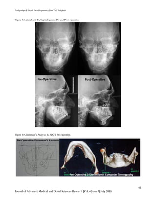
mandible.(Figure 2) Lateral Cephalogram and
Posteroanterior Cephalogram Confirmed the diagnosis of
facial asymmetry by deviation of jaw on the left side due to
underdevelopment of growth from childhood trauma
leading to TMJ ankylosis. (Figure 3)Grumman’s analysis
and Three-dimensional computed tomography elicited
mandibular deviation on left side. (Figure 4). After
confirmation of diagnosis first occlusal correction was
planned by performing a differential Le Fort I osteotomy as
the vertical height was more on the left side compared to
right side. So, the occlusal cant was corrected. After 6
months of correcting occlusion, facial asymmetry correction
was planned. An extended lateralsliding genioplasty was
done for correction of mandibular underdevelopment. We
achieved patients and our expectation by combining
orthognathic correction primarily and orthomorphic
correction secondarily after 6 months.
Figure 1: Clinical Picture & Occlusion Cant Pre and Post-
operative.
Figure 2: Deviation & Occlusion Pre and Post-operative.](https://image.slidesharecdn.com/42ndpublication-jamdsr-5thname-200113073924/85/42nd-publication-jamdsr-5th-name-2-320.jpg)
![Prathigudupu RS et al. Facial Asymmetry Post TMJ Ankylosis.
44
Journal of Advanced Medical and Dental Sciences Research |Vol. 6|Issue 7| July 2018
DISCUSSION:
Surgical correction of facial asymmetry is more challenging
than the correction of symmetrical deformities. Often,
unilateral mandibular deficiencies are associated with slight
downward growth of the maxilla on the affected side,
altering the position of midline structures of the middle-
third of the face and creating an occlusal cant. We resorted
to a line drawn perpendicular to the inter-inner canthal line
as the facial midline. [8] Inferior border osteotomy of the
mandible performed intraorally can be employed
independently or in conjunction with other procedures to
correct facial asymmetry of the mandible. Genioplasty is a
well-documented procedure to move the chin to a more
desirable position. The anterior chin can be moved in all 3
dimensions of space. Extended lateral sliding genioplasty is
the technique followed by the first author since 1992 for the
correction of facial asymmetry, especially for residual
deformities of unilateral TMJ ankylosis. In TMJ ankylosis,
because of the destruction of the growth center and the
missing functional stimulus, the mandible on the affected
side is short due to deficient growth. The opposite side of
the mandible is dragged towards the affected side. This
leads to facial asymmetry, with false fullness of the affected
side and deficiency of the unaffected side, and resultant
deviation of the mandible. The conventional technique of
aesthetic correction of the above-described deformity is by
ramus osteotomies and bone grafting. This technique often
jeopardizes the existing occlusion. In most patients, because
of the pre-existing trismus, the teeth are unhealthy, hence
orthodontic treatment may not be feasible. Benefits of the
‘long’ osteotomy cut extending to the gonial region
bilaterally have been put forward by Tessier. [9] They
include maintenance of a smooth natural inferior
mandibular border, noninterference with the occlusion, and
simultaneous increase in the width of the jaw line. Extended
lateral sliding genioplasty is an intraoral procedure where
the inferior mandibular order is cut from one gonial region
to the opposite gonial region below the inferior alveolar
canal. Wolfe estimated the position of the inferior alveolar
canal to be 6 mm below the mental foramen. [10] We made
the osteotomy cut on the deficient side approximately 5 mm
below the mandibular canal. However, the cut was slanted
downwards by approximately 45° towards the lingual side
so that the nerve canal was spared. Multiple holes were
made on the lingual cortex and the bone was split using
osteotomes and/ or a split spreader. None of the patients
experienced transection of the nerve inside the canal. The
divided bone segment was slid forward and laterally, so that
the chin and the sunken lateral aspect of the mandible were
augmented. The deficient side of the mandible was
lengthened and the deviation of the chin was corrected. The
same technique can be employed in conjunction with other
osteotomies such as sagittal ramus split, maxillary
osteotomies, etc., for correction of complex asymmetric
deformities. Stepping on the lateral mandibular contour is
often created after lateral sliding of the extended
genioplasty segment and, in such cases, autogenous bone
grafting can be used to interpose or obliterate the step
deformity. When aesthetic correction is performed
simultaneously with functional correction of ankylosis, the
resected bone may be used as the graft material thus
eliminating the need for donor site surgery. Management of
the mental nerve is an important issue. Hinds and Kent
favor dissection and protection of the mental nerve during
osteotomy. [11] Posnick et al also advocated dissection and
retraction during osteotomy and reported persistent sensory
deficit in 10% of their patients after 1 year. [12] Spear and
Kassan [13] and Lindquist and Obeid [14] advised
identification and protection of the mental nerve. These
authors reported permanent mental nerve deficit in 6% [13]
and 10% [13] of patients, respectively, and are against the
idea of dissecting the nerve. Converse and Wood-Smith
were of the opinion that the mental nerve may be divided if
it obstructs the osteotomy. [15] To obtain adequate space to
perform inferior border osteotomy of the mandible,
extensive dissection or traction is required on the mental
nerve. Moreover, inadvertent avulsion of the nerve is a
possibility and is a more serious problem than intentional
division of the nerve because it does not lend itself to
correction by microsurgical repair. The advantages of
extended lateral sliding genioplasty may be summarized as
follows: a simple procedure accomplished through an
intraoral degloving incision in the buccal vestibule, less
time-consuming than the conventional ramus osteotomies
for the correction of unilateral mandibular deficiencies,
existing occlusion is not disturbed, deficient side of the
mandible is lengthened, the chin is brought close to the
midline, the apparent deficiency on the unaffected side is
corrected by the lateral shift of the inferior border segment
and thus fullness is achieved, the procedure can be done
concomitantly with the correction of TMJ ankylosis, the
procedure can be combined with other orthognathic
procedures such as ramus sagittal split and maxillary
osteotomy, harmony and balance of the face (vertical and
horizontal proportions) are improved, psychological
rehabilitation of the patient is rapid.
CONCLUSION:
The surgical correction of facial asymmetry is extremely
challenging because the asymmetry may be centered at the
hard and/or soft tissue; any of a combination of dimensions;
and it may involve the maxilla, mandible, and symphysis or
any combination of the three. It is the effective treatment of
the hard tissues that brings about the most dramatic change,
as soft tissue defects are usually corrected after skeletal
correction.[16] Combination of orthognathic and
orthomorphic surgery is useful in correcting cases of
mandibular deformity which increases quality of life of
patients.](https://image.slidesharecdn.com/42ndpublication-jamdsr-5thname-200113073924/85/42nd-publication-jamdsr-5th-name-4-320.jpg)
![Prathigudupu RS et al. Facial Asymmetry Post TMJ Ankylosis.
45
Journal of Advanced Medical and Dental Sciences Research |Vol. 6|Issue 7| July 2018
REFERENCES:
1. Severt TR, Proffit WR. The prevalence of facial asymmetry
in thedentofacial deformities population at the University of
North Carolina.Int J Adult Orthod Orthognath Surg
1997;12:171-6.
2. Wong TY, Fang JJ, Chung CH, Huang JS, Lee JW.
Comparison of 2methods of making surgicalmodels for
correction of facial asymmetry.J Oral Maxillofac Surg
2005;63:200-8.
3. Stringer D, Brown B. Correction of mandibular asymmetry
using angledtitanium mesh. J Oral Maxillofac Surg
2009;67:1619-27.
4. El-Sheikh MM, Medra AM. Management of unilateral
temporomandibularankylosis associatedwith facial asymmetry.
J Craniomaxillofac Surg1997;25:109-15.
5. Cohen MM. Perspectives on craniofacial asymmetry. III.
Commonand/or well-known causes ofasymmetry. Int J Oral
Maxillofac Surg1995;24:127-33.
6. Salins PC, Venkatraman B, Kavarody M. Morphometric
basis fororthomorphic correction ofmandibular asymmetry. J
Oral MaxillofacSurg 2008;66:1523-31.
7. Kent JN, Craig MA. Secondary autogenous and alloplastic
reshapingprocedures for facialasymmetry. Atlas Oral
Maxillofac Surg Clin NorthAm 1996;4:83-105.
8. Mani V, Ranjith P. Efficacy of extended slidinggenioplasty
[thesis]. Calicut University, Kerala,India; 1993-1995.
9. Tessier P. Chin advancement as an aid in thecorrection of the
deformities of the mental and submentalregions: discussions.
Plast Reconstr Surg1981;67:630.
10. Wolfe S. Chin advancement as an aid in correctionof
deformities of the deformities of the mentaland submental
regions. Plast Reconstr Surg 1981;6:624-629.
11. Hinds EC, Kent JN. Genioplasty: the versatilityof horizontal
osteotomy. J Oral Surg 1969;27:690-700.
12. Posnick JC, AL-Qattan MM, Stepner NM.Alteration in facial
sensibility in adolescentsfollowingsagittal split and chin
osteotomies ofthe mandible. Plast Reconstr Surg 1996;97:920-
927.
13. Spear SL, Kassan M. Genioplasty. Clin Plast
Surg1989;48:210-216.
14. Lindquist CC, Obeid G. Complications of genioplastydone
alone or in combination with sagittalsplit osteotomy. Oral Surg
1988;6:13-16.
15. Converse JM, Wood-Smith D. Horizontal osteotomyof the
mandible. Plast Reconstr Surg1964;34:464-471.
16. Reyneke JP, Tsakiris P, Kienle F. A simple classification for
surgicaltreatment planning ofmaxillomandibular asymmetry.
Br J OralMaxillofac Surg 1997;35:349-51.
Source of support: Nil Conflict of interest: None declared
This work is licensed under CC BY: Creative Commons Attribution 3.0 License.](https://image.slidesharecdn.com/42ndpublication-jamdsr-5thname-200113073924/85/42nd-publication-jamdsr-5th-name-5-320.jpg)

This document describes a case report of a 23-year-old male patient who presented with facial asymmetry and flattening of the right side of the face due to childhood temporomandibular joint ankylosis. Treatment involved a two-stage procedure, first using orthognathic surgery (Le Fort I osteotomy) to correct occlusal cant, followed by orthomorphic surgery (extended lateral sliding genioplasty) 6 months later to correct the facial asymmetry and underdevelopment of the mandible. The combination of orthognathic and orthomorphic surgery successfully achieved functional and aesthetic goals by correcting the jaw deviation and restoring facial symmetry.


