
Recommended
More Related Content
What's hot
What's hot (20)
Similar to 2010 expedited correction of significant dentofacial
Similar to 2010 expedited correction of significant dentofacial (20)
Recently uploaded
Recently uploaded (20)
2010 expedited correction of significant dentofacial
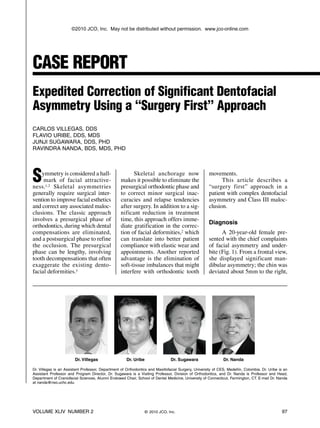
- 1. Symmetry is considered a hall- mark of facial attractive- ness.1,2 Skeletal asymmetries generally require surgical inter- vention to improve facial esthetics and correct any associated maloc- clusions. The classic approach in volves a presurgical phase of orthodontics, during which dental compensations are eliminated, and a postsurgical phase to refine the occlusion. The presurgical phase can be lengthy, involving tooth decompensations that often exaggerate the existing dento facial deformities.3 Skeletal anchorage now makes it possible to eliminate the presurgical orthodontic phase and to correct minor surgical inac- curacies and relapse tendencies after surgery. In addition to a sig- nificant reduction in treatment time, this approach offers imme- diate gratification in the correc- tion of facial deformities,2 which can translate into better patient compliance with elastic wear and appointments. Another reported advantage is the elimination of soft-tissue imbalances that might interfere with ortho dontic tooth movements. This article describes a “surgery first” approach in a patient with complex dentofacial asymmetry and Class III maloc- clusion. Diagnosis A 20-year-old female pre- sented with the chief complaints of facial asymmetry and under- bite (Fig. 1). From a frontal view, she displayed significant man- dibular asymmetry; the chin was deviated about 5mm to the right, VOLUME XLIV NUMBER 2 97 CASE REPORT Expedited Correction of Significant Dentofacial Asymmetry Using a “Surgery First” Approach CARLOS VILLEGAS, DDS FLAVIO URIBE, DDS, MDS JUNJI SUGAWARA, DDS, PHD RAVINDRA NANDA, BDS, MDS, PHD © 2010 JCO, Inc. Dr. Nanda Dr. Sugawara Dr. Uribe Dr. Villegas Dr. Villegas is an Assistant Professor, Department of Orthodontics and Maxillofacial Surgery, University of CES, Medellín, Colombia. Dr. Uribe is an Assistant Professor and Program Director, Dr. Sugawara is a Visiting Professor, Division of Orthodontics, and Dr. Nanda is Professor and Head, Department of Craniofacial Sciences, Alumni Endowed Chair, School of Dental Medicine, University of Connecticut, Farmington, CT. E-mail Dr. Nanda at nanda@nso.uchc.edu. ©2010 JCO, Inc. May not be distributed without permission. www.jco-online.com
- 2. Correction of Dentofacial Asymmetry Using a “Surgery First” Approach Fig. 1 20-year-old female with sig nificant mandibular asymmetry and skeletal Class III malocclusion be fore treatment. 98 JCO/FEBRUARY 2010
- 3. but there was no vertical compo- nent to the asymmetry. She had a consonant smile with 80% incisor display, but an asymmetrical lower lip line with less animation on the right. The maxillary dental mid- line was coincident with the cupid’s bow, while the mandibu- lar dental midline was deviated 5mm to the right. In the sagittal view, the patient had a slight con- cavity of the hard tissues due to a prognathic mandible. The soft- tissue profile was straight, how- ever, because of deficient chin thickness. The lower lip was slightly protrusive, and there was no mentolabial fold. The patient had a Class I molar occlusion on the right, a full-cusp Class III occlusion on the left, and a negative overjet of 4mm, with moderate anterior crowding in both arches. The maxillary incisor inclination was ideal, while the mandibular inci- sors were slightly upright. A uni- lateral crossbite was noted on the right side, and there was a mild anterior open bite. Because the patient was extremely self-conscious about her facial asymmetry, she was amenable to surgical options. She accepted a “surgery first” ap proach that would achieve an esthetic smile and normal occlu- sion while minimizing the time required in fixed appliances and addressing her main esthetic concern. Surgical Plan Although the patient had a slight paranasal deficiency, the maxillary anteroposterior posi- tion was adequate. Based on the etiology of the malocclusion, we decided on an asymmetrical sin- gle-jaw surgery, with the mandi- ble set back 7mm on the left and 3mm on the right, to address both the prognathism and the asym- metry. In addition, an anteriorly sliding genioplasty would main- tain soft-tissue convexity, reduce the lower lip protrusion, and accentuate the mentolabial fold (Fig. 2). The postsurgical occlusion was planned to exhibit excessive overjet and an end-to-end Class II Fig. 2 A. Model surgery showing asymmetrical mandibular setbacks of 7mm on left and 3mm on right, with menton and lower dental midline moved 3mm left to match facial and maxillary dental midlines. B. Expected postsurgical Class II occlusion, intended to maintain maxillary incisor inclination. VOLUME XLIV NUMBER 2 99 Villegas, Uribe, Sugawara, and Nanda A B
- 4. relationship. The buccal segments would then be distalized into a Class I occlusion, using maxillary miniplates as anchorage, to create the space needed to align the maxillary anterior teeth without affecting the ideal incisor inclina- tion. The excessive overjet would be resolved by labial movement of the lower incisors. Treatment Progress One week before ortho gnathic surgery, .022" preadjusted brackets were bonded, and bands were placed on the first and sec- ond molars. A bilateral sagittal split osteotomy was performed to achieve the required asymmetri- cal setback, accompanied by a sliding genioplasty with a 4mm advancement. All four third molars were extracted to avoid the need for later surgery.4,5 In addi- tion, four miniplates were placed on the infrazygomatic crest of the maxilla and in the external oblique ridge of the mandible (Fig. 3). After soft-tissue closure, .016" × .016" and .014" nickel titanium wires were inserted in the maxillary and mandibular arches, respectively, with the maxillary archwire bypassing the crowded central incisors and left lateral incisor (Fig. 4). Intermax illary elastics were worn from the maxillary first premolars to the mandibular canines and from the maxillary miniplate to the man- dibular canines. Two weeks after surgery, the patient exhibited moderate swelling, but an .016" × .016" stainless steel maxillary archwire could still be tied in, again bypass- ing the central incisors and the left lateral incisor. An elastomer- ic chain was extended from the miniplate to the canine on the right side and from the miniplate to the first premolar on the left side (Fig. 5). Two weeks later, the elastomeric chains were replaced by nickel titanium coil springs. Two months after surgery, the molars and canines were in Class I occlusion. Conveniently, a slight space had opened between the maxillary right central and lateral incisors, and this was used to match the midlines. Treatment Results Seven months after surgery, the fixed appliances were re moved. The final records showed good esthetic and occlusal results, and the superimpositions con- firmed the achievement of all treatment objectives. The miniplates were left in place for six months of retention. During this time, we evaluated the stability of the orthodontic treatment, and the miniplates could have been used if any post- operative orthodontic or surgical relapse had occurred. With no relapse evident, the miniplates were removed after six months (Fig. 6). Discussion Because orthodontic tooth movement generally has little ef 100 JCO/FEBRUARY 2010 Correction of Dentofacial Asymmetry Using a “Surgery First” Approach Fig. 3 Placement of four miniplates in infrazygomatic crest of maxilla and external oblique ridge of mandible during surgery.
- 5. fect on extraoral soft-tissue esthet- ics, camouflage treatment alone cannot be relied on to rectify severe dentofacial asymmetries. Surgical correction becomes complicated, however, when the soft- and hard-tissue discrepan- cies do not match. In this patient, although the hard-tissue profile was concave, the soft tissues were straight. Cor rection of the maloc- clusion therefore required an asymmetrical setback with an advancing genioplasty. The “surgery first” approach described by Nagasaka and col- leagues has two significant advan- tages: immediate correction of soft-tissue deformities and reduced treatment time.6-8 In addition, the placement of four miniplates pro- vides three-dimensional control for postsurgical correction of any relapse tendencies or slight dis- crepancies be tween the planned and actual surgical outcomes. If plates are inserted in all quad- rants regardless of the surgical procedure (one- or two-jaw), these vertical and anteroposterior ad justments in tooth position need not rely solely on elastics. Placing miniplates does increase the time required for surgery by an average 10-15 minutes per plate, but we have not encountered any intra- or postoperative complications with this surgical approach. In contrast to the technique described by Nagasaka and col- leagues, who placed passive stiff wires conforming to the maloc- VOLUME XLIV NUMBER 2 101 Villegas, Uribe, Sugawara, and Nanda Fig. 4 .016" .016" nickel titanium wire placed in maxillary arch, bypassing incisors to prevent flaring; .014" nickel titanium wire placed in mandibular arch for alignment. Fig. 5 Distalization of maxillary buccal segments into Class I relation ship, using anchorage from maxillary miniplates.
- 6. Fig. 6 A. Patient after seven months of treatment. B. Radiographs taken six months later, after removal of miniplates at end of retention period. C. Superimposition of cephalometric tracings before treatment (black), immediately after surgery (blue), and after six months of retention (red). B A 102 JCO/FEBRUARY 2010 Correction of Dentofacial Asymmetry Using a “Surgery First” Approach C
- 7. VOLUME XLIV NUMBER 2 103 Villegas, Uribe, Sugawara, and Nanda clusion,6 we inserted nickel tita- nium archwires after the soft tissues were sutured in the operat- ing room. This method could expedite tooth movement by tak- ing advantage of the increased cell turnover that occurs after mechanical alteration of bone.9 Such biological response has been noted with corticotomy-assisted tooth movement; bone turnover can also be accelerated in areas distant to the surgical site.10 In another departure from Nagasaka’s approach, we did not use a splint to stabilize the occlu- sion after surgery. Model surgery indicated that the occlusion would be stable in a cusp-to-cusp rela- tionship, and the maxillary teeth could then be distalized to relieve the anterior crowding while maintaining the incisor positions and inclination. The patient was delighted with the dramatic esthetic change achieved in such a short per iod of wearing fixed appliances. Conclusion The “surgery first” approach can be used to address complex dentofacial asymmetry, as shown in this case. Treatment time can be substantially reduced by elim- inating the presurgical phase and taking advantage of increased bone turnover, which in turn can accelerate tooth movement. ACKNOWLEDGMENTS: The authors thank Drs. Brett Holliday and Amirparviz Davoody for their collaboration on the manuscript. REFERENCES 1. Bashour, M.: History and current con- cepts in the analysis of facial attractive- ness, Plast. Reconstr. Surg. 118:741- 756, 2006. 2. Thornhill, R. and Gangestad, S.W.: Facial attractiveness, Trends Cogn. Sci. 3:452-460, 1999. 3. Proffit, W.R. and White, R.P.: Com bining surgery and orthodontics: Who does what, when? in Contemporary Treatment of Dentofacial Deformity, ed. W.R. Proffit, Mosby, St. Louis, 2003, pp. 245-267. 4. Mehra, P.; Castro, V.; Freitas, R.Z.; and Wolford, L.M.: Complications of the mandibular sagittal split ramus osteoto- my associated with the presence or absence of third molars, J. Oral Maxillofac. Surg. 59:854-858, 2001. 5. Precious, D.S.; Lung, K.E.; Pynn, B.R.; and Goodday, R.H.: Presence of impact- ed teeth as a determining factor of unfavorable splits in 1256 sagittal-split osteotomies, Oral Surg. Oral Med. Oral Pathol. Oral Radiol. Endod. 85:362- 365, 1998. 6. Nagasaka, H.; Sugawara, J.; Kawamura, H.; and Nanda, R.: “Surgery first” skel- etal Class III correction using the Skeletal Anchorage System, J. Clin. Orthod. 43:97-105, 2009. 7. Brachvogel, P.; Berten, J.L.; and Hausa men, J.E.: [Surgery before ortho dontic treatment: A concept for timing the combined therapy of skeletal dys- gnathias], Deutsche Zahn. Mund. Kieferheilk. Zentralb. 79:557-563, 1991. 8. Tsuruda, H. and Miyamoto, Y.: None or minimum pre-operative orthodontic treatment for orthognathic surgery in answer to patient’s request of immediate facial aspect change, J. Jap. Soc. Aesth. Plast. Surg. 25:79-86, 2003. 9. Frost, H.M.: The biology of fracture healing: An overview for clinicians, Part II, Clin. Orthop. Relat. Res. 248:294-309, 1989. 10. Mueller, M.; Schilling, T.; Minne, H.W.; and Ziegler, R.: A systemic acceleratory phenomenon (SAP) accompanies the regional acceleratory phenomenon (RAP) during healing of a bone defect in the rat, J. Bone Miner. Res. 6:401- 410, 1991.