Download as PDF, PPTX

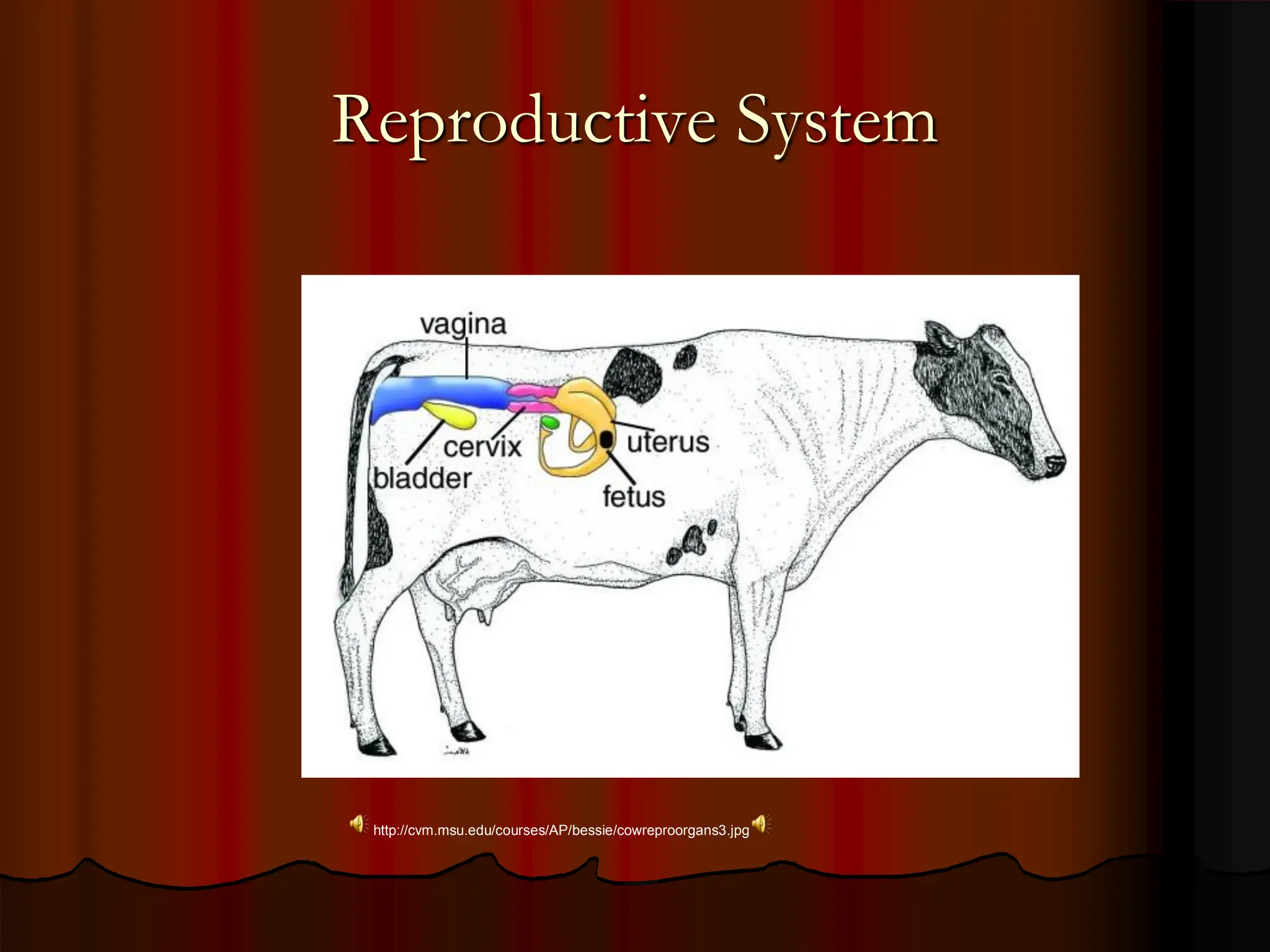
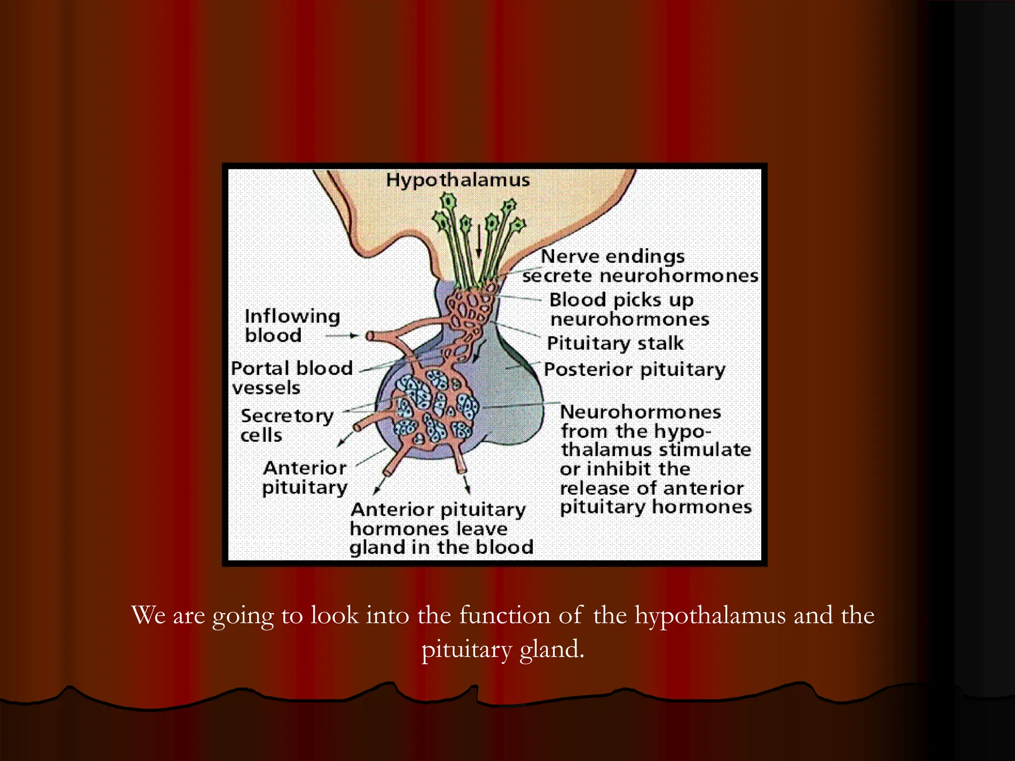


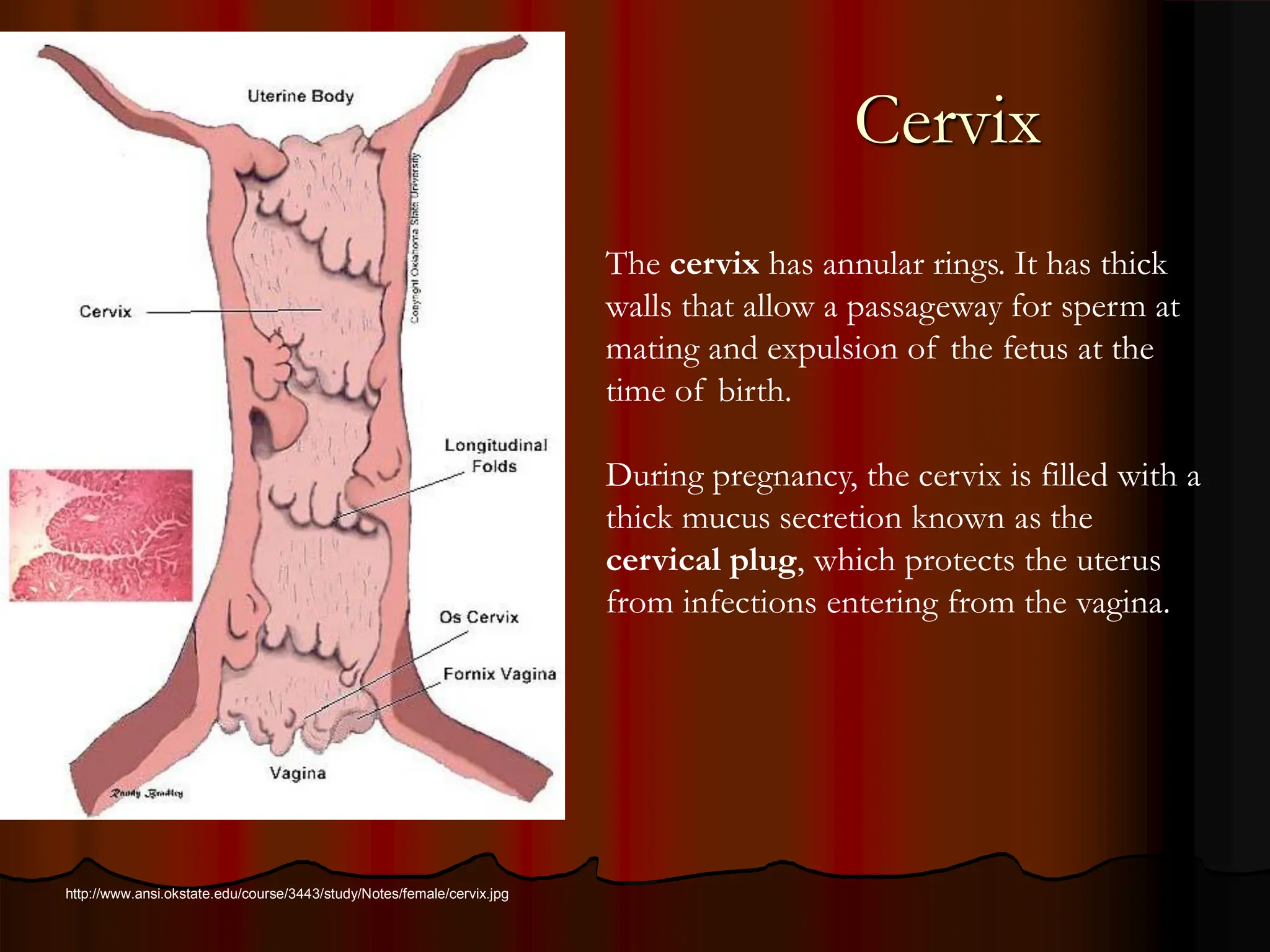
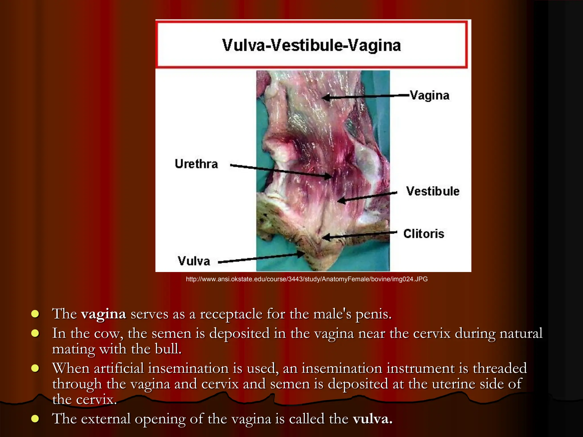


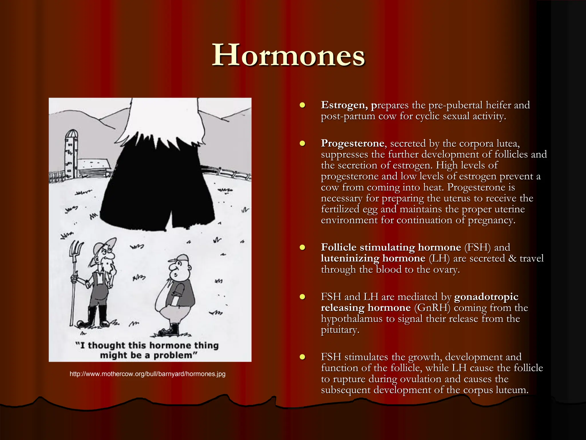


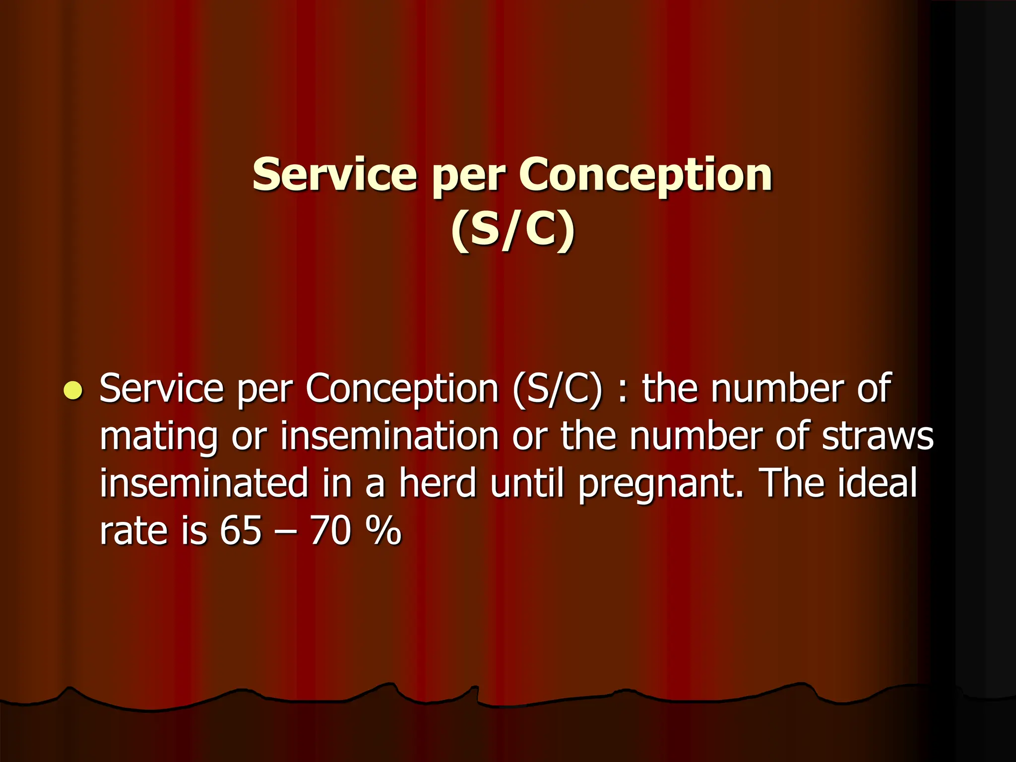
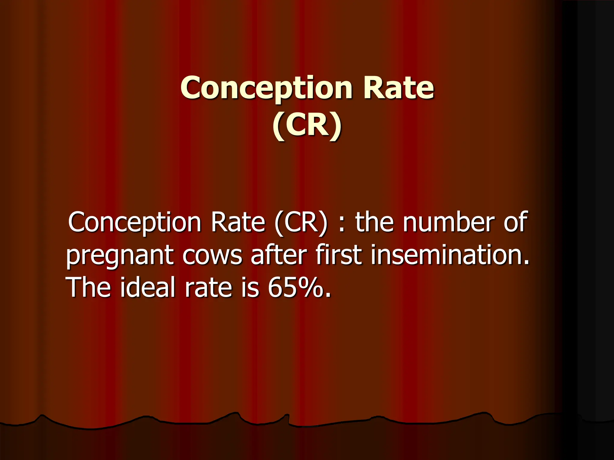


This document provides an introductory lecture on infertility and sterility for veterinary students. It discusses the key reproductive organs and hormones involved in the estrous cycle. It describes how the hypothalamus and pituitary gland regulate reproduction and the roles of the ovaries, uterus, cervix and other female organs. It then covers factors that can influence infertility and sterility, including endocrinological disorders, mismanagement, reproductive diseases, anatomical defects, and environmental defects. Finally, it discusses reproductive efficiency and various reproductive indices used to measure performance.


