#ismrm #ISMRM2022 #ISMRM22
3D-UTE Rosette Applications (MRI and MRSI) at UHF human (7T) and animal scanners (9.4T) using The Berkeley Advanced Reconstruction Toolbox (BART) toolbox
Uzay Emir
Afternoon Tea with BART
Time: 15:45-16:45
Unsupervised Deconvolution Neural Network for High Quality Ultrasound ImagingShujaat Khan
High quality US imaging demand large number of measurements that can increase the cost, size and power requirements. Therefore, low-powered, portable and 3D ultrasound imaging system require reconstruction algorithms that can produce high quality images using fewer receive measurements. Number of model specific methods has been proposed which doesn't work under perturbation. For instance, compressive deconvolution ultrasound which provide a reasonable quality with limited measurements however, it has its own down-sides such as high computation cost and accurate estimation of point spread function (PSF). An other major limitation of conventional methods is that they require RF or base-band signal which is difficult to obtain from portable US systems. To deal with the aforementioned issues, in this study we designed a novel deep deconvolution model for image domain-based deconvolution. The proposed deep deconvolution (DeepDeconv) model can be trained in an unsupervised fashion, alleviate the need of paired high and low quality images. The model was evaluated on both the phantom and in-vivo scans for various sampling configurations. The proposed DeepDeconv significantly enhance the details of anatomical structures and using unsupervised learning on average it achieved 2.14dB, 4.96dB and 0.01 units gain in CR, PSNR and SSIM values respectively, which are comparable to the supervised method.
While Phosphorous (31P) MRS (I) has been promising in experimental and clinical settings since the early 70s, it has been beset by prohibitively lower sensitivity, limited spectral-spatial resolution, and prolonged acquisition. This manuscript and proceedings of the annual scientific meeting of ISMRM in 2022 (REF1) and 2023 (REF2) demonstrate that our novel acquisition strategy, the novel Rosette Trajectory for fast and flexible MR(S)I contrast (Shen et al. 2023 (REF3), later we renamed it as PETALUTE after the translation to the preclinical scanners of 7T and 9.4T), enables operator-independent (1) rapid acquisition (~7 minutes), (2) reconstruction, and (3) processing pipeline, resulting in phosphorous metabolite ratio maps (10 x 10 x 10 mm3) of the whole brain.
In response to the “Repeat it with Me” challenge organized by the Reproducible Research study group of ISMRM, we demonstrated the power of this technique in 5 healthy volunteers at three different institutions with different experimental setups (2nd Place: UTE 31P 3D Rosette MRSI Reproducibility Team, REF4). Since the proposed acquisition/reconstruction/processing pipeline was operator/scanner/coil-independent, the Reproducer sub-teams successfully replicated the findings of the original proceeding in 2022 (REF1). As part of this challenge, we provided some MATLAB scripts and k-space data to reproduce some of the results described in this manuscript. The software and data can be downloaded from https://purr.purdue.edu/projects/ismrm31pmrsi.
These results will likely be of broad interest across clinical settings since the proposed acquisition strategy is not specific to any region, nuclei, or magnetic field and is operator-independent. This study's resolution and signal-to-noise ratios permit the metabolite maps in an experimentally and clinically feasible timeframe at 3 Tesla and 7T.
REF1 Bozymski B, Shen X, Ozen AC, Ibey S, Chiew M, Thomas A, Dydak U, Emir UE. Ultra-Short Echo Time 31P 3D MRSI at 3T with Novel Rosette k-space Trajectory. Proceedings 30th Scientific Meeting, International Society for Magnetic Resonance in Medicine, 2022.
REF2 Farley N, Bozymski B, Dydak U, Emir UE*. Fast 3D 31P MRSI Using Novel Rosette Petal Trajectory at 3T with x4 Accelerated Compressed Sensing. Proceedings 31st Scientific Meeting, International Society for Magnetic Resonance in Medicine, 2023.
REF3 Shen X, Özen AC, Sunjar A, Ilbey S, Sawiak S, Shi R, Chiew M, Emir UE. Ultrashort T2 components imaging of the whole brain using 3D dual-echo UTE MRI with rosette k-space pattern. Magnetic Resonance in Medicine. 2023;89(2):508–521.
REF4 https://challenge.ismrm.org/2023-24-reproducibility-challenge/results-22-23/
GABA spectroscopy
edited GABA 1H MEGA-PRESS spectra
GABA-edited
In this study, we have developed and demonstrated a non-water suppressed GABA editing Magnetic Resonance Spectroscopic Imaging technique using density-weighted concentric rings k-space trajectory that performs robustly within a clinically feasible acquisition time at 3T. The method has been validated in a series of phantom experiments and its feasibility assessed in a healthy volunteer with a high in-plane resolution of 7.5 × 7.5 mm. Experiments qualitatively demonstrate the advantage of the proposed method in terms of its improved resolution and reduced contamination of spectra from neighboring voxels.
Photoacoustic technology for biological tissues characterizationjournalBEEI
The existing photoacoustics (PA) imaging systems showed mixed performance in imaging characteristic and signal-to-noise ratio (SNR). This work presents the use of an in-house assembled PA system using a modulating laser beam of wavelength 633 nm for two-dimensional (2D) characterization of biological tissues. The differentiation of the tissues in this work is based on differences in their light absorption, wherein the produced photoacoustic signal detected by a transducer was translated into phase value that corresponds to the peak amplitude of optical absorption of tissue namely fat, liver and muscle. This work found fat tissue to produce the strongest PA signal with mean ± standard deviation (SD) phase value = 2.09 ± 0.31 while muscle produced the least signal with phase value = 1.03 ± 0.17. This work discovered the presence of stripes pattern in the reconstructed images of fat and muscle resulted from their structural properties. In addition, a comparison is made in an attempt to better assess the performance of the developed system with the related ones. This work concluded that the developed system may use as an alternative, noninvasive and label-free visualization method for characterization of biological tissues in the future.
Medical Imaging - Opportunities for Business Seminar
24/01/12
Session 2 Technology Showcase
Three technologies developed or enhances at the University of Leicester are presented
Modified Rosette PETALUTE for preclinical imagingUzay Emir
We have demonstrated a sequence, PETALUTE, for clinical and preclinical high (3T) and ultra-high fields (>7T) scanners.
The modified Rosette kspace dual-echo Acquisition/ PETALUTE strategy could further enhance integrated and translational research studies to understand human and non-human complex structures and physiologies better.
Universal plane wave compounding for high quality us imaging using deep learningShujaat Khan
Plane-wave compounding is to sum up several successive plane waves incident at different angles to form an image. By applying time-reversal of the received signals, transmit focusing can be synthesized. Unfortunately, to improve the temporal resolution, the number of plane waves should be reduced, which often degrades the image quality. To address this problem, an image domain learning method using neural networks has been proposed, but the network needs to be retrained when the number of plane waves changes. Herein, we propose, for the first time, a universal plane-wave compounding scheme using deep learning to directly process plane waves and RF data acquired at different view angles and sub-sampling rate to generate high quality US images.
Ultrasound Machine-A Revolution In Medical ImagingRAVI KANT
What is medical imaging?
Why ultrasound imaging is required?
History of ultrasound
What is ultrasound
Physical definition
Medical definition
Ultrasound production
The Returning echo
Doppler effect
What is Doppler ultrasound
Principles of instrumentation in ultrasonography
Transmitter and receiver circuits of ultrasound
Mechanical assembly of ultrasound machine
Manufacturing companies of USG
Sonoscape S40 color Doppler ultrasound system
Clinical applications of ultrasound
Future of ultraso
I reviewed 3 papers at 'SNU TF Study Group' in Korea.
3 papers tried to solve segmentation problems in medical images with Deep Learning.
Deep Learning 을 이용하여 의료 영상에서 Segmentation 문제를 풀고자 한 3가지 논문을 리뷰하였습니다. :)
In this work, we demonstrate the sodium magnetic resonance imaging (MRI) capabilities of a three-dimensional (3D) dual-echo ultrashort echo time (UTE) sequence with a novel rosette petal trajectory (PETALUTE), in comparison to the 3D density-adapted (DA) radial spokes UTE sequence. #mri #ismrm #msk
3D UTE 31P-MRSI with modified rosette k-space (PETALUTE): Comparison with con...Uzay Emir
Phosphorus-31 magnetic resonance spectroscopic imaging (31P-MRSI) provides valuable non-invasive in vivo information on tissue metabolism but is burdened by poor sensitivity and prolonged scan duration. Ultra-short echo time (UTE) acquisitions minimize signal loss when probing signals with relatively short spin-spin relaxation time (T2), while also preventing first-order dephasing. Here, a three-dimensional (3D) UTE sequence with a rosette k-space trajectory is applied to 31P-MRSI at 3T. Conventional chemical shift imaging (CSI) employs highly regular Cartesian k-space sampling, susceptible to substantial artifacts when accelerated via undersampling. In contrast, this novel sequence’s “petal-like” pattern offers incoherent sampling more suitable for compressed sensing (CS). These results showcase the competitive performance of UTE rosette 31P-MRSI against conventional weighted CSI with simulation, phantom, and in vivo leg muscle comparisons.
Rosette Applications (MRI and MRSI) at UHF human (7T) and animal scanners (9....Uzay Emir
MRI
MRSI
LEAF
PETAL
EGG
ROSETTE
MRI
siemens
bruker
his study aimed to develop a new 3D dual-echo rosette k-space trajectory, specifically for applications of ultra-short echo time (UTE) magnetic resonance imaging (MRI). The direct imaging of the myelin bilayer, which has ultra-short transverse relaxation time (uT2), was acquired to test the performance of the proposed UTE sequence.
Theory and Methods The rosette trajectory was developed based on rotations of a ‘petal-like’ pattern in the kx-ky plane, with oscillated extensions in kz-direction for 3D coverage. Five healthy volunteers were recruited and underwent ten dual-echo rosette UTE scans with varied echo times (TEs). Dual-exponential model fitting was performed to separate uT2 signals, with the output of uT2 fraction, uT2 value and long T2 value.
Results The reconstructed images’ signal contrast between white matter (WM) and grey matter (GM) increased with longer TEs. The WM regions had higher uT2 fraction values than GM (10.9%±1.9% vs. 5.7%±2.4%). The uT2 value was about 0.12 milliseconds in WM.
Conclusion The higher uT2 fraction value in WM compared to GM demonstrated the ability of the proposed sequence to capture rapidly decaying signals.
UTE 31P 3D Rosette MRSI reproducibility Team has demonstrated the Reproducibility of the method. The method has been challenged on #siemenshealthineers PET-MRI (mMR) and Prisma 3T scanners.ISMRM #spectroscopy ISMRM ChallengeISMRM RAPID Biomedical
https://lnkd.in/gnjCHtu2
Submillimeter fMRI Acquisition using a dual-echo Rosette kspace trajectory at 3TUzay Emir
n this study, we overcome the technological barrier against acquiring submillimeter resolution (~ 0.5 mm) fMRI data at 3T via a novel dual-echo Rosette k-space design. This design results in the fine representation of activation maps in two different functional tasks and might be a springboard in neuroimaging by providing very high-resolution spatiotemporal dynamics of neural networks. The method will be further evolved with the feedback from the MRI community via the GitHub platform as such for further acceleration, inflow saturation, and 3D coverage via 3D sampling and/or multiband approaches.
Simultaneous Measurement of functional MRI and MRS by Fast Non-water Suppress...Uzay Emir
The non-water suppressed magnetic resonance spectroscopic imaging (MRSI) sequence with concentric k-space trajectory was proposed to measure functional MRI and MRSI signals simultaneously. A right-hand finger-tapping task was performed at 3T MRI scanner to test the simultaneous hemodynamic and neurochemical measurements in the human primary motor cortex. The results showed a significant overlap betweenT2* and metabolite (glutamate) changes.
Quantitative Susceptibility Mapping (QSM) Using High-resolution Ultra-Short E...Uzay Emir
Karnik A, Shen X, Monsivais H, Sunjar A, Özen A, Ilbey S, Chiew M, Cakmak M, Rispoli J, Emir UE., Quantitative Susceptibility Mapping (QSM) Using High-resolution Ultra-Short Echo Time (UTE) MRI with Rosette k-space Pattern International Society for Magnetic Resonance in Medicine Annual Meeting 2022
High Resolution 3D Ultra-Short Echo Time MRI with Rosette k-Space Pattern for...Uzay Emir
High Resolution 3D Ultra-Short Echo Time MRI with Rosette k-Space Pattern for Brain Iron Content Mapping
Shen X, Özen A, Monsivais H, Ilbey S, Susnjar A, Karnik A, Chiew M, Emir UE. High Resolution 3D Ultra-Short Echo Time MRI with Rosette k-Space Pattern for Brain Iron Content Mapping, International Society for Magnetic Resonance in Medicine Annual Meeting 2022
Earliest Galaxies in the JADES Origins Field: Luminosity Function and Cosmic ...Sérgio Sacani
We characterize the earliest galaxy population in the JADES Origins Field (JOF), the deepest
imaging field observed with JWST. We make use of the ancillary Hubble optical images (5 filters
spanning 0.4−0.9µm) and novel JWST images with 14 filters spanning 0.8−5µm, including 7 mediumband filters, and reaching total exposure times of up to 46 hours per filter. We combine all our data
at > 2.3µm to construct an ultradeep image, reaching as deep as ≈ 31.4 AB mag in the stack and
30.3-31.0 AB mag (5σ, r = 0.1” circular aperture) in individual filters. We measure photometric
redshifts and use robust selection criteria to identify a sample of eight galaxy candidates at redshifts
z = 11.5 − 15. These objects show compact half-light radii of R1/2 ∼ 50 − 200pc, stellar masses of
M⋆ ∼ 107−108M⊙, and star-formation rates of SFR ∼ 0.1−1 M⊙ yr−1
. Our search finds no candidates
at 15 < z < 20, placing upper limits at these redshifts. We develop a forward modeling approach to
infer the properties of the evolving luminosity function without binning in redshift or luminosity that
marginalizes over the photometric redshift uncertainty of our candidate galaxies and incorporates the
impact of non-detections. We find a z = 12 luminosity function in good agreement with prior results,
and that the luminosity function normalization and UV luminosity density decline by a factor of ∼ 2.5
from z = 12 to z = 14. We discuss the possible implications of our results in the context of theoretical
models for evolution of the dark matter halo mass function.
Professional air quality monitoring systems provide immediate, on-site data for analysis, compliance, and decision-making.
Monitor common gases, weather parameters, particulates.
Seminar of U.V. Spectroscopy by SAMIR PANDASAMIR PANDA
Spectroscopy is a branch of science dealing the study of interaction of electromagnetic radiation with matter.
Ultraviolet-visible spectroscopy refers to absorption spectroscopy or reflect spectroscopy in the UV-VIS spectral region.
Ultraviolet-visible spectroscopy is an analytical method that can measure the amount of light received by the analyte.
Toxic effects of heavy metals : Lead and Arsenicsanjana502982
Heavy metals are naturally occuring metallic chemical elements that have relatively high density, and are toxic at even low concentrations. All toxic metals are termed as heavy metals irrespective of their atomic mass and density, eg. arsenic, lead, mercury, cadmium, thallium, chromium, etc.
This presentation explores a brief idea about the structural and functional attributes of nucleotides, the structure and function of genetic materials along with the impact of UV rays and pH upon them.
Salas, V. (2024) "John of St. Thomas (Poinsot) on the Science of Sacred Theol...Studia Poinsotiana
I Introduction
II Subalternation and Theology
III Theology and Dogmatic Declarations
IV The Mixed Principles of Theology
V Virtual Revelation: The Unity of Theology
VI Theology as a Natural Science
VII Theology’s Certitude
VIII Conclusion
Notes
Bibliography
All the contents are fully attributable to the author, Doctor Victor Salas. Should you wish to get this text republished, get in touch with the author or the editorial committee of the Studia Poinsotiana. Insofar as possible, we will be happy to broker your contact.
Richard's aventures in two entangled wonderlandsRichard Gill
Since the loophole-free Bell experiments of 2020 and the Nobel prizes in physics of 2022, critics of Bell's work have retreated to the fortress of super-determinism. Now, super-determinism is a derogatory word - it just means "determinism". Palmer, Hance and Hossenfelder argue that quantum mechanics and determinism are not incompatible, using a sophisticated mathematical construction based on a subtle thinning of allowed states and measurements in quantum mechanics, such that what is left appears to make Bell's argument fail, without altering the empirical predictions of quantum mechanics. I think however that it is a smoke screen, and the slogan "lost in math" comes to my mind. I will discuss some other recent disproofs of Bell's theorem using the language of causality based on causal graphs. Causal thinking is also central to law and justice. I will mention surprising connections to my work on serial killer nurse cases, in particular the Dutch case of Lucia de Berk and the current UK case of Lucy Letby.
Nutraceutical market, scope and growth: Herbal drug technologyLokesh Patil
As consumer awareness of health and wellness rises, the nutraceutical market—which includes goods like functional meals, drinks, and dietary supplements that provide health advantages beyond basic nutrition—is growing significantly. As healthcare expenses rise, the population ages, and people want natural and preventative health solutions more and more, this industry is increasing quickly. Further driving market expansion are product formulation innovations and the use of cutting-edge technology for customized nutrition. With its worldwide reach, the nutraceutical industry is expected to keep growing and provide significant chances for research and investment in a number of categories, including vitamins, minerals, probiotics, and herbal supplements.
Deep Behavioral Phenotyping in Systems Neuroscience for Functional Atlasing a...Ana Luísa Pinho
Functional Magnetic Resonance Imaging (fMRI) provides means to characterize brain activations in response to behavior. However, cognitive neuroscience has been limited to group-level effects referring to the performance of specific tasks. To obtain the functional profile of elementary cognitive mechanisms, the combination of brain responses to many tasks is required. Yet, to date, both structural atlases and parcellation-based activations do not fully account for cognitive function and still present several limitations. Further, they do not adapt overall to individual characteristics. In this talk, I will give an account of deep-behavioral phenotyping strategies, namely data-driven methods in large task-fMRI datasets, to optimize functional brain-data collection and improve inference of effects-of-interest related to mental processes. Key to this approach is the employment of fast multi-functional paradigms rich on features that can be well parametrized and, consequently, facilitate the creation of psycho-physiological constructs to be modelled with imaging data. Particular emphasis will be given to music stimuli when studying high-order cognitive mechanisms, due to their ecological nature and quality to enable complex behavior compounded by discrete entities. I will also discuss how deep-behavioral phenotyping and individualized models applied to neuroimaging data can better account for the subject-specific organization of domain-general cognitive systems in the human brain. Finally, the accumulation of functional brain signatures brings the possibility to clarify relationships among tasks and create a univocal link between brain systems and mental functions through: (1) the development of ontologies proposing an organization of cognitive processes; and (2) brain-network taxonomies describing functional specialization. To this end, tools to improve commensurability in cognitive science are necessary, such as public repositories, ontology-based platforms and automated meta-analysis tools. I will thus discuss some brain-atlasing resources currently under development, and their applicability in cognitive as well as clinical neuroscience.
Deep Behavioral Phenotyping in Systems Neuroscience for Functional Atlasing a...
3D-UTE Rosette MRI and MRSI at UHF human (7T) and animal scanners (9.4T) using The (BART) toolbox
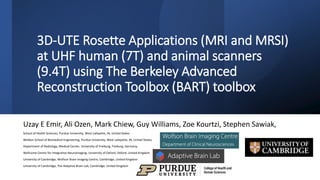
1. 3D-UTE Rosette Applications (MRI and MRSI)
at UHF human (7T) and animal scanners
(9.4T) using The Berkeley Advanced
Reconstruction Toolbox (BART) toolbox
Uzay E Emir, Ali Ozen, Mark Chiew, Guy Williams, Zoe Kourtzi, Stephen Sawiak,
School of Health Sciences, Purdue University, West Lafayette, IN, United States
Weldon School of Biomedical Engineering, Purdue University, West Lafayette, IN, United States,
Department of Radiology, Medical Center, University of Freiburg, Freiburg, Germany,
Wellcome Centre for Integrative Neuroimaging, University of Oxford, Oxford, United Kingdom
University of Cambridge, Wolfson Brain Imaging Centre, Cambridge, United Kingdom
University of Cambridge, The Adaptive Brain Lab, Cambridge, United Kingdom
2. A 3D-UTE Dual Echo Rosette MRI
and MRSI Sequence
2
Shen X, et al., doi: https://doi.org/10.1101/2021.09.18.460869
Normalized Radial UTE Reconstruction (RG) Normalized Rosette UTE Reconstruction (RG)
4. Day 1
Wednesday
Arrive at 3 pm: University of
Cambridge, Wolfson Brain Imaging
Centre, Cambridge, United
Kingdom
3:15 pm Installed the sequence
Of course, it did not work…
5. Day 2 Thursday
• Arrive at 1 pm University of Cambridge, Wolfson Brain
Imaging Centre, Cambridge, United Kingdom
• Phantom Experiments
• Did it work?
• Of course, it did work…. How do I know?
6. The Berkeley Advanced
Reconstruction Toolbox
(BART) toolbox
• Problems
• Non-Cartesian K-space
trajectory
• Giant data size
• Limited computational power
• Solution
• The Berkeley Advanced
Reconstruction Toolbox
(BART) toolbox
7. Day 3 k-Space Design 3D Rosette (high-resolution)
• Siemen 7T Terra
• 32 Channel Nova Coil
• Kmax=500/m
• 𝜔1=𝜔2=0.766 kHz
• Field of view (FOV):240x240x240 mm3
• Matrix size=256x256x256,
• Readout dwell time=10 𝜇𝑠
• Flip angle=7-degree
• TR=7 ms,
• Readout duration=4.2 ms
• RF pulse duration=20 𝜇𝑠
• TE: 30 𝜇𝑠 and 2.12 ms
• Number of petals: 71442 (40%)
• Total scan time: 8.7 minutes
Sequential k-space fill-
in
Kz
(/m)
First Last
8. Reconstruction
Two reconstruction methods were performed: compressed sensing (as described above)
and regular regridding applying a density compensated (Pipe&Menon) adjoint NUFFT.
Adjouint NUFFT Total Reconstruction time: ~ 2 minutes
Compressed sensing ~10 minutes.
9. Day 3 ~ 1 mm isotropic Resolution 7T
~ 0.5 mm isotropic Resolution 7T
10. Day 3 How About 7T MRSI
• Siemen 7T Terra
• 32 Channel Nova Coil
• Field of view (FOV):240x240x240 mm3
• Matrix size=48x48x48,
• Readout dwell time=10 𝜇𝑠
• Flip angle=30-degree
• TR=500 ms,
• Readout duration=~460 us
• RF pulse duration=20 𝜇𝑠
• TE: 30 𝜇𝑠, Maximum
• Spectral Bandwidth ~4.4 Khz
• Number of petals: 1444
• Total scan time: ~10 minutes
11. How about 9.4T Bruker animal Scanner (PetalUTE, Stephen Sawiak)
With this in mind, we sought out to study the validity of a novel UTE 3D MRSI sequence with a rosette k-space trajectory for use in phosphorus-31 spectroscopic imaging. This sequence has been successfully tested in proton phantoms and phosphorus localization phantoms. The rosette trajectory’s multiple crossings of k-space origin theoretically lead to smooth transitions in temporal repetitions for spectra. Additionally, the unprecedently short UTE intrinsically avoids all baseline and phasing issues. More details can be found in the linked pre-printed paper below.
To study this sequence, we recruited 5 healthy volunteers for uninterrupted, back-to-back leg muscle scanning with the novel MRSI sequence and an existing conventional MRSI sequence. We hypothesize that the novel UTE rosette sequence can simplify first-order phase and baseline corrections while providing competitive SNR relative to the conventional sequence.