Download to read offline
![POTHESIS OpenAccess
Granuloma central bilateral de
células gigantes del ángulo
mandibular en tres mujeres de la
misma familia.
Main text
Central giant cell granuloma (CGCG) is defined by the
World Health Organization as an intraosseous le- sion
consisting of cellular fibrous tissue that contains multiple
foci of haemorrhage, aggregations of multiple nucleated
giant cells, and occasionally trabeculae of woven bone
[1].
It is uncommon (7% of all benign jaw lesions), and the
biologic behaviour ranges from quiescent to ag- gressive,
with pain, root resorption and a tendency to recurrence
after excision [1]. In the great part of cases,
CGCG lesion is unilateral. Sometime the lesion is located
in a mandibular angle. And very few rare cases are
reported in literature of bilateral CGCG lo- cated at the
two angles of the mandible [2–4].
A case of bilateral CGCG of the mandibular angle has
been reported in a 12 years old female, and clas- sified as
idiopathic, as none of the family members of the young
girl presented with a similar lesion [2]. An- other sporadic
case has been reported in an 18 years
old girl, associated with neurofibromatosis type 1 [3].
Finally, another case of bilateral CGCG of the man-
dibular angle was reported in a 8 years old female with
Noonan’s syndrome [4].
In this cases series, we describe the first report in
literature of a repetitive bilateral CGCG of the two
mandibular angles, in three females from the same
family. These rare presentations of CGCG may be de-
fined as hereditary bilateral CGCG of the mandibular
angles or also cherubism-like lesions.
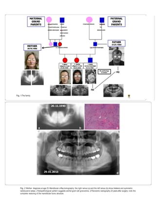
In 1990, a 24-year-old young athlete was exposed to
clinical observation at the maxillofacial surgery of the
University of L’Aquila, central Italy, for the appearance of
two osteolytic lesions at branches and mandibular an- gles
(Fig. 1).
These lesions appeared symmetrical to radiological
examinations (Fig. 2a, b).
The patient underwent surgical intervention and histo-
logical examination (Fig. 2c) revealed a case of GCGC. The
patient was then subjected to regular follow-up over the
years.
We currently have an x-ray performed after 23 years
from surgery, which confirms the absence of relapses and
a good mandibular bone restructuring.
PathologymaxilarMedicine (2019)10.12](https://image.slidesharecdn.com/2-200613061455/85/2-granuloma-upagu-gb-1-320.jpg)
![POTHESIS OpenAccess
Granuloma central bilateral de
células gigantes del ángulo
mandibular en tres mujeres de la
misma familia.
Main text
Central giant cell granuloma (CGCG) is defined by the
World Health Organization as an intraosseous le- sion
consisting of cellular fibrous tissue that contains multiple
foci of haemorrhage, aggregations of multiple nucleated
giant cells, and occasionally trabeculae of woven bone
[1].
It is uncommon (7% of all benign jaw lesions), and the
biologic behaviour ranges from quiescent to ag- gressive,
with pain, root resorption and a tendency to recurrence
after excision [1]. In the great part of cases,
CGCG lesion is unilateral. Sometime the lesion is located
in a mandibular angle. And very few rare cases are
reported in literature of bilateral CGCG lo- cated at the
two angles of the mandible [2–4].
A case of bilateral CGCG of the mandibular angle has
been reported in a 12 years old female, and clas- sified as
idiopathic, as none of the family members of the young
girl presented with a similar lesion [2]. An- other sporadic
case has been reported in an 18 years
old girl, associated with neurofibromatosis type 1 [3].
Finally, another case of bilateral CGCG of the man-
dibular angle was reported in a 8 years old female with
Noonan’s syndrome [4].
In this cases series, we describe the first report in
literature of a repetitive bilateral CGCG of the two
mandibular angles, in three females from the same
family. These rare presentations of CGCG may be de-
fined as hereditary bilateral CGCG of the mandibular
angles or also cherubism-like lesions.
In 1990, a 24-year-old young athlete was exposed to
clinical observation at the maxillofacial surgery of the
University of L’Aquila, central Italy, for the appearance of
two osteolytic lesions at branches and mandibular an- gles
(Fig. 1).
These lesions appeared symmetrical to radiological
examinations (Fig. 2a, b).
The patient underwent surgical intervention and histo-
logical examination (Fig. 2c) revealed a case of GCGC. The
patient was then subjected to regular follow-up over the
years.
We currently have an x-ray performed after 23 years
from surgery, which confirms the absence of relapses and
a good mandibular bone restructuring.
PathologymaxilarMedicine (2019)10.12](https://image.slidesharecdn.com/2-200613061455/75/2-granuloma-upagu-gb-1-2048.jpg)
![After getting married in 1995 she had three children: a
son in 1996 and two daughters, respectively, in 1999 and
2006.
The mother, due to her previous pathological lesion,
had made radiological controls in childhood to the male
child, with negative results.
On the contrary, at the age of 9 years, two symmetrical
bilateral osteolytic lesions of the jaw were observed in the
first female daughter, in the same sites as the mother (Figs.
3 and 4).
Subsequently, Cone beam CT scans showed the same
lesions to the second female daughter, but earlier, at age of
6 years [5].
All the lesions were surgically removed (Figs. 4a, 5, 6
and 7a, b, and f ) and the histopathologic diagnosis was
always identical (Fig. 8): giant cell central granulomas,
with patterns that showed an absolute correspondence
between them and with the mother (compare Figs. 2c,3,
4b, c, 5, 6 and 7c, d, g, h).
After the surgery, radiological follow-up examinations
showed no relapses and good restructuring of the
mandibular bone structure (Figs. 2c,3, 4, 5, 6, 7 and 9).
The father was free from this disease. Periodical yearly
follow-up was suggested for the two sisters until the end
of puberty.
To the best of the authors’ knowledge, this is the first
report of three cases of bilateral CGCG of the
mandibular angles in three females from the same
family. Considering the repetition of the lesion in sub-
jects belonging to the same family, considering the
particular location of the lesions (the mandibular angles
in all three subjects), this situation may be attributed to
the presence of a grade I (low level) Cherubism, or to the
occurrence of cherubism-like le- sions, as the cases did
not show the other peculiar characteristics of
Cherubism [6–8]. Table 1 shows the summary of the
differences between Cherubism and idiopathic CGCG
lesions that was followed in order to classify the lesion of
the present cases. CGCG lesions may be associated with
other disorders like Neuro- fibromatosis type 1 [3],
gingival fibromatosis as well as Noonan’s syndrome [4],
all of them are Rasopathies.
Fig. 3 TA,female, diagnosis at age 9 (4-gen-2008)-a Panoramic radiography shows on both side of the mandible two symmetric large multilocular
radiolucent lesions involving the angle and the ramus regions (white arrows). In the lower dental arch, there are only the first molar at right side (b)
and the first and second molars at left side (c) CTCBstudy of the mandible, respectively, of the right and the left site, shows the extension of the
lesions. Note their critical relationship with the mandibular canal and its neurovascular structures, in particular the inferior alveolar nerves
Page 3 of 8](https://image.slidesharecdn.com/2-200613061455/85/2-granuloma-upagu-gb-3-320.jpg)
![Noonan syndrome is an autosomal dominantly inher-
ited syndrome with variable expressivity. And multiple
CGCG lesions in Noonan’s syndrome may be aggres-
sive and cause complications. For these reasons, the
diagnosis of Noonan’s syndrome was firstly taken in
consideration. But the physical examination of these
subjects contributed to discard the diagnosis of Noo-
nan’s syndrome, that is characterized by short stature
and atypical face like a broad or webbed neck, low set
and posteriorly angulated ears, ptosis, hypertelorism,
and downward-slanting eyes [9].
Cherubism is an autosomal dominantly inherited
condition, with variable expressivity, that is character-
ized by multi-quadrant radiolucent lesions of the jaws
and a progressive and clinically, symmetrical enlarge-
ment of the mandible and/or the maxilla [10–12].
There is usually a familial history of similarly affected
family members and the regression of the lesions is
often seen following puberty [8]. In the present family
the mother was 24-year-old at the time of the first
diagnosis, consequently she could probably be consid-
ered as a missed diagnosis until that age.
From a cellular point of view, the cherubism-like le-
sions appear microscopically generally indistinguish-
able from CGCG, except occasionally, when a fairly
characteristic condensation of perivascular collagen is
evident [10]. Consequently, the clinical aspects pro-
vide helpful clues to distinguish cherubism from
CGCGs. CGCGs mainly affect patients between 10 and
30 years (while cherubism is more prevalent in children)
and are typically found unilaterally in the frontal region
of the mandible, whereas symmetrical
Fig. 4 TA, female, diagnosis at age 9 – a-c and d-f: respectively the lesion of the right mandibular side and the left mandibular side. Note for
each one the intraoperative aspect, and e.e. 10× and e.e. 20× histopatological speciments that show a moderately cellular and partially
collagenized stroma, characterized by melted cells with dense nuclei and giant cells osteoclast like
Fig. 5 TA, female, diagnosis at age 9 – Follow-up at 5 years (16-avr-
2013).Panoramic radiography shows the good aspect of the bone
mandibular structures and the absence of relapse
Page 4 of 8](https://image.slidesharecdn.com/2-200613061455/85/2-granuloma-upagu-gb-4-320.jpg)
![lesions are found in cherubism [13]. The present cases
show cherubism-like lesions.
Cherubism originates from genetic alteration in the
SH3BP2 gene, and currently, it is believed to be caused
by a gain-of-function mutation in the gene coding a c-
Abltyrosine kinase-binding protein (SH3BP2) located
on the short arm of chromosome 4 [14]. Only a sporadic
case of CGCG with mutation of this gene was previously
published [11, 15]. While another study conducted on a
group of patients with an aggressive CGCG did not show
any mutations, indicating that Cherubism is indeed a
distinct entity from CGCG [16].
Fig. 6 TC,female, diagnosis at age 6 (29-mar-2012) - a Panoramic radiography shows on both side of the mandible two symmetric large
multilocular radiolucent lesions involving the angle and the ramus regions and the second molars. (white arrows). In the lower dental arch there
are the first and the second molars (b, c) CTCBstudy of the mandible, respectively, of the right and the left site, shows the extension of the lesions.
Note their critical relationship with the mandibular canal and its neurovascular structures, in particular theinferior alveolar nerves
Fig. 7 TA, female, diagnosis at age 9 – a-d and e-h: respectively the lesion of the right mandibular side and the left mandibular side. Note for
each one the intraoperative aspect, the excised tissue, e.e. 10× and e.e. 20× histopatological speciments that show a moderately cellular and
partially collagenized stroma, characterized by melted cells with dense nuclei and giant cells osteoclast like
Pag 5 of 8](https://image.slidesharecdn.com/2-200613061455/85/2-granuloma-upagu-gb-5-320.jpg)
![In the present cases, the patients do not present the
typical swelling of bilateral mandibular angle region,
typical of Cherubism (accompanied by hypertelorism1).
But the repetition of the same cherubin-like lesions in
three female subjects belonging to the same family, is
suggestive for this diagnosis. Unfortunately, the family
refused to perform genetic analysis to investigate the
mutation in the SH3BP2 gene.
Dental findings in Cherubism include marked dis-
placement of developing or agenesia of second and third
molars as well as premature exfoliation of primary teeth
[17]. In addition, in Cherubism a marked cervical
lymphadenopathy is common.
In the present cases, there were not all common clin-
ical aspects of Cherubism and only females were charac-
terized by lesions. While Cherubism, in the scientific
literature, is reported to be more common in males or
equally distributed between males and females [17]. For
the CGCG lesions, instead, the predominant distribution
among females, respect to males, is certain [2], corre- lated
to the hormonal influence due to ovarian hor- mones,
oestrogen and progesterone, which are supposed
to be responsible for the development of CGCG as for
other pathologies [18–21].
For example, some cases of central giant cell lesion in
pregnant patients have showed a proliferation, and also in
subjects during a hormonal therapy [18]. But an im-
munostaining research, aimed to the detection of of es-
trogen and progesterone receptor proteins in 10 CGCG
lesions, failed to evidence estrogen receptor protein, ex-
cept for an occasional mononuclear cell stained weakly
positive for estrogen receptor protein. [In other cases,
estrogen receptor positivity was found in stromal
cells. In ten of these, osteoclast-type giant cells also ex-
hibited estrogen receptor immunostaining [22]. Due to
the different results in literature, the direct influence of the
ovarian hormones, estrogen and progesterone, in the
development and growth of these lesions is still to be
considered only a hypothesis.
For the present three cases, therefore, the hypothesis may
be a hereditary form of bilateral CGCG of the man-
dibular angles - lesions that could be defined as cherubism-
like lesions - or a rare manifestation of grade I
Cherubism. CGCGs of the jaws are commonly treated by
surgical curettage. And the management generally involves
long-term follow-up, with the assumption that these le-
sions will stabilize during puberty. Thus, a yearly follow-up
was suggested to the patients until the end of puberty.
Conclusion
DISCUSIÓN
POSIBLE TRATAMIENTO
DESCRIPCION DE LA LESION
ESTO ES SU TAREA
Fig. 8 Absolute correspondence of the histological aspect of the lesions in the two sisters: the histological framework consisted of a moderately
cellular and partially collagenized stroma, characterized by melted cells with dense nuclei and giant cells osteoclast like
Fig. 9 TA, female, diagnosis at age 9. Follow-up at 8 months years
(16-oct-2013).Panoramic radiography shows the good aspect of the
bone mandibular structures and the absence of relapse
Page 6 of 8](https://image.slidesharecdn.com/2-200613061455/85/2-granuloma-upagu-gb-6-320.jpg)

- This document reports on a case series of three females from the same family who presented with bilateral central giant cell granulomas (CGCG) of the mandibular angles. - The first case was a 24-year-old woman who was diagnosed with bilateral CGCG lesions of the mandibular branches and angles. She later gave birth to three children, two daughters who also developed bilateral CGCG lesions of the mandibular angles. - The repetitive bilateral CGCG lesions in these three related females, considering the specific location in the mandibular angles, suggests a possible diagnosis of cherubism-like lesions or low-grade cherubism inherited in an autosomal dominant manner,


![Crimson Publishers-Ewing’s Sarcoma (E.S) [Endothelial Myeloma]](https://cdn.slidesharecdn.com/ss_thumbnails/ooij-181109124921-thumbnail.jpg?width=640&height=640&fit=bounds)


