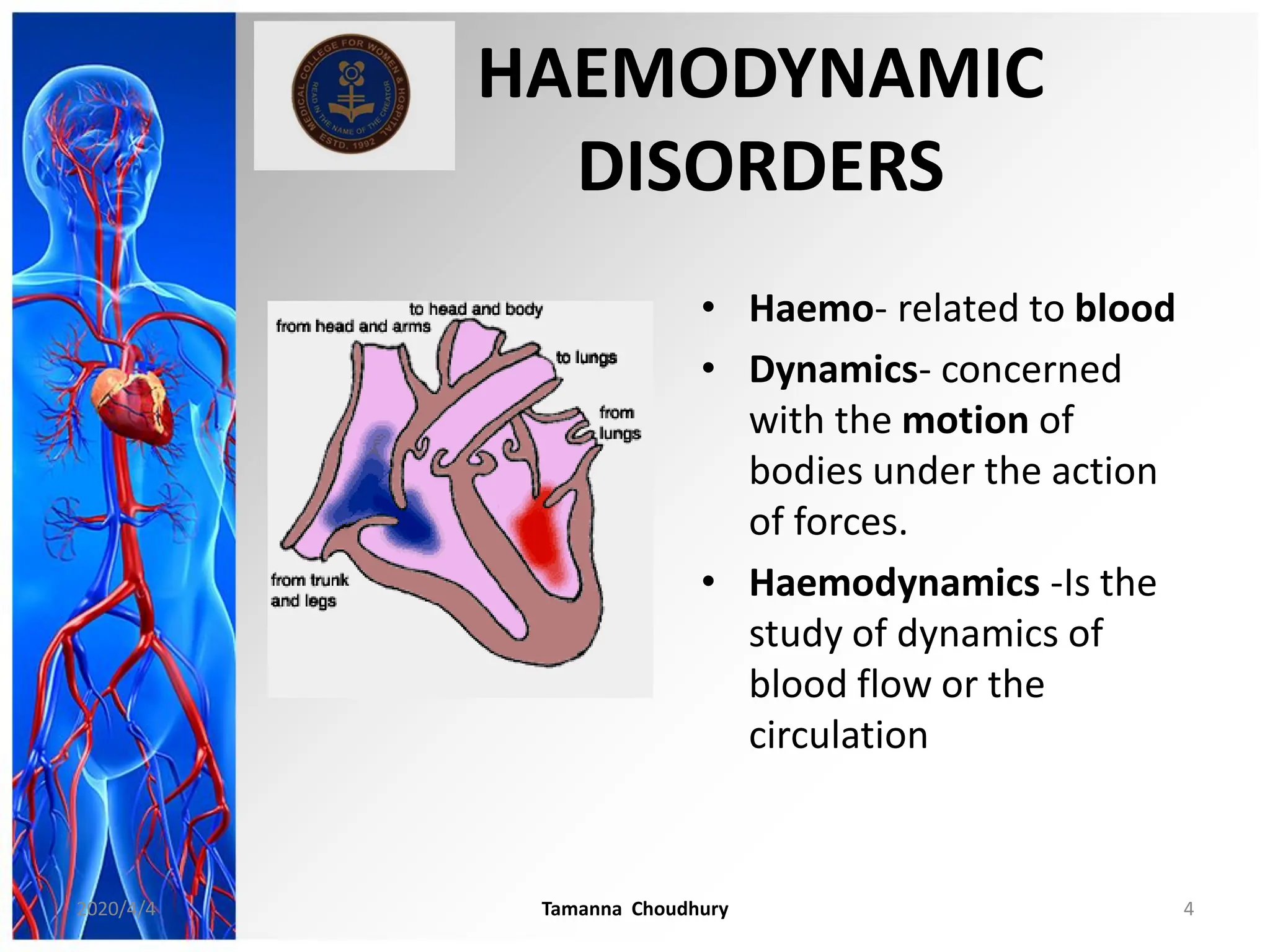
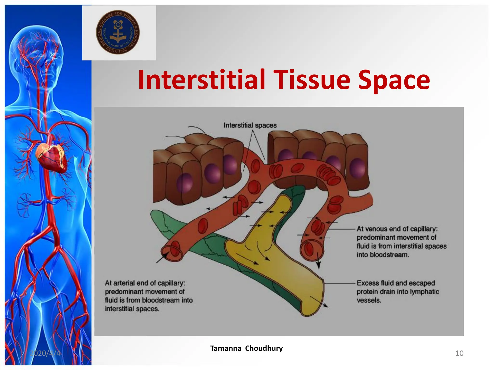
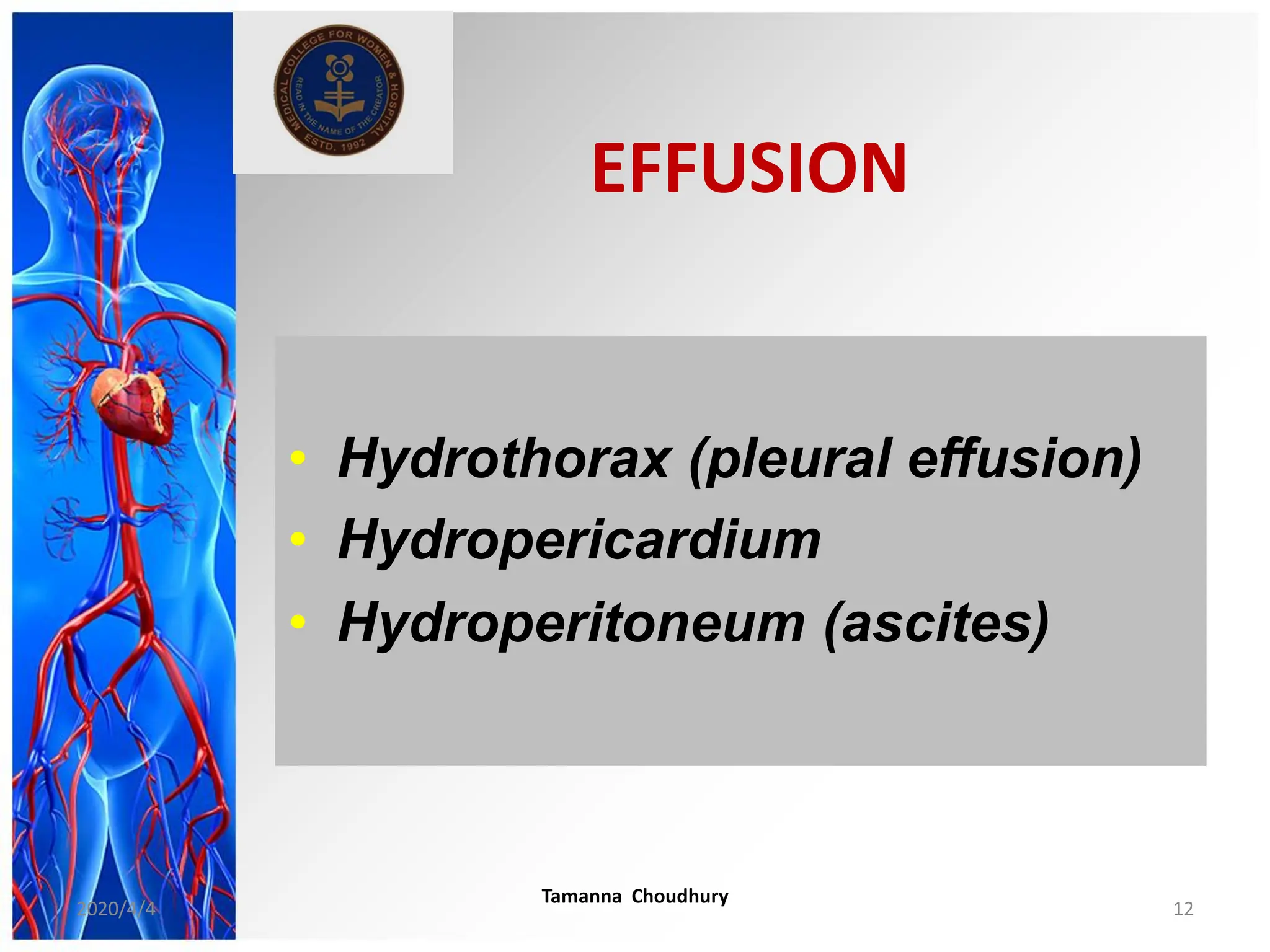
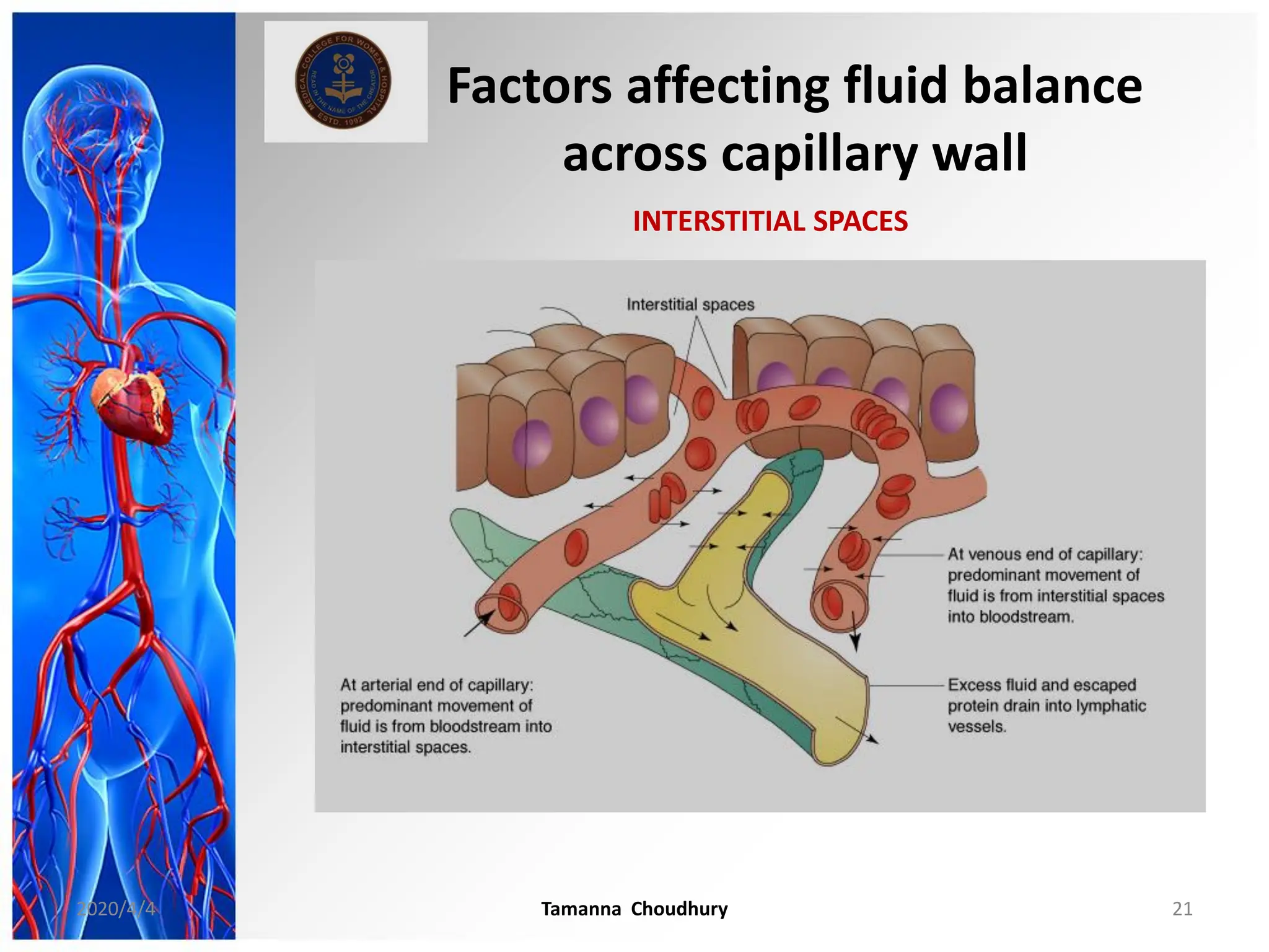
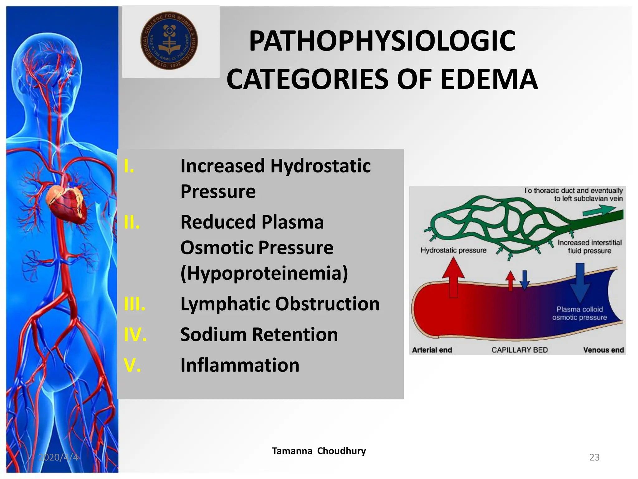
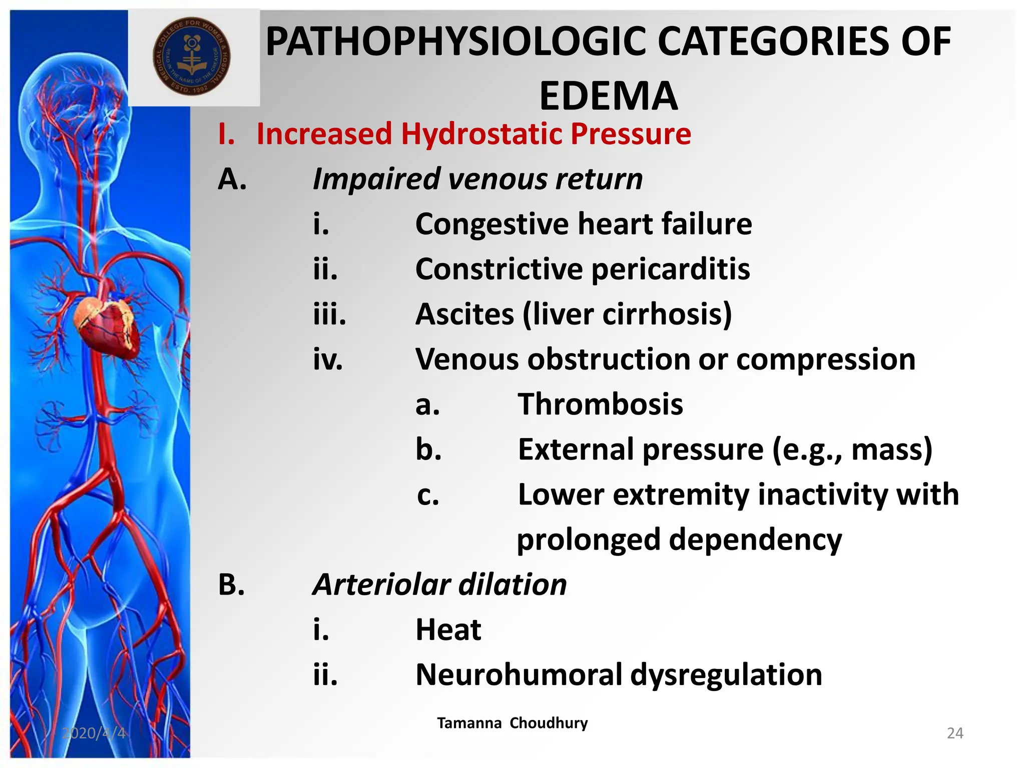
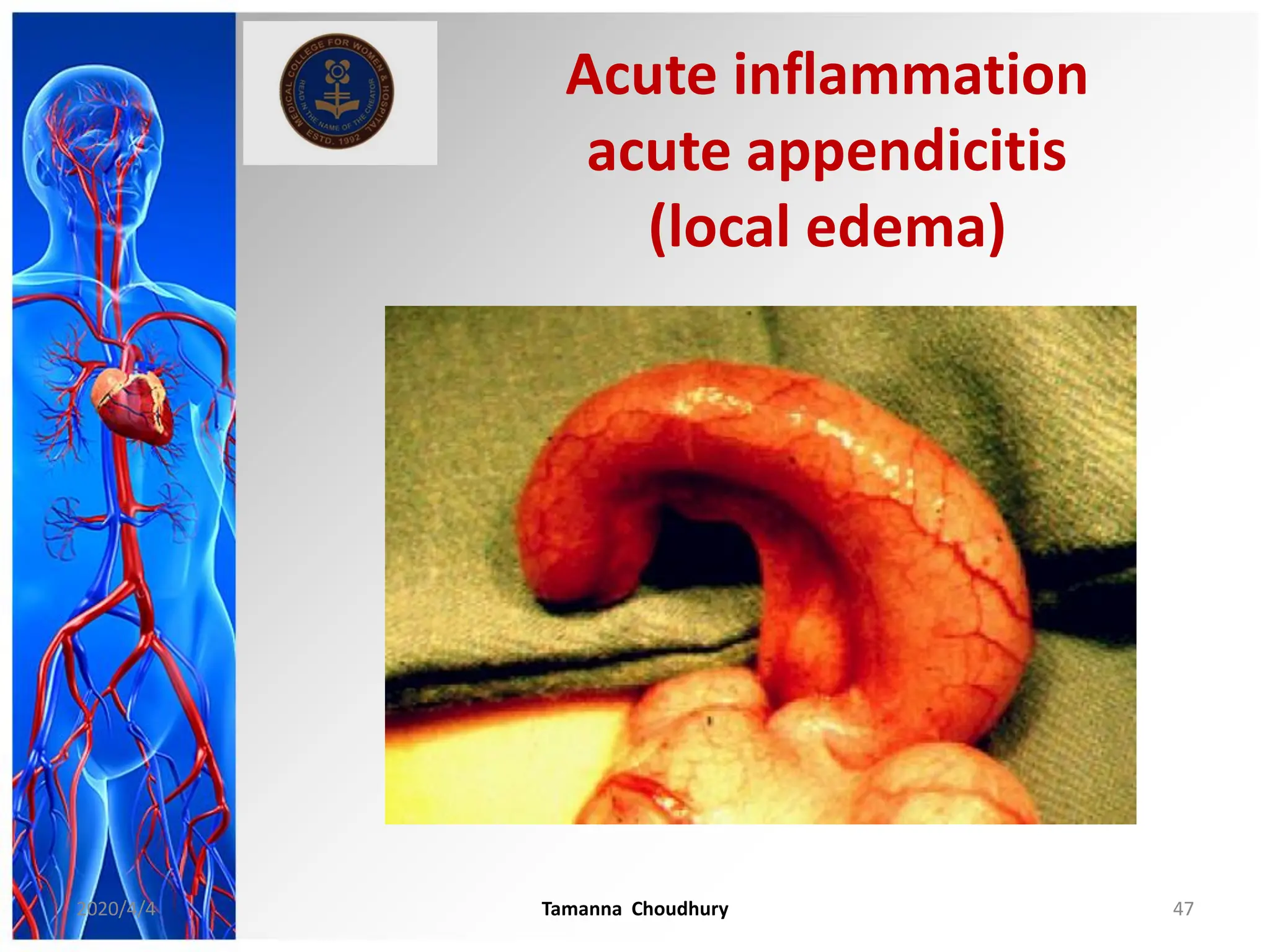
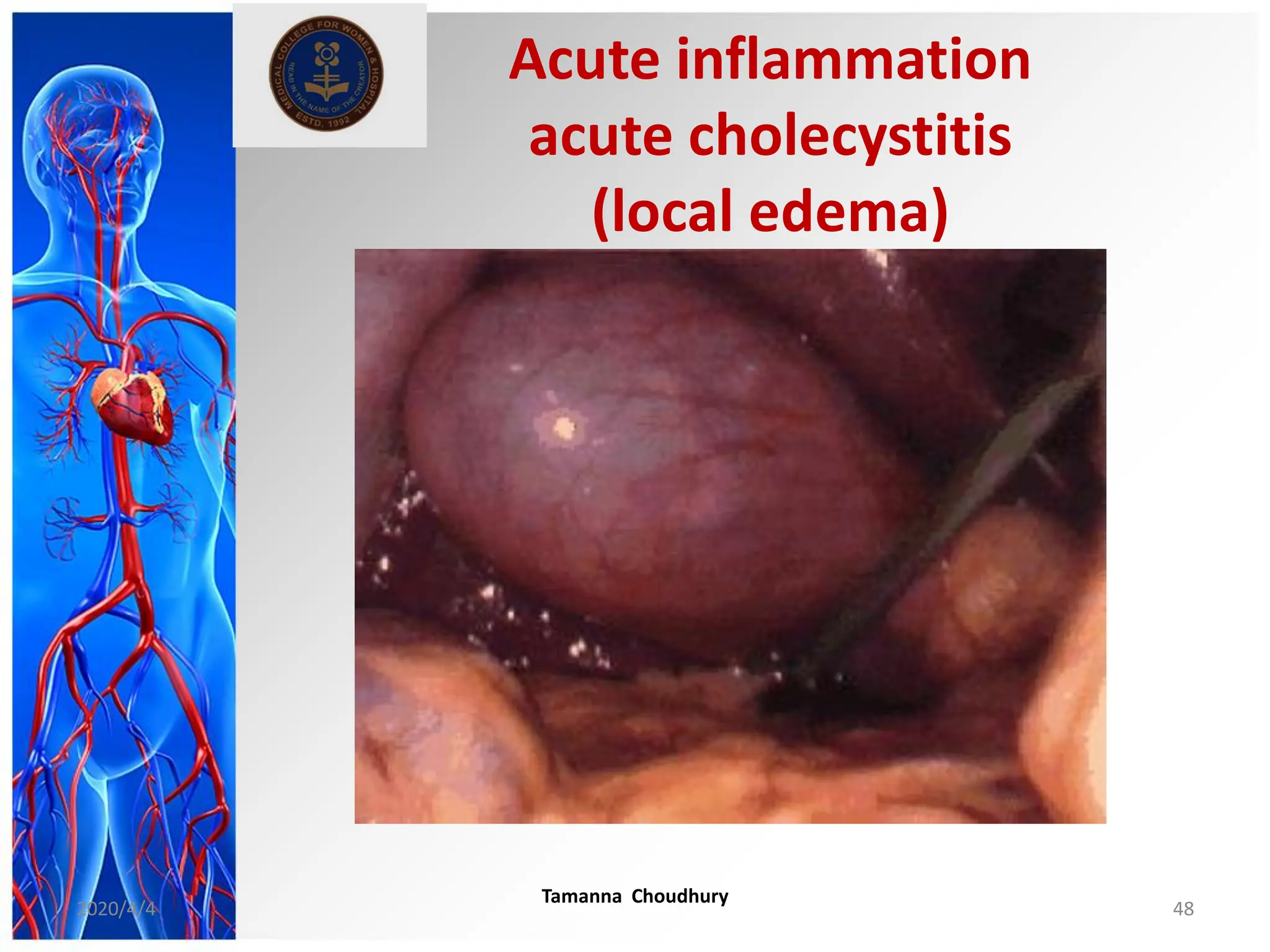
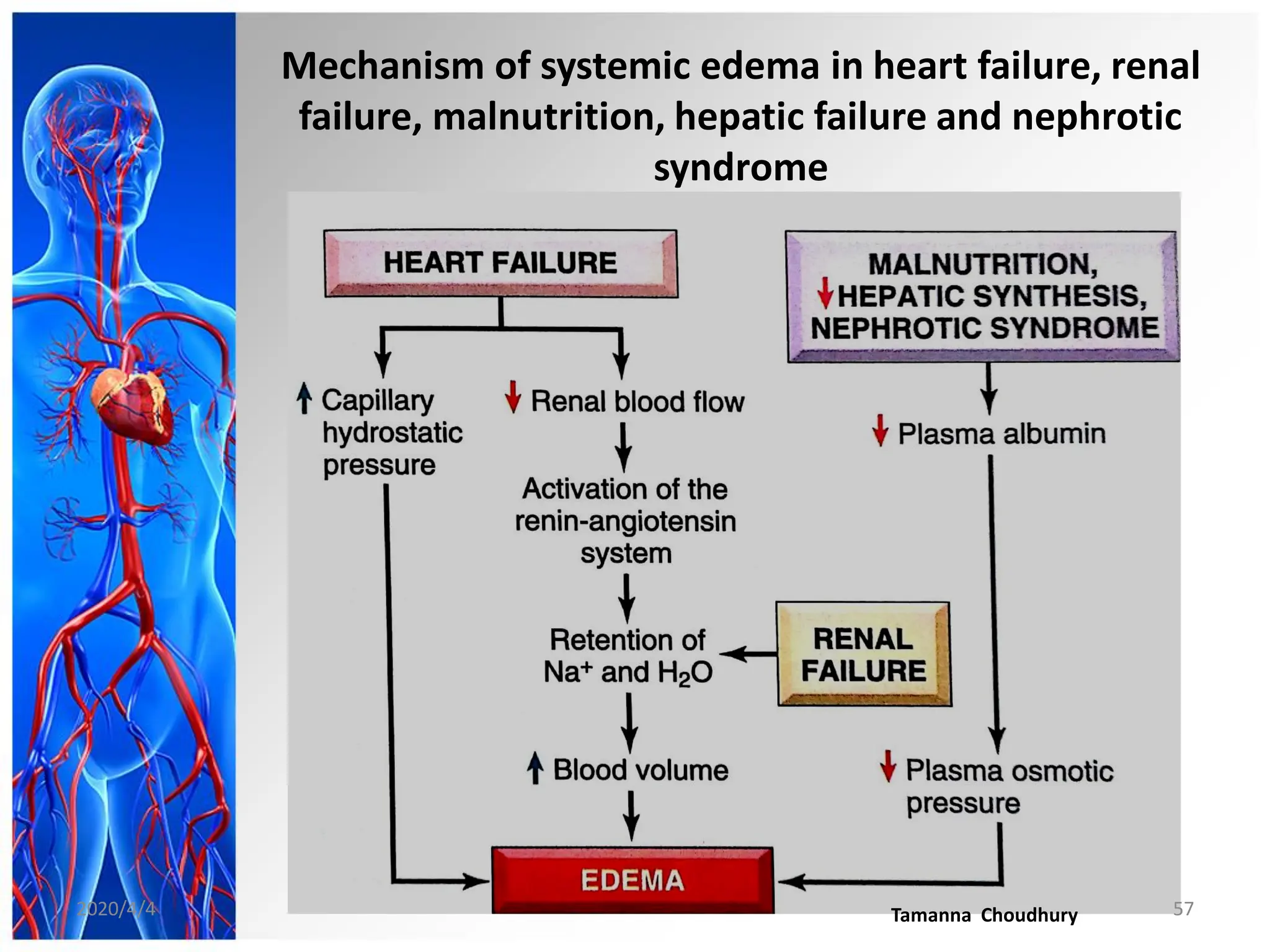
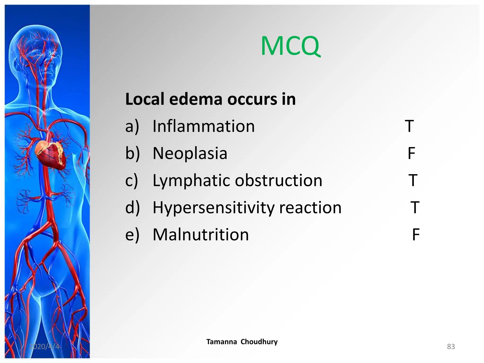
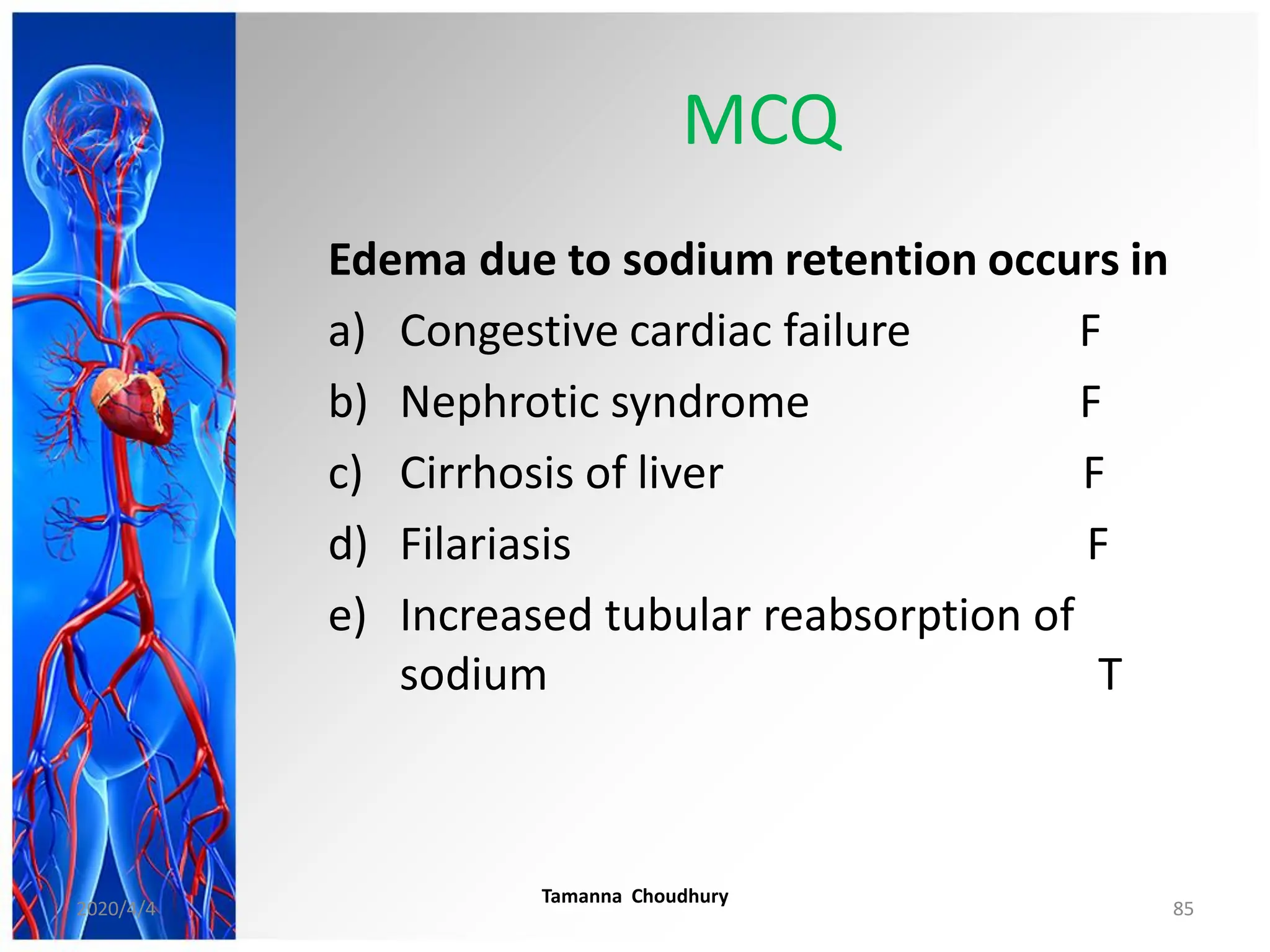
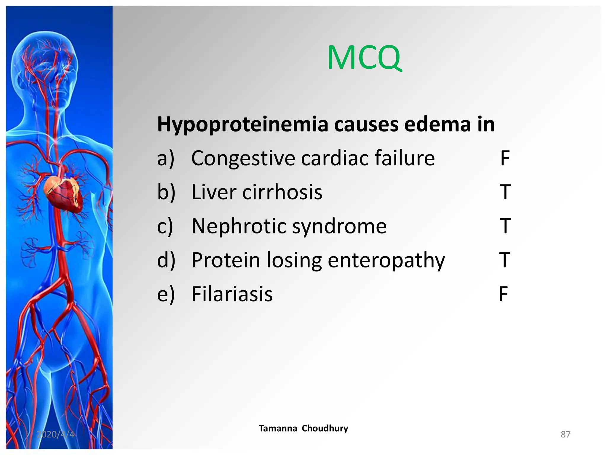
HAEMODYNAMIC DISORDERS,
THROMBOEMBOLIC DISEASE,
and SHOCK
Professor Tamanna Choudhury
HOD, Pathology
MCWH
2020/4/4 1
TOPIC 1
EDEMA & EFFUSION
2020/4/4 2
References:
• Robbins & Cotran Pathologic Basis of
Disease- 9th edition
• Davidson’s Principles and Practice of
Medicine-23rd edition
• IMAGES- Above mentioned books &
internet
TOPICS
1. EDEMA & EFFUSION
2. THROMBOSIS
3. EMBOLISM
4. INFARCTION
5. SHOCK
6. HYPEREMIA & CONGESTION
7. HEAMORRHAGE
8. DISSEMINATED INTRAVASCULAR
COAGULATION (DIC)
2020/4/4
Tamanna Choudhury
3
HAEMODYNAMIC
DISORDERS
• Haemo- related to blood
• Dynamics- concerned
with the motion of
bodies under the action
of forces.
• Haemodynamics -Is the
study of dynamics of
blood flow or the
circulation
2020/4/4 Tamanna Choudhury 4
HAEMODYNAMIC
DISORDERS
• The health and well being of cells and
tissues depends not only on the circulation
of blood which delivers oxygen but also on
normal fluid balance
• Alteration of any of these results in some
disorders known as haemodynamic
disorders
2020/4/4 5
HAEMODYNAMIC DISORDERS,
THROMBOEMBOLIC DISEASE and
SHOCK
Disorders of haemodynamics
 Edema & Effusion
 Hyperemia & Congestion
 Shock
Disorders of abnormal bleeding and clotting
 Hemorrhage
 Thrombosis
 Embolism
 Infarction
2020/4/4
Tamanna Choudhury
6
EDEMA & EFFUSION
2020/4/4
Tamanna Choudhury
7
TOPICS
• Definition
• Pathophysiology
• Types and examples
• Morphology
• Clinical significance
• Renal edema
• Cardiac edema
• Hepatic edema
• Pulmonary edema
• Cerebral edema
2020/4/4 Tamanna Choudhury 8
EDEMA- DEFINITION
Accumulation of fluid in the interstitial
tissue spaces
2020/4/4
Tamanna Choudhury
9
Interstitial Tissue Space
2020/4/4
Tamanna Choudhury
10
EFFUSION
DEFINITION
Accumulation of fluid in the body
cavities
2020/4/4
Tamanna Choudhury
11
EFFUSION
• Hydrothorax (pleural effusion)
• Hydropericardium
• Hydroperitoneum (ascites)
2020/4/4
Tamanna Choudhury
12
Pleural effusion
This is a right sided pleural effusion (in a baby)
The fluid is clear, pale yellow in appearance (serous
effusion)
2020/4/4
Tamanna Choudhury
13
Pleural effusions
Bilateral pleural effusions.
The fluid appears reddish, because there has been
hemorrhage into the effusion
2020/4/4 14
ASCITES
• It is clinically detectable
when at least 500 mL
have accumulated
CAUSES:
• Malignant disease
• Cardiac failure
• Hepatic cirrhosis
2020/4/4 Tamanna Choudhury 15
Body fluid compartments
• 60% of body weight is water
(in a healthy adult)
• 2/3rd (40%) is INTRACELLULAR
• 15% is INTERSTITIAL
• Only 5% is INTRA-VASCULAR
(plasma)
Three major fluid compartments
 Intravascular
 Interstitial and
 Intracellular
2020/4/4
Tamanna Choudhury
16
MECHANISM OF NORMAL
CONTROL IN SYSTEMIC
CIRCULATION
2020/4/4 Tamanna Choudhury 17
Starling’s Hypothesis
• states that the fluid movement across
the capillary wall is dependent on the
balance between
• the hydrostatic pressure gradient and
• the oncotic pressure gradient across
the capillary
2020/4/4 Tamanna Choudhury 18
Fluid balance across capillary
wall
Hydrostatic Pressure
Osmotic pressure 25-28 mm
32 mm
12 mm
Arterial End Venular End
Lymphatic Drainage
2020/4/4
Tamanna Choudhury
19
Factors affecting fluid balance
across capillary wall
2020/4/4
Tamanna Choudhury
20
Factors affecting fluid balance
across capillary wall
2020/4/4 Tamanna Choudhury 21
INTERSTITIAL SPACES
PATHOPHYSIOLOGIC
CATEGORIES OF EDEMA
2020/4/4 Tamanna Choudhury 22
PATHOPHYSIOLOGIC
CATEGORIES OF EDEMA
I. Increased Hydrostatic
Pressure
II. Reduced Plasma
Osmotic Pressure
(Hypoproteinemia)
III. Lymphatic Obstruction
IV. Sodium Retention
V. Inflammation
2020/4/4
Tamanna Choudhury
23
PATHOPHYSIOLOGIC CATEGORIES OF
EDEMA
I. Increased Hydrostatic Pressure
A. Impaired venous return
i. Congestive heart failure
ii. Constrictive pericarditis
iii. Ascites (liver cirrhosis)
iv. Venous obstruction or compression
a. Thrombosis
b. External pressure (e.g., mass)
c. Lower extremity inactivity with
prolonged dependency
B. Arteriolar dilation
i. Heat
ii. Neurohumoral dysregulation
2020/4/4
Tamanna Choudhury
24
PATHOPHYSIOLOGIC CATEGORIES OF
EDEMA
II. Reduced Plasma Osmotic Pressure
(Hypoproteinemia)
A. Protein-losing glomerulopathies
(nephrotic syndrome)
B. Liver cirrhosis (ascites)
C. Malnutrition
D. Protein -losing gastroenteropathy
2020/4/4 Tamanna Choudhury 25
PATHOPHYSIOLOGIC CATEGORIES OF
EDEMA
III. Lymphatic Obstruction
A.Inflammatory
B.Neoplastic
C.Postsurgical
D.Post irradiation
2020/4/4
Tamanna Choudhury
26
PATHOPHYSIOLOGIC CATEGORIES
OF EDEMA
IV. Sodium Retention
A. Excessive salt intake with
renal insufficiency
B. Increased tubular reabsorption of
sodium
i. Renal Hypoperfusion
ii. Increased secretion of
renin- angiotensin- aldosterone
2020/4/4
Tamanna Choudhury
27
PATHOPHYSIOLOGIC CATEGORIES OF
EDEMA
V.Inflammation
A. Acute inflammation
B. Chronic inflammation
C. Angiogenesis
2020/4/4
Tamanna Choudhury
28
TYPES OF EDEMA
I. Pathophysiology
II. Extent of involvement
III. Clinical basis
2020/4/4 Tamanna Choudhury 29
TYPES OF EDEMA
I. On the basis of pathophysiology
 INFLAMMATORY - Due to increased vascular
permeability (exudate)
 NON-INFLAMMATORY - Due to alterations in
haemodynamic forces across the capillary wall
(transudate)
2020/4/4 Tamanna Choudhury 30
TYPES OF EDEMA
II. On the basis of extent of involvement
 LOCAL – pulmonary, cerebral, effusions
 GENERALISED- anasarca
2020/4/4
Tamanna Choudhury
31
TYPES OF EDEMA
III. On clinical basis
 PITTING
 NON PITTING
2020/4/4
Tamanna Choudhury
32
Comparison of an exudate with a transudate
EXUDATE TRANSUDATE
Total protein
content
High, as in
plasma>3g/dl
Low, <3 g/dl
Distribution of
protein
As in plasma Nearly all albumin
Fibrinogen Present, as it clots
spontaneously
Not present.
No tendency to
coagulate
Specific gravity High
(over 1.018)
Low
(about 1.012)
Inflammatory cells Plentiful;
(poly,lympho)
Few present;
nearly all
mesothelial cell
2020/4/4
Tamanna Choudhury
33
Pitting edema of the leg
A. In a patient with congestive heart failure, severe
edema of the leg is demonstrated by applying pressure
with a finger.
B. The resulting “pitting” reflects the inelasticity of
the fluid-filled tissue.
2020/4/4
Tamanna Choudhury
34
Non pitting edema
(Pretibial myxedema)
2020/4/4 Tamanna Choudhury 35
MORPHOLOGY OF
EDEMA
• Edema is most easily
recognized grossly
• Microscopically,
clearing and
separation of the
extracellular matrix
elements.
2020/4/4
Tamanna Choudhury
36
MORPHOLOGY OF EDEMA
• Although any organ or tissue in the body
may be involved,
• Most commonly encountered in
 subcutaneous tissues
 the lungs and
 the brain
2020/4/4
Tamanna Choudhury
37
MORPHOLOGY OF EDEMA
Subcutaneous
Variable distribution
• Diffuse
• Dependent :
Cardiac failure
( legs and sacral area)
• Periorbital :
Nephrotic syndrome
Dependent edema
Periorbital edema
2020/4/4
Tamanna Choudhury
38
 Severe, generalized edema is
called anasarca
MORPHOLOGY OF EDEMA
2020/4/4
Tamanna Choudhury
39
CLINICAL FEATURES
Merely annoying to fatal
2020/4/4
Tamanna Choudhury
40
Subcutaneous edema
 In cardiac & renal failure subcutaneous
edema signals underlying disease
 May impair wound healing &
clearance of infection
CLINICAL FEATURES
2020/4/4
Tamanna Choudhury
41
CLINICAL FEATURES
Pulmonary edema
• Interferes with normal ventilatory
function
• Favours bacterial infection
2020/4/4 Tamanna Choudhury 42
CLINICAL FEATURES
Brain edema
• Can be rapidly fatal
• Brain substance can herniate through
foramen magnum
• Brain stem vascular supply can be
compressed
• Both the conditions can injure
medullary centre & cause death
2020/4/4 Tamanna Choudhury 43
Laryngeal edema
Fatal
2020/4/4 Tamanna Choudhury 44
LOCAL EDEMA
2020/4/4
Tamanna Choudhury
45
Local edema- causes
• Acute Inflammation
• Hypersensitivity reaction
• Venous obstruction
• Lymphatic obstruction
2020/4/4 Tamanna Choudhury 46
Acute inflammation
acute appendicitis
(local edema)
2020/4/4 Tamanna Choudhury 47
Acute inflammation
acute cholecystitis
(local edema)
2020/4/4
Tamanna Choudhury
48
Local edema (Cont)
Hypersensitivity edema
Vascular permeability seen in
 type I
 type III &
 type IV hypersensitivity reaction
2020/4/4 Tamanna Choudhury 49
Local edema
Edema due to venous obstruction
Examples
• Venous thrombosis
• A tumour / growth compressing a vein
Increased hydrostatic pressure proximal
to the obstruction
2020/4/4
Tamanna Choudhury
50
Local edema (Cont)
Edema due to lymphatic obstruction
Examples :-
(a) Filariasis
(b) Recurrent bacterial lymphangitis
(c) Lymphatic edema of arm & peau d'
orange appearance of the breast in
breast carcinoma
2020/4/4
Tamanna Choudhury
51
Filariasis
 W. bancrofti parasites are
mainly transmitted by Culex
quinquefasciatus mosquitoes
and some species of
Anopheles
 Brugia parasites are mainly
transmitted by Mansonia
mosquitoes
2020/4/4
Tamanna Choudhury
52
Edema secondary to lymphatic obstruction.
Massive edema of the right lower extremity
(elephantiasis) in a patient with obstruction of lymphatic
drainage.
2020/4/4
Tamanna Choudhury
53
Peau d’ orange appearance in
breast carcinoma
• Peau d Orange is a
French term, which
means “skin of an
orange.”
2020/4/4
Tamanna Choudhury
54
GENERALIZED EDEMA
2020/4/4
Tamanna Choudhury
55
Generalized edema
CAUSES:
 Heart failure
 Renal failure
 Nephrotic syndrome
 Cirrhosis of liver
 Malnutrition
2020/4/4
Tamanna Choudhury
56
Mechanism of systemic edema in heart failure, renal
failure, malnutrition, hepatic failure and nephrotic
syndrome
2020/4/4 Tamanna Choudhury 57
Renin Angiotensin Aldosterone axis
2020/4/4
Tamanna Choudhury
58
Nephrotic Syndrome
• Massive proteinuria (daily loss of
3.5gm or more of protein)
• Hypoalbuminaemia- plasma albumin
level less than 3gm/L
• Generalized edema
• Hyperlipidemia and lipiduria
2020/4/4
Tamanna Choudhury
59
Edema in Nephrotic
Syndrome
• Nephrotic syndrome is caused by a
derangement in glomerular capillary
walls resulting in increased
permeability to plasma proteins
2020/4/4
Tamanna Choudhury
60
NEPHROTIC SYNDROME-
pathogenesis
Leaky glomerular capillary wall
Heavy proteinuria
Hypoproteinaemia
Decreased colloidal
osmotic pressure
EDEMA
2020/4/4
Tamanna Choudhury
61
Edema in Nephrotic
Syndrome
Direct consequence
Aggravates edema
2020/4/4 Tamanna Choudhury 62
EDEMA IN CIRRHOSIS OF
LIVER
2020/4/4
Tamanna Choudhury
63
EDEMA (ascites) IN LIVER CIRRHOSIS
 Increased hydrostatic pressure: elevated
pressure in the veins running through the liver
(sinusoidal hypertension)
 Percolation of hepatic lymph into the
peritoneal cavity
 Splanchnic vasodilation and hyperdynamic
circulation
 Hypoalbuminemia: a decrease in liver
function caused by scarring of the liver,
i.e., cirrhosis.
 Failure in hepatic inactivation of aldosterone
2020/4/4
Tamanna Choudhury
64
Systemic, portal,
hepatic circulation
Splanchnic circulation
2020/4/4
Tamanna Choudhury
65
1. Increased hydrostatic
pressure
2. Percolation of hepatic
lymph into the peritoneal
cavity
3. Splanchnic vasodilation
and hyperdynamic
circulation
4. Hypoalbuminemia
5. Failure in hepatic
inactivation of
aldosterone
EDEMA IN CIRRHOSIS OF LIVER
1.
3.
4. 5.
2.
Ref: Davidson’s Principle & Practice of
Medicine. 23rd ed. Chapter 22
2020/4/4
Tamanna Choudhury
66
PULMONARY EDEMA
2020/4/4
Tamanna Choudhury
67
CLASSIFICATION OF PULMONARY
OEDEMA
• A. HEMODYNAMIC EDEMA
• B. EDEMA DUE TO MICROVASCULAR
INJURY
• C. EDEMA OF UNDETERMINED
ORIGIN
2020/4/4
Tamanna Choudhury
68
CAUSES OF PULMONARY OEDEMA (Cont)
EDEMA DUE TO HAEMODYNAMIC CAUSE
Increased Hydrostatic pressure
 Left heart failure
 Volume overload
Decreased oncotic pressure
 Nephrotic syndrome
 Liver disease
Lymphatic obstruction
2020/4/4 Tamanna Choudhury 69
CAUSES OF PULMONARY OEDEMA(Cont)
EDEMA DUE TO MICROVASCULAR INJURY
Infectious agents : viruses, mycoplasma,
other
Inhaled gases : oxygen, sulfur dioxide,
cyanates, smoke
Liquid aspiration : gastric contents, near-
drowning
Drugs and chemicals:
2020/4/4
Tamanna Choudhury
70
CAUSES OF PULMONARY
OEDEMA(Cont)
EDEMA OF UNDETERMINED ORIGIN
High altitude
Neurogenic
2020/4/4 Tamanna Choudhury 71
Grossly: Weight
Frothy, blood tinged fluid – on section
M/E : Alveolar capillaries engorged
Intra-alveolar precipitate
Alveolar microhaemorrhage
Hemosiderin laden macrophages
(heart failure cell)
Fluid is expressed
from the cut
surfaces of both
lungs
Pulmonary edema
2020/4/4 Tamanna Choudhury 72
CEREBRAL EDEMA
2020/4/4
Tamanna Choudhury
73
Cerebral edema- causes
 Primary or metastatic tumour
 Infarction
 Contusion
 Abscess
 Encephalitis
2020/4/4
Tamanna Choudhury
74
MORPHOLOGY OF CEREBRAL EDEMA
Cerebral edema:
 Swollen
 Softer
 Gyri – Flattened
 Sulci – Narrowed
 Ventricular cavities
– compressed
2020/4/4
Tamanna Choudhury
75
• Definition- edema, effusion
• Pathophysiological categories of edema
• Types of edema
• Mechanism of edema- heart failure, renal failure,
nephrotic syndrome, hepatic cause (Edema in
cirrhosis)
• Morphology of edema
• Local edema- causes & examples
• Pulmonary edema
• Cerebral edema
What we have learnt in this class
2020/4/4
Tamanna Choudhury
76
Practice questions
1. Define edema.
2. What are the types of edema ? What is anasarca?
3. What are the pathophysiological categories of edema?
4. What are the causes of generalized edema?
5. What are the causes of localized edema? Give few examples of
localized edema.
6. What are the differences between exudates & transudates?
7. What is the clinical significance of edema?
8. Discuss the mechanism of edema in heart failure.
9. Discuss the mechanism of edema in nephrotic syndrome.
10. What is the mechanism of edema in liver cirrhosis?
11. What are the causes of pulmonary edema? What is heart failure
cell?
12. Mention few causes of cerebral edema.
2020/4/4
Tamanna Choudhury
77
MCQ
Edema occurs due to
a) Decreased hydrostatic pressure
b) Lymphatic obstruction
c) Increased plasma colloidal osmotic
pressure
d) Sodium retention
e) inflammation
2020/4/4
Tamanna Choudhury
78
MCQ
Edema occurs due to
a) Decreased hydrostatic pressure F
b) Lymphatic obstruction T
c) Increased plasma colloidal osmotic
pressure F
d) Sodium retention T
e) Inflammation T
2020/4/4
Tamanna Choudhury
79
MCQ
Exudate is associated with
a) Low protein content
b) Inflammation
c) Low specific gravity
d) Increased tendency to clot
e) High cellular content
2020/4/4
Tamanna Choudhury
80
MCQ
Exudate is associated with
a) Low protein content F
b) Inflammation T
c) Low specific gravity F
d) Increased tendency to clot T
e) High cellular content T
2020/4/4
Tamanna Choudhury
81
MCQ
Local edema occurs in
a) Inflammation
b) Neoplasia
c) Lymphatic obstruction
d) Hypersensitivity reaction
e) Malnutrition
2020/4/4
Tamanna Choudhury
82
MCQ
Local edema occurs in
a) Inflammation T
b) Neoplasia F
c) Lymphatic obstruction T
d) Hypersensitivity reaction T
e) Malnutrition F
2020/4/4
Tamanna Choudhury
83
MCQ
Edema due to sodium retention occurs in
a) Congestive cardiac failure
b) Nephrotic syndrome
c) Cirrhosis of liver
d) Filariasis
e) Increased tubular reabsorption of
sodium
2020/4/4
Tamanna Choudhury
84
MCQ
Edema due to sodium retention occurs in
a) Congestive cardiac failure F
b) Nephrotic syndrome F
c) Cirrhosis of liver F
d) Filariasis F
e) Increased tubular reabsorption of
sodium T
2020/4/4
Tamanna Choudhury
85
MCQ
Hypoproteinemia causes edema in
a) Congestive cardiac failure
b) Liver cirrhosis
c) Nephrotic syndrome
d) Protein losing enteropathy
e) Filariasis
2020/4/4
Tamanna Choudhury
86
MCQ
Hypoproteinemia causes edema in
a) Congestive cardiac failure F
b) Liver cirrhosis T
c) Nephrotic syndrome T
d) Protein losing enteropathy T
e) Filariasis F
2020/4/4
Tamanna Choudhury
87
2020/4/4 88

1.MBBS-Edema-Copy.pdf

  • 1.
    HAEMODYNAMIC DISORDERS, THROMBOEMBOLIC DISEASE, andSHOCK Professor Tamanna Choudhury HOD, Pathology MCWH 2020/4/4 1 TOPIC 1 EDEMA & EFFUSION
  • 2.
    2020/4/4 2 References: • Robbins& Cotran Pathologic Basis of Disease- 9th edition • Davidson’s Principles and Practice of Medicine-23rd edition • IMAGES- Above mentioned books & internet
  • 3.
    TOPICS 1. EDEMA &EFFUSION 2. THROMBOSIS 3. EMBOLISM 4. INFARCTION 5. SHOCK 6. HYPEREMIA & CONGESTION 7. HEAMORRHAGE 8. DISSEMINATED INTRAVASCULAR COAGULATION (DIC) 2020/4/4 Tamanna Choudhury 3
  • 4.
    HAEMODYNAMIC DISORDERS • Haemo- relatedto blood • Dynamics- concerned with the motion of bodies under the action of forces. • Haemodynamics -Is the study of dynamics of blood flow or the circulation 2020/4/4 Tamanna Choudhury 4
  • 5.
    HAEMODYNAMIC DISORDERS • The healthand well being of cells and tissues depends not only on the circulation of blood which delivers oxygen but also on normal fluid balance • Alteration of any of these results in some disorders known as haemodynamic disorders 2020/4/4 5
  • 6.
    HAEMODYNAMIC DISORDERS, THROMBOEMBOLIC DISEASEand SHOCK Disorders of haemodynamics  Edema & Effusion  Hyperemia & Congestion  Shock Disorders of abnormal bleeding and clotting  Hemorrhage  Thrombosis  Embolism  Infarction 2020/4/4 Tamanna Choudhury 6
  • 7.
  • 8.
    TOPICS • Definition • Pathophysiology •Types and examples • Morphology • Clinical significance • Renal edema • Cardiac edema • Hepatic edema • Pulmonary edema • Cerebral edema 2020/4/4 Tamanna Choudhury 8
  • 9.
    EDEMA- DEFINITION Accumulation offluid in the interstitial tissue spaces 2020/4/4 Tamanna Choudhury 9
  • 10.
  • 11.
    EFFUSION DEFINITION Accumulation of fluidin the body cavities 2020/4/4 Tamanna Choudhury 11
  • 12.
    EFFUSION • Hydrothorax (pleuraleffusion) • Hydropericardium • Hydroperitoneum (ascites) 2020/4/4 Tamanna Choudhury 12
  • 13.
    Pleural effusion This isa right sided pleural effusion (in a baby) The fluid is clear, pale yellow in appearance (serous effusion) 2020/4/4 Tamanna Choudhury 13
  • 14.
    Pleural effusions Bilateral pleuraleffusions. The fluid appears reddish, because there has been hemorrhage into the effusion 2020/4/4 14
  • 15.
    ASCITES • It isclinically detectable when at least 500 mL have accumulated CAUSES: • Malignant disease • Cardiac failure • Hepatic cirrhosis 2020/4/4 Tamanna Choudhury 15
  • 16.
    Body fluid compartments •60% of body weight is water (in a healthy adult) • 2/3rd (40%) is INTRACELLULAR • 15% is INTERSTITIAL • Only 5% is INTRA-VASCULAR (plasma) Three major fluid compartments  Intravascular  Interstitial and  Intracellular 2020/4/4 Tamanna Choudhury 16
  • 17.
    MECHANISM OF NORMAL CONTROLIN SYSTEMIC CIRCULATION 2020/4/4 Tamanna Choudhury 17
  • 18.
    Starling’s Hypothesis • statesthat the fluid movement across the capillary wall is dependent on the balance between • the hydrostatic pressure gradient and • the oncotic pressure gradient across the capillary 2020/4/4 Tamanna Choudhury 18
  • 19.
    Fluid balance acrosscapillary wall Hydrostatic Pressure Osmotic pressure 25-28 mm 32 mm 12 mm Arterial End Venular End Lymphatic Drainage 2020/4/4 Tamanna Choudhury 19
  • 20.
    Factors affecting fluidbalance across capillary wall 2020/4/4 Tamanna Choudhury 20
  • 21.
    Factors affecting fluidbalance across capillary wall 2020/4/4 Tamanna Choudhury 21 INTERSTITIAL SPACES
  • 22.
  • 23.
    PATHOPHYSIOLOGIC CATEGORIES OF EDEMA I.Increased Hydrostatic Pressure II. Reduced Plasma Osmotic Pressure (Hypoproteinemia) III. Lymphatic Obstruction IV. Sodium Retention V. Inflammation 2020/4/4 Tamanna Choudhury 23
  • 24.
    PATHOPHYSIOLOGIC CATEGORIES OF EDEMA I.Increased Hydrostatic Pressure A. Impaired venous return i. Congestive heart failure ii. Constrictive pericarditis iii. Ascites (liver cirrhosis) iv. Venous obstruction or compression a. Thrombosis b. External pressure (e.g., mass) c. Lower extremity inactivity with prolonged dependency B. Arteriolar dilation i. Heat ii. Neurohumoral dysregulation 2020/4/4 Tamanna Choudhury 24
  • 25.
    PATHOPHYSIOLOGIC CATEGORIES OF EDEMA II.Reduced Plasma Osmotic Pressure (Hypoproteinemia) A. Protein-losing glomerulopathies (nephrotic syndrome) B. Liver cirrhosis (ascites) C. Malnutrition D. Protein -losing gastroenteropathy 2020/4/4 Tamanna Choudhury 25
  • 26.
    PATHOPHYSIOLOGIC CATEGORIES OF EDEMA III.Lymphatic Obstruction A.Inflammatory B.Neoplastic C.Postsurgical D.Post irradiation 2020/4/4 Tamanna Choudhury 26
  • 27.
    PATHOPHYSIOLOGIC CATEGORIES OF EDEMA IV.Sodium Retention A. Excessive salt intake with renal insufficiency B. Increased tubular reabsorption of sodium i. Renal Hypoperfusion ii. Increased secretion of renin- angiotensin- aldosterone 2020/4/4 Tamanna Choudhury 27
  • 28.
    PATHOPHYSIOLOGIC CATEGORIES OF EDEMA V.Inflammation A.Acute inflammation B. Chronic inflammation C. Angiogenesis 2020/4/4 Tamanna Choudhury 28
  • 29.
    TYPES OF EDEMA I.Pathophysiology II. Extent of involvement III. Clinical basis 2020/4/4 Tamanna Choudhury 29
  • 30.
    TYPES OF EDEMA I.On the basis of pathophysiology  INFLAMMATORY - Due to increased vascular permeability (exudate)  NON-INFLAMMATORY - Due to alterations in haemodynamic forces across the capillary wall (transudate) 2020/4/4 Tamanna Choudhury 30
  • 31.
    TYPES OF EDEMA II.On the basis of extent of involvement  LOCAL – pulmonary, cerebral, effusions  GENERALISED- anasarca 2020/4/4 Tamanna Choudhury 31
  • 32.
    TYPES OF EDEMA III.On clinical basis  PITTING  NON PITTING 2020/4/4 Tamanna Choudhury 32
  • 33.
    Comparison of anexudate with a transudate EXUDATE TRANSUDATE Total protein content High, as in plasma>3g/dl Low, <3 g/dl Distribution of protein As in plasma Nearly all albumin Fibrinogen Present, as it clots spontaneously Not present. No tendency to coagulate Specific gravity High (over 1.018) Low (about 1.012) Inflammatory cells Plentiful; (poly,lympho) Few present; nearly all mesothelial cell 2020/4/4 Tamanna Choudhury 33
  • 34.
    Pitting edema ofthe leg A. In a patient with congestive heart failure, severe edema of the leg is demonstrated by applying pressure with a finger. B. The resulting “pitting” reflects the inelasticity of the fluid-filled tissue. 2020/4/4 Tamanna Choudhury 34
  • 35.
    Non pitting edema (Pretibialmyxedema) 2020/4/4 Tamanna Choudhury 35
  • 36.
    MORPHOLOGY OF EDEMA • Edemais most easily recognized grossly • Microscopically, clearing and separation of the extracellular matrix elements. 2020/4/4 Tamanna Choudhury 36
  • 37.
    MORPHOLOGY OF EDEMA •Although any organ or tissue in the body may be involved, • Most commonly encountered in  subcutaneous tissues  the lungs and  the brain 2020/4/4 Tamanna Choudhury 37
  • 38.
    MORPHOLOGY OF EDEMA Subcutaneous Variabledistribution • Diffuse • Dependent : Cardiac failure ( legs and sacral area) • Periorbital : Nephrotic syndrome Dependent edema Periorbital edema 2020/4/4 Tamanna Choudhury 38
  • 39.
     Severe, generalizededema is called anasarca MORPHOLOGY OF EDEMA 2020/4/4 Tamanna Choudhury 39
  • 40.
    CLINICAL FEATURES Merely annoyingto fatal 2020/4/4 Tamanna Choudhury 40
  • 41.
    Subcutaneous edema  Incardiac & renal failure subcutaneous edema signals underlying disease  May impair wound healing & clearance of infection CLINICAL FEATURES 2020/4/4 Tamanna Choudhury 41
  • 42.
    CLINICAL FEATURES Pulmonary edema •Interferes with normal ventilatory function • Favours bacterial infection 2020/4/4 Tamanna Choudhury 42
  • 43.
    CLINICAL FEATURES Brain edema •Can be rapidly fatal • Brain substance can herniate through foramen magnum • Brain stem vascular supply can be compressed • Both the conditions can injure medullary centre & cause death 2020/4/4 Tamanna Choudhury 43
  • 44.
  • 45.
  • 46.
    Local edema- causes •Acute Inflammation • Hypersensitivity reaction • Venous obstruction • Lymphatic obstruction 2020/4/4 Tamanna Choudhury 46
  • 47.
    Acute inflammation acute appendicitis (localedema) 2020/4/4 Tamanna Choudhury 47
  • 48.
    Acute inflammation acute cholecystitis (localedema) 2020/4/4 Tamanna Choudhury 48
  • 49.
    Local edema (Cont) Hypersensitivityedema Vascular permeability seen in  type I  type III &  type IV hypersensitivity reaction 2020/4/4 Tamanna Choudhury 49
  • 50.
    Local edema Edema dueto venous obstruction Examples • Venous thrombosis • A tumour / growth compressing a vein Increased hydrostatic pressure proximal to the obstruction 2020/4/4 Tamanna Choudhury 50
  • 51.
    Local edema (Cont) Edemadue to lymphatic obstruction Examples :- (a) Filariasis (b) Recurrent bacterial lymphangitis (c) Lymphatic edema of arm & peau d' orange appearance of the breast in breast carcinoma 2020/4/4 Tamanna Choudhury 51
  • 52.
    Filariasis  W. bancroftiparasites are mainly transmitted by Culex quinquefasciatus mosquitoes and some species of Anopheles  Brugia parasites are mainly transmitted by Mansonia mosquitoes 2020/4/4 Tamanna Choudhury 52
  • 53.
    Edema secondary tolymphatic obstruction. Massive edema of the right lower extremity (elephantiasis) in a patient with obstruction of lymphatic drainage. 2020/4/4 Tamanna Choudhury 53
  • 54.
    Peau d’ orangeappearance in breast carcinoma • Peau d Orange is a French term, which means “skin of an orange.” 2020/4/4 Tamanna Choudhury 54
  • 55.
  • 56.
    Generalized edema CAUSES:  Heartfailure  Renal failure  Nephrotic syndrome  Cirrhosis of liver  Malnutrition 2020/4/4 Tamanna Choudhury 56
  • 57.
    Mechanism of systemicedema in heart failure, renal failure, malnutrition, hepatic failure and nephrotic syndrome 2020/4/4 Tamanna Choudhury 57
  • 58.
    Renin Angiotensin Aldosteroneaxis 2020/4/4 Tamanna Choudhury 58
  • 59.
    Nephrotic Syndrome • Massiveproteinuria (daily loss of 3.5gm or more of protein) • Hypoalbuminaemia- plasma albumin level less than 3gm/L • Generalized edema • Hyperlipidemia and lipiduria 2020/4/4 Tamanna Choudhury 59
  • 60.
    Edema in Nephrotic Syndrome •Nephrotic syndrome is caused by a derangement in glomerular capillary walls resulting in increased permeability to plasma proteins 2020/4/4 Tamanna Choudhury 60
  • 61.
    NEPHROTIC SYNDROME- pathogenesis Leaky glomerularcapillary wall Heavy proteinuria Hypoproteinaemia Decreased colloidal osmotic pressure EDEMA 2020/4/4 Tamanna Choudhury 61
  • 62.
    Edema in Nephrotic Syndrome Directconsequence Aggravates edema 2020/4/4 Tamanna Choudhury 62
  • 63.
    EDEMA IN CIRRHOSISOF LIVER 2020/4/4 Tamanna Choudhury 63
  • 64.
    EDEMA (ascites) INLIVER CIRRHOSIS  Increased hydrostatic pressure: elevated pressure in the veins running through the liver (sinusoidal hypertension)  Percolation of hepatic lymph into the peritoneal cavity  Splanchnic vasodilation and hyperdynamic circulation  Hypoalbuminemia: a decrease in liver function caused by scarring of the liver, i.e., cirrhosis.  Failure in hepatic inactivation of aldosterone 2020/4/4 Tamanna Choudhury 64
  • 65.
    Systemic, portal, hepatic circulation Splanchniccirculation 2020/4/4 Tamanna Choudhury 65
  • 66.
    1. Increased hydrostatic pressure 2.Percolation of hepatic lymph into the peritoneal cavity 3. Splanchnic vasodilation and hyperdynamic circulation 4. Hypoalbuminemia 5. Failure in hepatic inactivation of aldosterone EDEMA IN CIRRHOSIS OF LIVER 1. 3. 4. 5. 2. Ref: Davidson’s Principle & Practice of Medicine. 23rd ed. Chapter 22 2020/4/4 Tamanna Choudhury 66
  • 67.
  • 68.
    CLASSIFICATION OF PULMONARY OEDEMA •A. HEMODYNAMIC EDEMA • B. EDEMA DUE TO MICROVASCULAR INJURY • C. EDEMA OF UNDETERMINED ORIGIN 2020/4/4 Tamanna Choudhury 68
  • 69.
    CAUSES OF PULMONARYOEDEMA (Cont) EDEMA DUE TO HAEMODYNAMIC CAUSE Increased Hydrostatic pressure  Left heart failure  Volume overload Decreased oncotic pressure  Nephrotic syndrome  Liver disease Lymphatic obstruction 2020/4/4 Tamanna Choudhury 69
  • 70.
    CAUSES OF PULMONARYOEDEMA(Cont) EDEMA DUE TO MICROVASCULAR INJURY Infectious agents : viruses, mycoplasma, other Inhaled gases : oxygen, sulfur dioxide, cyanates, smoke Liquid aspiration : gastric contents, near- drowning Drugs and chemicals: 2020/4/4 Tamanna Choudhury 70
  • 71.
    CAUSES OF PULMONARY OEDEMA(Cont) EDEMAOF UNDETERMINED ORIGIN High altitude Neurogenic 2020/4/4 Tamanna Choudhury 71
  • 72.
    Grossly: Weight Frothy, bloodtinged fluid – on section M/E : Alveolar capillaries engorged Intra-alveolar precipitate Alveolar microhaemorrhage Hemosiderin laden macrophages (heart failure cell) Fluid is expressed from the cut surfaces of both lungs Pulmonary edema 2020/4/4 Tamanna Choudhury 72
  • 73.
  • 74.
    Cerebral edema- causes Primary or metastatic tumour  Infarction  Contusion  Abscess  Encephalitis 2020/4/4 Tamanna Choudhury 74
  • 75.
    MORPHOLOGY OF CEREBRALEDEMA Cerebral edema:  Swollen  Softer  Gyri – Flattened  Sulci – Narrowed  Ventricular cavities – compressed 2020/4/4 Tamanna Choudhury 75
  • 76.
    • Definition- edema,effusion • Pathophysiological categories of edema • Types of edema • Mechanism of edema- heart failure, renal failure, nephrotic syndrome, hepatic cause (Edema in cirrhosis) • Morphology of edema • Local edema- causes & examples • Pulmonary edema • Cerebral edema What we have learnt in this class 2020/4/4 Tamanna Choudhury 76
  • 77.
    Practice questions 1. Defineedema. 2. What are the types of edema ? What is anasarca? 3. What are the pathophysiological categories of edema? 4. What are the causes of generalized edema? 5. What are the causes of localized edema? Give few examples of localized edema. 6. What are the differences between exudates & transudates? 7. What is the clinical significance of edema? 8. Discuss the mechanism of edema in heart failure. 9. Discuss the mechanism of edema in nephrotic syndrome. 10. What is the mechanism of edema in liver cirrhosis? 11. What are the causes of pulmonary edema? What is heart failure cell? 12. Mention few causes of cerebral edema. 2020/4/4 Tamanna Choudhury 77
  • 78.
    MCQ Edema occurs dueto a) Decreased hydrostatic pressure b) Lymphatic obstruction c) Increased plasma colloidal osmotic pressure d) Sodium retention e) inflammation 2020/4/4 Tamanna Choudhury 78
  • 79.
    MCQ Edema occurs dueto a) Decreased hydrostatic pressure F b) Lymphatic obstruction T c) Increased plasma colloidal osmotic pressure F d) Sodium retention T e) Inflammation T 2020/4/4 Tamanna Choudhury 79
  • 80.
    MCQ Exudate is associatedwith a) Low protein content b) Inflammation c) Low specific gravity d) Increased tendency to clot e) High cellular content 2020/4/4 Tamanna Choudhury 80
  • 81.
    MCQ Exudate is associatedwith a) Low protein content F b) Inflammation T c) Low specific gravity F d) Increased tendency to clot T e) High cellular content T 2020/4/4 Tamanna Choudhury 81
  • 82.
    MCQ Local edema occursin a) Inflammation b) Neoplasia c) Lymphatic obstruction d) Hypersensitivity reaction e) Malnutrition 2020/4/4 Tamanna Choudhury 82
  • 83.
    MCQ Local edema occursin a) Inflammation T b) Neoplasia F c) Lymphatic obstruction T d) Hypersensitivity reaction T e) Malnutrition F 2020/4/4 Tamanna Choudhury 83
  • 84.
    MCQ Edema due tosodium retention occurs in a) Congestive cardiac failure b) Nephrotic syndrome c) Cirrhosis of liver d) Filariasis e) Increased tubular reabsorption of sodium 2020/4/4 Tamanna Choudhury 84
  • 85.
    MCQ Edema due tosodium retention occurs in a) Congestive cardiac failure F b) Nephrotic syndrome F c) Cirrhosis of liver F d) Filariasis F e) Increased tubular reabsorption of sodium T 2020/4/4 Tamanna Choudhury 85
  • 86.
    MCQ Hypoproteinemia causes edemain a) Congestive cardiac failure b) Liver cirrhosis c) Nephrotic syndrome d) Protein losing enteropathy e) Filariasis 2020/4/4 Tamanna Choudhury 86
  • 87.
    MCQ Hypoproteinemia causes edemain a) Congestive cardiac failure F b) Liver cirrhosis T c) Nephrotic syndrome T d) Protein losing enteropathy T e) Filariasis F 2020/4/4 Tamanna Choudhury 87
  • 88.