
Recommended
Recommended
More Related Content
What's hot
What's hot (20)
Similar to 1.Hemodynamic and electrophysiology [Autosaved].pptx
Similar to 1.Hemodynamic and electrophysiology [Autosaved].pptx (20)
Recently uploaded
Recently uploaded (20)
1.Hemodynamic and electrophysiology [Autosaved].pptx
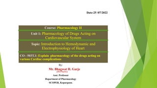
- 1. Course: Pharmacology II Unit 1: Pharmacology of Drugs Acting on Cardiovascular System Topic: Introduction to Hemodynamic and Electrophysiology of Heart CO - 503T.1: Explain pharmacology of the drugs acting on various Cardiac complications By: Mr. Bhagwat H. Garje (M.Pharm). Asst. Professor Department of Pharmacology SCOPER, Kopargaon. Date:25 /07/2022
- 2. Topic Covers a. Introduction to Hemodynamic and Electrophysiology of Heart b. Drugs used in Congestive Heart Failure c. Anti-Hypertensive Drugs d. Anti-Anginal Drugs e. Anti-Arrhythmic Drugs f. Anti-Hyperlipidemic Drugs 2
- 3. a. Introduction to Hemodynamic and Electrophysiology of Heart 3 The Human Heart
- 4. CONTENT A. HEMODYNAMIC - Introduction - Composition of Blood - Mechanism of hemodynamic B. Electrophysiology of Heart - Conduction System of Heart - ECG 4
- 5. Hemodynamic 5 • Hemodynamic= hemo (Blood) + dynamic (rate / motion) • Hemodynamics is a physical and physiological principles of blood flow (circulatory system) in the body. • It includes the circulation of blood through the heart and other parts of the body.
- 7. Mechanism of Hemodynamic The Heart is a muscular organ that acts as pump for circulation of blood throughout the body. Human heart has Four chambers namely Two Auricles and Two Ventricles. Auricles and ventricles are separated by valves that ensure unidirectional flow of blood. The Right Auricle/Atrium and Right Ventricles are separated by Tricuspid valve & Left Auricle and Left ventricles by Bicuspid/Mitral Valve 7
- 8. The Superior and Inferior vena cava brings deoxygenated blood to the right auricle from where it is emptied to right ventricle. The deoxygenated blood from right ventricle passes to lungs through pulmonary artery. Oxygenation of impure blood takes place in lungs where alveoli are the functional units. The oxygenated blood is then transferred to left auricle by pulmonary vein It then passes to left ventricle from where the blood is circulated to the systemic circulation through aorta 8
- 9. Properties of blood itself that affect its flow: 1. Viscosity 2. Inertial mass (mass of an object measured by its resistance to acceleration) 3. Volume of blood to be moved Factors that affect the motion of blood through the vascular channels include: 1. Size of blood vessel 2. Condition of blood vessel 3. Smoothness of lumen 4. Elasticity of muscular layer (tunica media) 5. Destination of blood (distal vascular bed) 9
- 10. Cardiac Output Flow of blood is usually measured in L/min Total amount of blood flowing through the circulation Cardiac Output (CO) Cardiac Output = Stroke Vol. x Heart Rate = 5 L/min Influenced by Blood Pressure & Resistance Force of blood -Blood viscosity against vessel wall -Vessel Length ↑ with water retention with dehydration, haemorrhage -Vessel Elasticity -Vasoconstriction/Vasodilation 10
- 12. 12 Conduction System of Heart
- 13. 13
- 14. 14 The heart is able to contract on its own because it contains specialized cardiac muscle tissue that spontaneously forms impulses and transmits them to the myocardium to initiate contraction. • The conducting system of the heart is composed of the following 5 components: 1. Sinoatrial node (SA node) 2. Atrioventricular node (AV node) 3. Atrioventricular bundle of His) 4. Left and right branches of bundle (of His) 5. Subendocardial Purkinje fibers. Components
- 15. 15 • The sequence of electrical events during one full contraction of the heart muscle: • An excitation signal (an action potential) is created by the sinoatrial (SA) node. • The wave of excitation spreads across the atria, causing them to contract. • Upon reaching the atrioventricular (AV) node, the signal is delayed. • It is then conducted into the bundle of His, down the interventricular septum. • The bundle of His and the Purkinje fibers spread the wave impulses along the ventricles, causing them to contract. Conduction System of Heart
- 16. 16 Electro Cardio Graph (ECG)
- 17. 17 • ECG is defined as “recording of electrical activity of heart on a graph paper.” Or • Graphical representation of electrical activity of heart.
- 18. 18 • ECG gives information about rate and rhythm of the heart. • The physical orientation of heart i.e. axis. • Its a diagnostic tool for various heart conditions like hypertrophies, ischemia, infarction, arrhythmias conduction. problems and pace maker activity. • ECG does not provide information about mechanical activity. Significance of ECG
- 19. 19 Normal ECG
- 20. 20 • P wave-Atrial depolarization • QRS complex-Ventricular depolarization • T wave-Ventricular repolarization Wave Forms
- 21. 21 • P Wave shows atrial depolarization. • Its duration is 0.1 sec (2 and half small sqr) and height is 2.5 mv (2 and half small sqr). • Presence of p waves in ECG strip shows the sinus rhythm. P- Wave
- 22. 22 • QRS complex represent the ventricular depolarization. • its normal duration is about 0. 08 seconds.(less than 2 small sqr) and height is about 5 to 20 small sqrs. • It is a wide complex because it mask the atrial repolarization. • Q wave is first wave of this complex but often absent. QRS-Complex
- 23. 23 • It represent the ventricular repolarization. • It is repolarizing wave but shows the upward deflection because the part depolarized in the last is first to be repolarized,, that is base of heart depolarized in the last but is first to be repolarized. T- Wave • T wave should not be more than one third of R wave. • T wave inversion represent ischemia of heart. • Tall and peaked R wave is present in hyperkalemia. • Flattened R waves in pericarditis and myocarditis.
- 24. 24