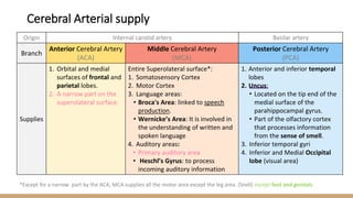
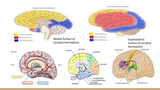
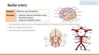
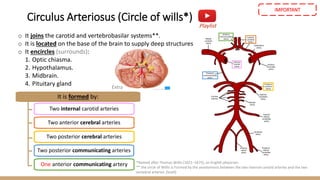
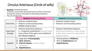
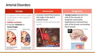
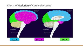
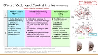
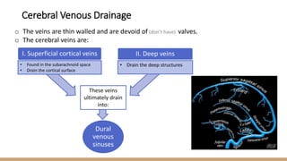
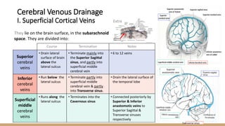
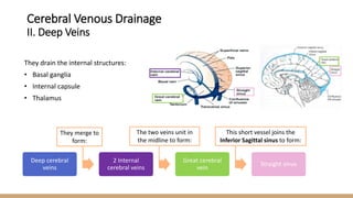
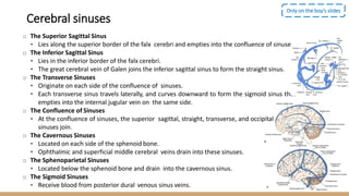
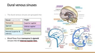
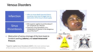
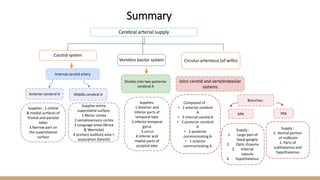
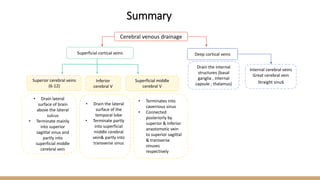
Please check our Editing File
{ ْ
‫ن‬َ‫م‬َ‫و‬
ْ َ
‫ّك‬َ‫و‬َ‫ت‬َ‫ي‬
َْ
‫ل‬َ‫ع‬
ْ‫ه‬ َ
‫اّلل‬
َْ‫ُو‬‫ه‬َ‫ف‬
َْ‫ح‬
ُُُُْْ‫س‬ }
Lecture (16)
▪ Important
▪ Doctors Notes
▪ Notes/Extra explanation
Cerebral Blood
Circulation
‫دفعة‬ ‫عمل‬ ‫عىل‬ ‫ي‬
‫أساس‬ ‫بشكل‬ ‫ي‬
‫مبن‬ ‫العمل‬ ‫هذا‬
436
‫اجعة‬‫ر‬‫الم‬ ‫مع‬
‫للمذاكرة‬ ‫ي‬
‫األساس‬‫المصدر‬ ‫عن‬ ‫ي‬
‫يغن‬ ‫وال‬ ‫المالحظات‬ ‫وإضافة‬ ‫والتدقيق‬
▪ Objectives
At the end of the lecture, students should be able to:
✓ List the cerebral arteries.
✓ Describe the cerebral arterial supply regarding the origin,
distribution and branches.
✓ Describe the arterial Circle of Willis .
✓ Describe the cerebral venous drainage and its termination.
✓ Describe arterial & venous vascular disorders and their clinical
manifestations.
Cerebral Circulation
o It is the movement of blood through the network of blood vessels to
supply the brain.
o The movement of blood in the cerebral circulation is called cerebral
blood flow.
o The arteries carry oxygenated blood and other nutrients to the brain.
o The veins carry deoxygenated blood back to the heart removing carbon
dioxide and other metabolic products.
o The two anterior cerebral circulations (left and right) are connected to
each other by ONE anterior communicating artery.
o The anterior circulation (carotid) is interconnected to the posterior
circulation (basilar) via bilateral TWO posterior communicating arteries.
o Posterior communicating arteries are part of Circle of Willis.
Only on the boy’s slides
Extra
The arterial supply of the cerebrum is
composed of/provided by 2 arterial systems:
1) Carotid System (Posterior cerebral circulation)
Composed of:
• Internal carotid artery and its branches:
• Anterior cerebral artery* (smaller)
• Middle cerebral artery* (bigger)
Supply anterior portion of brain
The two vertebral arteries (from the subclavian
artery) unite to form basilar artery*.
It divides at the upper border of pons into two
posterior cerebral arteries*
Supply posterior portion of brain
Extra
*We will talk about these in more detail later.
Cerebral Arterial supply
Extra
2) Vertebro-Basilar System (Posterior cerebral circulation)
Origin Internal carotid artery Basilar artery
Branch
Anterior Cerebral Artery
(ACA)
Middle Cerebral Artery
(MCA)
Posterior Cerebral Artery
(PCA)
Supplies
1. Orbital and medial
surfaces of frontal and
parietal lobes.
2. A narrow part on the
superolateral surface.
Entire Superolateral surface*:
1. Somatosensory Cortex
2. Motor Cortex
3. Language areas:
• Broca's Area: linked to speech
production.
• Wernicke’s Area: It is involved in
the understanding of written and
spoken language
4. Auditory areas:
• Primary auditory area
• Heschl’s Gyrus: to process
incoming auditory information
1. Anterior and inferior temporal
lobes
2. Uncus:
• Located on the tip end of the
medial surface of the
parahippocampal gyrus.
• Part of the olfactory cortex
that processes information
from the sense of smell.
3. Inferior temporal gyri
4. Inferior and Medial Occipital
lobe (visual area)
*Except for a narrow part by the ACA, MCA supplies all the motor area except the leg area. (Snell) except foot and genitals
Cerebral Arterial supply
Medial Surface of
Cerebral Hemisphere
Superolateral
Surface of Cerebral
Hemisphere
Basilar artery
o The vertebral arteries unite at the lower border of the pons to form the basilar artery.
o Pontine: related to the pons.
o Its formed by 2 vertebral artery
Only on the boy’s slides
Supplies: Mid brain and cerebellum
Branches: • Anterior inferior cerebellar artery.
• Pontine branches.
• Superior cerebellar artery.
Extra
o It joins the carotid and vertebrobasilar systems**.
o It is located on the base of the brain to supply deep structures
o It encircles (surrounds):
1. Optic chiasma.
2. Hypothalamus.
3. Midbrain.
4. Pituitary gland
*Named after Thomas Willis (1621–1675), an English physician.
** the circle of Willis is Formed by the anastomosis between the two internal carotid arteries and the two
vertebral arteries. (Snell)
Playlist
Extra
It is formed by:
Two internal carotid arteries
Two anterior cerebral arteries
Two posterior cerebral arteries
Two posterior communicating arteries
One anterior communicating artery
Circulus Arteriosus (Circle of wills*)
IMPORTANT
o Branches: Perforating arteries
numerous small vessels that penetrate the surface of the brain
through the anterior and posterior perforating substances.
o They are divided into:
Anterior Perforating Arteries Posterior Perforating Arteries
Arise from:
• Anterior cerebral artery.
• Anterior communicating artery.
• Middle cerebral artery.
• Posterior cerebral artery .
• Posterior communicating artery.
Enter brain
through:
• Anterior perforated substance
(an irregularly quadrilateral (having 4 sides)
area in front of the optic tract and behind
the olfactory trigone).
• Posterior Perforated substance.
(perforated substance: a layer of grey
matter, that is pierced to allow blood
vessels to pass)
Supplies
1. Large part of basal ganglia.
2. Optic chiasma.
3. Internal capsule ( a white matter
structure).
4. Hypothalamus .
1. Ventral portion of Midbrain.
2. Parts of Subthalamus and
Hypothalamus.
Circulus Arteriosus (Circle of wills)
Angioma
Aneurysm
Stroke
Arterial Disorders
• It happens when blood supply
to the brain is interrupted or
reduced.
• Sudden occlusion
• It can be: Ischemic or
hemorrhagic.
• localized, blood-filled balloon-
like bulge in the wall of
a blood vessel.
• Dilation of the wall (may take
years)
• benign tumors derived from
cells of the vascular or
lymphatic vessel walls
(epithelium) or derived from
cells of the tissues surrounding
these vessels.
Effects of Occlusion of Cerebral Arteries
Effects of Occlusion of Cerebral Arteries (Manifestations)
(Important may come as scenario)
Optic radiation: axons from the neurons in the lateral geniculate nucleus to the primary visual cortex.
Hemianopsia: decreased vision or blindness in half the visual field.
Homonymous hemianopsia: the loss of half of the visual field on the same side in both eyes (see picture on right).
Cortical blindness: total or partial vision loss because of damage to the occipital cortex.
Anterior
cerebral artery
Extra
Artery
Anterior Cerebral
Artery (ACA)
Middle Cerebral Artery
(MCA)
Posterior Cerebral
Artery
(PCA)
Effect
1- Motor disturbance in
contralateral distal leg
2- Difficulty in
prefrontal lobe
functions:
• Cognitive thinking
• Judgement
• Motor initiation
• Self monitoring
1- Contralateral weakness of:
face, arm & hands “more than legs”
• 2- Contralateral sensory loss of:
face, arm, and hands more than
legs
• Visual field cut (damage to optic
radiation)
4- Aphasia (language disturbance):
• In Broca’s area: production
• In Wernicke’s area: comprehension
1- Visual disturbances:
• Unilateral lesion:
contralateral
homonymous
hemianopsia
• Bilateral lesions: cortical
blindness, patients
unaware they cannot
see (Anton’s Syndrome)
2- Memory impairment: if
temporal lobe is affected
IMPORTANT
o The veins are thin walled and are devoid of (don’t have) valves.
o The cerebral veins are:
• Found in the subarachnoid space
• Drain the cortical surface
I. Superficial cortical veins II. Deep veins
• Drain the deep structures
These veins
ultimately drain
into:
Dural
venous
sinuses
Cerebral Venous Drainage
Extra
They lie on the brain surface, in the subarachnoid
space. They are divided into:
Superior
anastomotic vein
Transverse sinus
Course Termination Notes
Superior
cerebral
veins
• Drain lateral
surface of brain
above the
lateral sulcus
• Terminate mainly into
the Superior Sagittal
sinus, and partly into
superficial middle
cerebral vein
• 6 to 12 veins
Inferior
cerebral
veins
• Run below the
lateral sulcus
• Terminate partly into
superficial middle
cerebral vein & partly
into Transverse sinus.
• Drain the lateral surface of
the temporal lobe
Superficial
middle
cerebral
veins
• Runs along the
lateral sulcus
• Terminates into the
Cavernous sinus
• Connected posteriorly by
Superior & Inferior
anastomotic veins to
Superior Sagittal &
Transverse sinuses
respectively
Cerebral Venous Drainage
I. Superficial Cortical Veins
Deep cerebral
veins
2 Internal
cerebral veins
Great cerebral
vein
Straight sinus
They merge to
form:
The two veins unit in
the midline to form:
This short vessel joins the
Inferior Sagittal sinus to form:
They drain the internal structures:
• Basal ganglia
• Internal capsule
• Thalamus
Cerebral Venous Drainage
II. Deep Veins
o The Superior Sagittal Sinus
• Lies along the superior border of the falx cerebri and empties into the confluence of sinuses.
o The Inferior Sagittal Sinus
• Lies in the inferior border of the falx cerebri.
• The great cerebral vein of Galen joins the inferior sagittal sinus to form the straight sinus.
o The Transverse Sinuses
• Originate on each side of the confluence of sinuses.
• Each transverse sinus travels laterally, and curves downward to form the sigmoid sinus that
empties into the internal jugular vein on the same side.
o The Confluence of Sinuses
• At the confluence of sinuses, the superior sagittal, straight, transverse, and occipital .
sinuses join.
o The Cavernous Sinuses
• Located on each side of the sphenoid bone.
• Ophthalmic and superficial middle cerebral veins drain into these sinuses.
o The Sphenoparietal Sinuses
• Located below the sphenoid bone and drain into the cavernous sinus.
o The Sigmoid Sinuses
• Receive blood from posterior dural venous sinus veins.
Only on the boy’s slides
Cerebral sinuses
o Formed by the union of the right and left brachiocephalic veins.
o Brachiocephalic veins are formed by the union
o Of internal jugular and subclavian veins.
o Drains venous blood Superior vena cava
o from:
o Head, neck, thoracic wall & upper limbs
o It passes downward and enter the right atrium.
Only on the boy’s slides
Superior vena cava
o The dural venous sinuses are grouped into:
o Blood flows from transverse & sigmoid
sinuses into IJV Internal Jugular Vein.
Single:
Superior sagittal
Inferior sagittal
Straight
Occipital
Paired: (right & left)
Transverse
Sigmoid
Cavernous
Petrosal (superior &
inferior)
Dural venous sinuses
o Obstruction of venous drainage of the brain leads to
Cerebral swelling (edema) and raised Intracranial
Pressure.
• refers to tissue death (necrosis) that is
caused by a local lack of oxygen due to
obstruction of the tissue's blood supply
Infarction
• SSS (superior sagittal sinus) thrombosis*:
can complicate ear infection
• Cavernous sinus thrombosis: As a
complication of infection in the dangerous
area of the face**
Sinus
thrombosis
*Superior sagittal sinus thrombosis (SSST) is the most common type
of dural venous sinus thrombosis and is potentially devastating
**
Extra
Venous Disorders
These 2 videos summarize the lecture:
(to view them download the ppt version)
Duration: 01:42 Duration: 01:18
Only on the boy’s slides
Cerebral arterial supply
Carotid system
Vertebro basilar system Circulus arteriosus (of willis)
Divides into two posterior
cerebral A
Middle cerebral A
Anterior cerebral A
Supplies : 1.orbital
& medial surfaces of
frontal and parietal
lobes
3.Narrow part on
the superolateral
surface
Supplies entire
superolatral surface:
1.Motor cortex
2.somatosensory cortex
3.Language areas (Broca
& Wernicke)
4.primary auditory area +
association (heschl)
Supplies:
1.Anterior and
inferior parts of
temporal lobe
2.Inferior temporal
gyrus
3.uncus
4.Inferior and
medial parts of
occipital lobe
Internal carotid artery
Joins carotid and vertebrobasilar
systems
Composed of :
• 2 anterior cerebral
A
• 2 internal carotid A
• 2 posterior cerebral
A
• 2 posterior
communicating A
• 1 anterior
communicating A
Branches :
APA
Supply :
1. Large part of
basal ganglia
2. Optic chiasma
3. Internal
capsule
4. hypothalamus
PPA
Supply :
1. Ventral portion
of midbrain
2. Parts of
subthalamus and
hypothalamus
Summary
Cerebral venous drainage
Superficial cortical veins Deep cortical veins
Superior cerebral veins
(6-12)
• Drain lateral
surface of brain
above the lateral
sulcus
• Terminate mainly
into superior
sagittal sinus and
partly into
superficial middle
cerebral vein
Inferior
cerebral V
• Drain the lateral
surface of the
temporal lobe
• Terminate partly
into superficial
middle cerebral
vein& partly into
transverse sinus
Superficial middle
cerebral V
• Terminates into
cavernous sinus
• Connected
posteriorly by
superior & inferior
anastomotic vein
to superior sagittal
& transverse
sinuses
respectively
Drain the internal
structures (basal
ganglia , internal
capsule , thalamus)
Internal cerebral veins
Great cerebral vein
Straight sinus
Summary
MCQs
(1) What does the basilar artery supply?
A) Mid brain B) Cerebellum
C) A&B D) Motor cortex
(2) Which of the following are paired Dural sinuses?
A) Inferior sagittal B) Straight
C) Cavernous D) Occipital
(3) Which of the following is the one of the effects that occur
when the posterior cerebral artery is occluded?
A) Visual disturbances
B) Language disturbances
C) Motor disturbance in the contralateral distal leg
D) Contralateral sensory loss of face
(4) Which of the following supplies the optic chiasma?
A) Anterior perforating arteries
B) Posterior perforating arteries
C) Middle perforating arteries
D) Anterior cerebral artery
(5) What does the the circle of willis not encircle?
A) Optic chiasma B) Hypothalamus
C) Midbrain D) Cerebellum
(6) Which of the following is not a branch of the basilar artery
A) Anterior inferior cerebellar artery.
B) Pontine branches
C) Superior cerebellar artery
D) Middle cerebral artery
(7) What do the cerebral veins NOT drain?
A) Basal ganglia B) Internal capsule
C) Thalamus D) Hypothalamus
(8) Which of the following runs below the lateral sulcus:
A) Superficial cerebral veins B) Inferior cerebral veins
C) Superficial cerebral artery D) Inferior cerebral artery
(9) A patient Came to the hospital and his family noticed
difference in his personality and he cant make decisions,
what is the most likely diagnosis?
A) Middle cerebral artery occlusion B) Superior sagittal sinus
C) Thrombosis anterior cerebral artery
D) Right temporal and right nasal Fibers
(10) Blood filled bulge in the wall of a blood vessel?
A) Stroke B) Aneurysm
C) Angioma D) Sinus thrombosis
Answers
(1) C (6) D
(2) C (7) D
(3) A (8) B
(4) A (9) C
(5) D (10) B
SAQ
(1) From which arteries do the anterior perforating arteries arise fro,
• Anterior cerebral artery
• Anterior communicating artery
• Middle cerebral artery
(2) Which parts do the posterior perforating arteries supply?
• Ventral portion of Midbrain
• Parts of Subthalamus and Hypothalamus
(3) A 35 year old man came to the hospital complaining of impaired
memory and cortical blindness.
(A) Which cerebral artery is affected?
Posterior cerebral artery
(B) Which side is most likely affected?
Bilateral lesion (affecting both sides) which will lead to Anton’s syndrome
Team Leaders:
Faisal Fahad Alsaif
Rawan Mohammad Alharbi
Team Members:
Abdulaziz Aldukhayel
Abdulrahman Alduhayyim
Rinad Alghoraiby
Rawan Mishal
Good luck
Special thank for team436
▪ References:
1.Girls’ & Boys’ Slides
2.Greys Anatomy for Students
3.TeachMeAnatomy.com
Twitter.com/Anatomy437
Anatomyteam.437@gmail.com

16- Cerebral Circulation. (Edited).pdf

  • 1.
    Please check ourEditing File { ْ ‫ن‬َ‫م‬َ‫و‬ ْ َ ‫ّك‬َ‫و‬َ‫ت‬َ‫ي‬ َْ ‫ل‬َ‫ع‬ ْ‫ه‬ َ ‫اّلل‬ َْ‫ُو‬‫ه‬َ‫ف‬ َْ‫ح‬ ُُُُْْ‫س‬ } Lecture (16) ▪ Important ▪ Doctors Notes ▪ Notes/Extra explanation Cerebral Blood Circulation ‫دفعة‬ ‫عمل‬ ‫عىل‬ ‫ي‬ ‫أساس‬ ‫بشكل‬ ‫ي‬ ‫مبن‬ ‫العمل‬ ‫هذا‬ 436 ‫اجعة‬‫ر‬‫الم‬ ‫مع‬ ‫للمذاكرة‬ ‫ي‬ ‫األساس‬‫المصدر‬ ‫عن‬ ‫ي‬ ‫يغن‬ ‫وال‬ ‫المالحظات‬ ‫وإضافة‬ ‫والتدقيق‬
  • 2.
    ▪ Objectives At theend of the lecture, students should be able to: ✓ List the cerebral arteries. ✓ Describe the cerebral arterial supply regarding the origin, distribution and branches. ✓ Describe the arterial Circle of Willis . ✓ Describe the cerebral venous drainage and its termination. ✓ Describe arterial & venous vascular disorders and their clinical manifestations.
  • 3.
    Cerebral Circulation o Itis the movement of blood through the network of blood vessels to supply the brain. o The movement of blood in the cerebral circulation is called cerebral blood flow. o The arteries carry oxygenated blood and other nutrients to the brain. o The veins carry deoxygenated blood back to the heart removing carbon dioxide and other metabolic products. o The two anterior cerebral circulations (left and right) are connected to each other by ONE anterior communicating artery. o The anterior circulation (carotid) is interconnected to the posterior circulation (basilar) via bilateral TWO posterior communicating arteries. o Posterior communicating arteries are part of Circle of Willis. Only on the boy’s slides Extra
  • 4.
    The arterial supplyof the cerebrum is composed of/provided by 2 arterial systems: 1) Carotid System (Posterior cerebral circulation) Composed of: • Internal carotid artery and its branches: • Anterior cerebral artery* (smaller) • Middle cerebral artery* (bigger) Supply anterior portion of brain The two vertebral arteries (from the subclavian artery) unite to form basilar artery*. It divides at the upper border of pons into two posterior cerebral arteries* Supply posterior portion of brain Extra *We will talk about these in more detail later. Cerebral Arterial supply Extra 2) Vertebro-Basilar System (Posterior cerebral circulation)
  • 5.
    Origin Internal carotidartery Basilar artery Branch Anterior Cerebral Artery (ACA) Middle Cerebral Artery (MCA) Posterior Cerebral Artery (PCA) Supplies 1. Orbital and medial surfaces of frontal and parietal lobes. 2. A narrow part on the superolateral surface. Entire Superolateral surface*: 1. Somatosensory Cortex 2. Motor Cortex 3. Language areas: • Broca's Area: linked to speech production. • Wernicke’s Area: It is involved in the understanding of written and spoken language 4. Auditory areas: • Primary auditory area • Heschl’s Gyrus: to process incoming auditory information 1. Anterior and inferior temporal lobes 2. Uncus: • Located on the tip end of the medial surface of the parahippocampal gyrus. • Part of the olfactory cortex that processes information from the sense of smell. 3. Inferior temporal gyri 4. Inferior and Medial Occipital lobe (visual area) *Except for a narrow part by the ACA, MCA supplies all the motor area except the leg area. (Snell) except foot and genitals Cerebral Arterial supply
  • 6.
    Medial Surface of CerebralHemisphere Superolateral Surface of Cerebral Hemisphere
  • 7.
    Basilar artery o Thevertebral arteries unite at the lower border of the pons to form the basilar artery. o Pontine: related to the pons. o Its formed by 2 vertebral artery Only on the boy’s slides Supplies: Mid brain and cerebellum Branches: • Anterior inferior cerebellar artery. • Pontine branches. • Superior cerebellar artery. Extra
  • 8.
    o It joinsthe carotid and vertebrobasilar systems**. o It is located on the base of the brain to supply deep structures o It encircles (surrounds): 1. Optic chiasma. 2. Hypothalamus. 3. Midbrain. 4. Pituitary gland *Named after Thomas Willis (1621–1675), an English physician. ** the circle of Willis is Formed by the anastomosis between the two internal carotid arteries and the two vertebral arteries. (Snell) Playlist Extra It is formed by: Two internal carotid arteries Two anterior cerebral arteries Two posterior cerebral arteries Two posterior communicating arteries One anterior communicating artery Circulus Arteriosus (Circle of wills*) IMPORTANT
  • 9.
    o Branches: Perforatingarteries numerous small vessels that penetrate the surface of the brain through the anterior and posterior perforating substances. o They are divided into: Anterior Perforating Arteries Posterior Perforating Arteries Arise from: • Anterior cerebral artery. • Anterior communicating artery. • Middle cerebral artery. • Posterior cerebral artery . • Posterior communicating artery. Enter brain through: • Anterior perforated substance (an irregularly quadrilateral (having 4 sides) area in front of the optic tract and behind the olfactory trigone). • Posterior Perforated substance. (perforated substance: a layer of grey matter, that is pierced to allow blood vessels to pass) Supplies 1. Large part of basal ganglia. 2. Optic chiasma. 3. Internal capsule ( a white matter structure). 4. Hypothalamus . 1. Ventral portion of Midbrain. 2. Parts of Subthalamus and Hypothalamus. Circulus Arteriosus (Circle of wills)
  • 10.
    Angioma Aneurysm Stroke Arterial Disorders • Ithappens when blood supply to the brain is interrupted or reduced. • Sudden occlusion • It can be: Ischemic or hemorrhagic. • localized, blood-filled balloon- like bulge in the wall of a blood vessel. • Dilation of the wall (may take years) • benign tumors derived from cells of the vascular or lymphatic vessel walls (epithelium) or derived from cells of the tissues surrounding these vessels.
  • 11.
    Effects of Occlusionof Cerebral Arteries
  • 12.
    Effects of Occlusionof Cerebral Arteries (Manifestations) (Important may come as scenario) Optic radiation: axons from the neurons in the lateral geniculate nucleus to the primary visual cortex. Hemianopsia: decreased vision or blindness in half the visual field. Homonymous hemianopsia: the loss of half of the visual field on the same side in both eyes (see picture on right). Cortical blindness: total or partial vision loss because of damage to the occipital cortex. Anterior cerebral artery Extra Artery Anterior Cerebral Artery (ACA) Middle Cerebral Artery (MCA) Posterior Cerebral Artery (PCA) Effect 1- Motor disturbance in contralateral distal leg 2- Difficulty in prefrontal lobe functions: • Cognitive thinking • Judgement • Motor initiation • Self monitoring 1- Contralateral weakness of: face, arm & hands “more than legs” • 2- Contralateral sensory loss of: face, arm, and hands more than legs • Visual field cut (damage to optic radiation) 4- Aphasia (language disturbance): • In Broca’s area: production • In Wernicke’s area: comprehension 1- Visual disturbances: • Unilateral lesion: contralateral homonymous hemianopsia • Bilateral lesions: cortical blindness, patients unaware they cannot see (Anton’s Syndrome) 2- Memory impairment: if temporal lobe is affected IMPORTANT
  • 13.
    o The veinsare thin walled and are devoid of (don’t have) valves. o The cerebral veins are: • Found in the subarachnoid space • Drain the cortical surface I. Superficial cortical veins II. Deep veins • Drain the deep structures These veins ultimately drain into: Dural venous sinuses Cerebral Venous Drainage
  • 14.
    Extra They lie onthe brain surface, in the subarachnoid space. They are divided into: Superior anastomotic vein Transverse sinus Course Termination Notes Superior cerebral veins • Drain lateral surface of brain above the lateral sulcus • Terminate mainly into the Superior Sagittal sinus, and partly into superficial middle cerebral vein • 6 to 12 veins Inferior cerebral veins • Run below the lateral sulcus • Terminate partly into superficial middle cerebral vein & partly into Transverse sinus. • Drain the lateral surface of the temporal lobe Superficial middle cerebral veins • Runs along the lateral sulcus • Terminates into the Cavernous sinus • Connected posteriorly by Superior & Inferior anastomotic veins to Superior Sagittal & Transverse sinuses respectively Cerebral Venous Drainage I. Superficial Cortical Veins
  • 15.
    Deep cerebral veins 2 Internal cerebralveins Great cerebral vein Straight sinus They merge to form: The two veins unit in the midline to form: This short vessel joins the Inferior Sagittal sinus to form: They drain the internal structures: • Basal ganglia • Internal capsule • Thalamus Cerebral Venous Drainage II. Deep Veins
  • 16.
    o The SuperiorSagittal Sinus • Lies along the superior border of the falx cerebri and empties into the confluence of sinuses. o The Inferior Sagittal Sinus • Lies in the inferior border of the falx cerebri. • The great cerebral vein of Galen joins the inferior sagittal sinus to form the straight sinus. o The Transverse Sinuses • Originate on each side of the confluence of sinuses. • Each transverse sinus travels laterally, and curves downward to form the sigmoid sinus that empties into the internal jugular vein on the same side. o The Confluence of Sinuses • At the confluence of sinuses, the superior sagittal, straight, transverse, and occipital . sinuses join. o The Cavernous Sinuses • Located on each side of the sphenoid bone. • Ophthalmic and superficial middle cerebral veins drain into these sinuses. o The Sphenoparietal Sinuses • Located below the sphenoid bone and drain into the cavernous sinus. o The Sigmoid Sinuses • Receive blood from posterior dural venous sinus veins. Only on the boy’s slides Cerebral sinuses
  • 17.
    o Formed bythe union of the right and left brachiocephalic veins. o Brachiocephalic veins are formed by the union o Of internal jugular and subclavian veins. o Drains venous blood Superior vena cava o from: o Head, neck, thoracic wall & upper limbs o It passes downward and enter the right atrium. Only on the boy’s slides Superior vena cava
  • 18.
    o The duralvenous sinuses are grouped into: o Blood flows from transverse & sigmoid sinuses into IJV Internal Jugular Vein. Single: Superior sagittal Inferior sagittal Straight Occipital Paired: (right & left) Transverse Sigmoid Cavernous Petrosal (superior & inferior) Dural venous sinuses
  • 19.
    o Obstruction ofvenous drainage of the brain leads to Cerebral swelling (edema) and raised Intracranial Pressure. • refers to tissue death (necrosis) that is caused by a local lack of oxygen due to obstruction of the tissue's blood supply Infarction • SSS (superior sagittal sinus) thrombosis*: can complicate ear infection • Cavernous sinus thrombosis: As a complication of infection in the dangerous area of the face** Sinus thrombosis *Superior sagittal sinus thrombosis (SSST) is the most common type of dural venous sinus thrombosis and is potentially devastating ** Extra Venous Disorders
  • 20.
    These 2 videossummarize the lecture: (to view them download the ppt version) Duration: 01:42 Duration: 01:18 Only on the boy’s slides
  • 21.
    Cerebral arterial supply Carotidsystem Vertebro basilar system Circulus arteriosus (of willis) Divides into two posterior cerebral A Middle cerebral A Anterior cerebral A Supplies : 1.orbital & medial surfaces of frontal and parietal lobes 3.Narrow part on the superolateral surface Supplies entire superolatral surface: 1.Motor cortex 2.somatosensory cortex 3.Language areas (Broca & Wernicke) 4.primary auditory area + association (heschl) Supplies: 1.Anterior and inferior parts of temporal lobe 2.Inferior temporal gyrus 3.uncus 4.Inferior and medial parts of occipital lobe Internal carotid artery Joins carotid and vertebrobasilar systems Composed of : • 2 anterior cerebral A • 2 internal carotid A • 2 posterior cerebral A • 2 posterior communicating A • 1 anterior communicating A Branches : APA Supply : 1. Large part of basal ganglia 2. Optic chiasma 3. Internal capsule 4. hypothalamus PPA Supply : 1. Ventral portion of midbrain 2. Parts of subthalamus and hypothalamus Summary
  • 22.
    Cerebral venous drainage Superficialcortical veins Deep cortical veins Superior cerebral veins (6-12) • Drain lateral surface of brain above the lateral sulcus • Terminate mainly into superior sagittal sinus and partly into superficial middle cerebral vein Inferior cerebral V • Drain the lateral surface of the temporal lobe • Terminate partly into superficial middle cerebral vein& partly into transverse sinus Superficial middle cerebral V • Terminates into cavernous sinus • Connected posteriorly by superior & inferior anastomotic vein to superior sagittal & transverse sinuses respectively Drain the internal structures (basal ganglia , internal capsule , thalamus) Internal cerebral veins Great cerebral vein Straight sinus Summary
  • 23.
    MCQs (1) What doesthe basilar artery supply? A) Mid brain B) Cerebellum C) A&B D) Motor cortex (2) Which of the following are paired Dural sinuses? A) Inferior sagittal B) Straight C) Cavernous D) Occipital (3) Which of the following is the one of the effects that occur when the posterior cerebral artery is occluded? A) Visual disturbances B) Language disturbances C) Motor disturbance in the contralateral distal leg D) Contralateral sensory loss of face (4) Which of the following supplies the optic chiasma? A) Anterior perforating arteries B) Posterior perforating arteries C) Middle perforating arteries D) Anterior cerebral artery (5) What does the the circle of willis not encircle? A) Optic chiasma B) Hypothalamus C) Midbrain D) Cerebellum (6) Which of the following is not a branch of the basilar artery A) Anterior inferior cerebellar artery. B) Pontine branches C) Superior cerebellar artery D) Middle cerebral artery (7) What do the cerebral veins NOT drain? A) Basal ganglia B) Internal capsule C) Thalamus D) Hypothalamus (8) Which of the following runs below the lateral sulcus: A) Superficial cerebral veins B) Inferior cerebral veins C) Superficial cerebral artery D) Inferior cerebral artery (9) A patient Came to the hospital and his family noticed difference in his personality and he cant make decisions, what is the most likely diagnosis? A) Middle cerebral artery occlusion B) Superior sagittal sinus C) Thrombosis anterior cerebral artery D) Right temporal and right nasal Fibers (10) Blood filled bulge in the wall of a blood vessel? A) Stroke B) Aneurysm C) Angioma D) Sinus thrombosis
  • 24.
    Answers (1) C (6)D (2) C (7) D (3) A (8) B (4) A (9) C (5) D (10) B
  • 25.
    SAQ (1) From whicharteries do the anterior perforating arteries arise fro, • Anterior cerebral artery • Anterior communicating artery • Middle cerebral artery (2) Which parts do the posterior perforating arteries supply? • Ventral portion of Midbrain • Parts of Subthalamus and Hypothalamus (3) A 35 year old man came to the hospital complaining of impaired memory and cortical blindness. (A) Which cerebral artery is affected? Posterior cerebral artery (B) Which side is most likely affected? Bilateral lesion (affecting both sides) which will lead to Anton’s syndrome
  • 26.
    Team Leaders: Faisal FahadAlsaif Rawan Mohammad Alharbi Team Members: Abdulaziz Aldukhayel Abdulrahman Alduhayyim Rinad Alghoraiby Rawan Mishal Good luck Special thank for team436 ▪ References: 1.Girls’ & Boys’ Slides 2.Greys Anatomy for Students 3.TeachMeAnatomy.com Twitter.com/Anatomy437 Anatomyteam.437@gmail.com