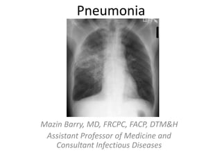
1. Pneumonia
Mazin Barry, MD, FRCPC, FACP, DTM&H
Assistant Professor of Medicine and
Consultant Infectious Diseases
2. Terminology
• Merriam Webster Dictionary
– pneu·mo·nia noun nu̇-mō-nyə, nyu̇-
• Origin of PNEUMONIA
– New Latin, from Greek, from pneumōn lung,
alteration of pleumōn
– First Known Use: 1603
3. Historical Points
• Referred to pneumonia as a
disease "named by the
ancients."
• “If sweats come out about
the neck and head, for such
sweats are bad, as
proceeding from the
suffocation, rales, and the
violence of the disease
which is obtaining the
upper hand”
Hippocrates Ancient Greek
Physician known as the “Father of
Medicine” (c. 460 BC – 370 BC)
4. Historical Points
• “the most widespread
and fatal of all acute
diseases, pneumonia, is
now Captain of the Men
of Death.”
The Principles and Practice of Medicine;
4th ed. New York, Appleton, 1901
Sir William Osler
5. What is Pneumonia?
• Pneumonia is an
inflammatory condition of the
lung
• characterized by
inflammation of the
parenchyma of the lung
(alveoli)
• Abnormal alveolar filling with
fluid causing Air space disease
(consolidation and exudation)
6.
7. Pneumonia: Definitions
• Community-acquired pneumonia (CAP)
Cough/fever/sputum production + infiltrate, related to community
• Healthcare-associated pneumonia (HCAP)
Pneumonia that develops within 48 hours of admission in pts with:
– Hospitalization in acute care hospital for >2 d in past 90 d
– Residence in NH or LTC facility
– Chronic dialysis within 30 days
– Home IV therapy, home wound care in past 30 days
– Family member with MDR pathogen
• Hospital-acquired pneumonia (HAP)
Pneumonia > 48 hours after admission
• Ventilator-associated pneumonia (VAP)
pneumonia > 48 hours after intubation
8. Epidemiology
• Unclear Few population-based statistics on the
condition alone
• Pneumonia & influenza = 6th leading causes of death in
the world
• Single most common cause of infection-related
mortality
• Age-adjusted death rate = 22 per 100,000 per year
• Mortality rate: 1-5% out-Pt, 12% In-Pt, 40% ICU
• Death rates increase with comorbidity and age
• Affects race and sex equally
9. Pathogenesis
• Inhalation, aspiration and hematogenous
spread are the 3 main mechanisms by which
bacteria reaches the lungs
• Primary inhalation: when organisms bypass
normal respiratory defense mechanisms or
when the Pt inhales organisms that colonize
the upper respiratory tract or respiratory
support equipment
10. Pathogenesis
• Aspiration: occurs when the Pt aspirates
colonized upper respiratory tract secretions
– Stomach: reservoir of GNR that can ascend,
colonizing the respiratory tract.
• Hematogenous: originate from a distant
source and reach the lungs via the blood
stream.
11. Pathogenesis
• Microaspiration from nasopharynx: S. Pneumonia
• Inhalation: TB, viruses, Legionella
• Aspiration: anaerobes
• Bloodborne: Staph endocarditis, septic emboli
• Direct extension: trauma
12. Pathogens
• CAP usually caused by a single organism
• Even with extensive diagnostic testing, most
investigators cannot identify a specific etiology
for CAP in ≥ 50% of patients.
• Caused by a variety of Bacteria, Viruses, Fungi
• Streptococcus pneumoniae is the most
common pathogen 60-70% of the time
15. Clinical Signs Positive LR Negative LR
General appearance
Cachexia 4.0 NS
Abnormal mental status 2.2 NS
Vital signs
Temp >37.9 C 2.2 0.7
RR > 28/min 2.2 0.8
HR >100 bpm 1.6 0.7
Lung findings
Percussion dullness 3.0 NS
Reduced breath sounds 2.3 0.8
Bronchial breath sounds 3.3 NS
Aegophony 4.1 NS
Crackles 2.0 0.8
Wheezes NS NS
NS= not significant. LR= Likelihood Ratio
From McGee S, Evidence-based physical diagnosis, 2nd edition. St Louis: Saunders, 2007.
16. Triaging Patients with Pneumonia
• Febrile respiratory illness (FRI) should be
placed on droplet and contact precautions
(Single room, use mask, gown and gloves)
• Upgrade to Airborne Infection Isolation (AII)-
Negative pressure room with HEPA-Filter, with
use of fit-tested respirator (e.g. N-95), in
addition to contact precautions for:
– Sick patients anticipating intubation
– Aerosolizing procedures e.g. suctioning,
nebulization
18. Clinical Diagnosis: CXR
• Demonstrable infiltrate by CXR or other
imaging technique
– Establish Dx and presence of complications
(pleural effusion, multilobar disease)
– May not be possible in some outpatient settings
– CXR: classically thought of as the gold standard
19. Infiltrate Patterns
Pattern Possible Diagnosis
Lobar S. pneumo, Kleb, H. flu,
GN
Patchy Atypicals, viral, Legionella
Interstitial Viral, PCP, Legionella
Cavitary Anaerobes, Kleb, TB, S.
aureus, fungi
Large effusion Staph, anaerobes, Kleb
20. A chest X-ray showing a very prominent wedge shaped pneumonia in the right lung
24. Empiric outpt Management in Previously
Healthy Pt
• Organisms: S. pneumoniae, Mycoplasma pneumoniae,
viral, Chlamydophila pneumoniae, H.influenzae
• Recommended abx:
– Advanced generation macrolide (azithromycin or
clarithromycin); or doxycycline
• If abx within past 3 months:
– Respiratory quinolone (moxifloxacin, levofloxacin), OR
– Advanced macrolide + amoxicillin, OR
– Advanced macrolide + amoxicillin-clavulanate
IDSA/ATS Guidelines 2007
25. Empiric outpt Management in Pt with
comorbidities
• Comorbidities: cardiopulmonary dz or
immunocompromised state
• Organisms: S. pneumoniae, viral, H. ifluenzae, aerobic
GN rods, S.aureus
• Recommended Abx:
– Respiratory quinolone, OR advanced macrolide
• Recent Abx:
– Respiratory quinolone OR
– Advanced macrolide + beta-lactam
IDSA/ATS Guidelines 2007
26. Empiric Inpt Management-Medical
Ward
• Organisms: all of the above plus polymicrobial
infections (+/- anaerobes), Legionella
• Recommended Parenteral Abx:
– Respiratory fluoroquinolone, OR
– Advanced macrolide plus a beta-lactam
• Recent Abx:
– As above. Regimen selected will depend on nature of
recent antibiotic therapy.
IDSA/ATS Guidelines 2007
27. Complications of Pneumonia
• Bacteremia
• Respiratory and circulatory failure
• Pleural effusion (Parapneumonic effusion),
empyema, and abscess
– Pleural fluid always needs analysis in setting of
pneumonia (do a thoracocentisis)
– Always needs drainage: Chest tube, surgical
28. Streptococcus pneumonia
• Most common cause of CAP
• Gram positive diplococci
• “Typical” symptoms (e.g. malaise, shaking
chills, fever, rusty sputum, pleuritic chest pain,
cough)
• Lobar infiltrate on CXR
• 25% bacteremic
29. Risk factors for S.pneumonia
• Splenectomy (Asplenia)
• Sickle cell disease, hematologic diseases
• Smoking
• Bronchial Asthma and COPD
• HIV
• ETOH
30. S. Pneumonia Prevention
• Pneumococcal conjugate vaccine (PCV) is a vaccine used to
protect infants and young children
– 13 serotypes of Streptococcus
• Pneumococcal polysaccharide vaccine (PPSV)
– 23 serotypes of Streptococcus
• For both children and adults in special risk categories:
– Serious pulmonary problems, eg. Asthma, COPD
– Serious cardiac conditions, eg., CHF
– Severe Renal problems
– Long term liver disease
– DM requiring medication
– Immunosuppression due to disease (e.g. HIV or SLE) or treatment (e.g.
chemotherapy or radio therapy, long-term steroid use
– Asplenia
31. Haemophilus influenzae
• Nonmotile, Gram negative rod
• Secondary infection on top of Viral disease,
immunosuppression, splecnectomy patients
• Encapsulated type b (Hib)
– The capsule allows them to resist phagocytosis
and complement-mediated lysis in the
nonimmune host
• Hib conjugate vaccine
32. Specific Treatment
• Guided by susceptibility testing when available
• S. pneumonia:
– β-lactams Cephalosporins, eg Ceftriaxone,
Penicillin G
– Macrolides eg.Azithromycin
– Fluoroquinolone (FQ) eg.levofloxacin
– Highly Penicillin Resistant: Vancomycin
• H. influenzae:
– Ceftriaxone, Amoxocillin/Clavulinic Acid
(Augmentin), FQ, TMP-SMX
33. CAP: Influenza
• More common cause in children
– RSV, influenza, parainfluenza
• Influenza most important viral cause in adults,
especially during winter months
• Preventable with annual vaccination
• Inhale small aerosolized particles from coughing, sneezing1-4
day incubation ‘uncomplicated influenza’ (fever, myalgia,
malaise, rhinitis)Pneumonia
• Adults > 65 account for 63% of annual influenza-associated
hospitalizations and 85% of influenza-related deaths
.
34. CAP: Influenza
• First worlwide pandemic of H1N1 Influenza A (2009-2010)
• Ongoing epidemic in Saudi Arabia
• H1N1 risk factors
– pregnant, obesity, cardipulmonary disease, chronic renal disease,
chronic liver disease
• CXR findings often subtle, to full blown ARDS
• Respiratory (or Droplet) isolation for suspected or
documented influenza (Wear mask and gloves)
• NP swab for, Rapid Ag test Influ A,B. H1N1 PCR RNA
• Current Seasonal Influenza Vaccine prevents disease (given
every season)
• Bacterial pnemonia (S. pneumo, S. aureus) may follow viral
pneumonia
35. Influenza: Therapy
Neuraminidase
inhibitors
Oseltamivir /
Tamiflu
75mg po bid Influenza A, B
Zanamivir /
Relenza
10mg (2 inhalations) BID
Adamantanes Amantadine /
Symmetrel
100mg po bid Influenza A
Rimantadine /
Flumadine
100mg po qd
• H1N1 resistant to Adamantanes
• Neuraminidase inhibitors:
– 70-90% effective for prophylaxis
– Give within 48h of symptom onset to reduce duration/severity of
illness, and viral shedding
– Osteltamivir dose in severe disease 150mg bid
36. CAP: MERS-CoV
• New novel Corona Virus first described in September 2012 in Saudi
Arabia
• Titled Middle East Respiratory Syndrome Corona Virus (MERS-CoV)
• Causes severe disease, with high mortality rate reaching 40%
• Clinically indistinguishable from any other FRI
• 1643 laboratory-confirmed cases with 702 deaths (in KSA alone)
• Mostly related to hospital outbreaks
– Early recognition and immediate placement on airborne and contact
isolation vital in controlling spread of disease
• Camels well established as reservoirs of virus
37.
38. CAP: Atypicals
• Mycoplasma pneumoniae, Chlamydophila pneumoniae, Legionella;
Coxiella burnetii (Q fever), Francisella tularensis (tularemia),
Chlamydia psittaci (psittacosis)
• Approximately 15% of all CAP
• ‘Atypical’: not detectable on gram stain; won’t grow on standard
media
• Unlike bacterial CAP, often extrapulmonary manifestations:
– Mycoplasma: otitis, nonexudative pharyngitis, watery diarrhea, erythema
multiforme, increased cold agglutinin titre
– Chlamydophila: laryngitis
• Most don’t have a bacterial cell wall Don’t respond to β-lactams
• Therapy: macrolides, tetracyclines, quinolones (intracellular
penetration, interfere with bacterial protein synthesis)
39. Q fever
• Coxiella burnetti
• Exposure to farm animals or parturient cats
• Epidemic in Middle east, recent large outbreaks in
Iraq, and Occupied territories (Israel)
• Acute Pneumonia, severe headache, hepatitis
• Diagnosis: complement fixation, new NAAT
• Chronic: endocarditis, FUO, granuloma in liver
• Treatment: Doxycycline, Rifampin,
hydroxychloroquine
42. Who is at risk for Pseudomonal
Pneumonia?
• Immunocompromised pts (HIV, solid organ or bone marrow
transplant, neutropenic, chronic oral steroids)
• Alcoholics
• Frequent prior antibiotic use
• Recent hospital admission
• Structural lung abnormalities
– Cystic fibrosis, bronchiectasis, severe COPD
– Prophylaxis with tobramycin nebs
• Rare in previously healthy pts
**Gram stain/sputum culture (if good quality) is usually
adequate to exclude need for empiric coverage
*** Treatment: Ceftazidime, cefepime, pip/tazo, amikacin,
tobramycin, aztreonam, ciprofloxacin, carbapenems,
Polymixin B
43. Who is at risk for Acinetobacter
Pneumonia?
• CAP
– Alcoholics
– Smoking
– Chronic lung disease
– DM
– Residence in tropical developing country
• HAP
– Admission to burns unit or ICU
– Mechanical ventilation
– Length of hospital stay
– Surgey
– Wounds
– Previous infection (independent of previous Abx use)
– Fecal colonization with Acinetobacter
– Treatment with broad spectrum antibiotics
– Indwelling central intravenous or urinary catheters
– Parenteral nutrition
• Treatment: Polymixin B (colistin), tigecycline
44. Who is at risk for which pathogens?
• Pnemonia in nursing home/long term care
facility residents similar to pneumonia in
hospitalized pts:
– Pseudomonas, Acinetobacter, MRSA
• Chronic hemodialysis:
– Increased risk of MRSA (not Pseudomonas or
Acinetobacter)
• COPD:
– Increased risk for Pseudomonas (not MRSA)
45. Remember these associations:
• Asplenia: Strep pneumo, H. influ.
• Alcoholism: Strep pneumo, oral anaerobes, K. pneumo.,
Acinetobacter, MTB
• COPD/smoking: H. influenzae, Pseudomonas, Legionella,
Strep pneumo, Moraxella catarrhalis, Chlamydophila
pneumoniae
• Aspiration: Klebsiella, E. Coli, oral anaerobes
• HIV: S. pneumo, H. influ, P. aeruginosa, MTB, PCP, Crypto,
Histo, Aspergillus, atypical mycobacteria
• Recent hotel, cruise ship: Legionella
• Structural lung disease (bronchiectasis): Pseudomonas
aerogenosa, Burkholderia cepacia, Staph. aureus
46. Pneumonia: Outpatient or Inpatient?
• CURB-65
– 5 indicators of increased mortality: confusion, BUN >7, RR
>30, SBP <90 or DBP <60, age >65
– Mortality: 2 factors9%, 3 factors15%, 5 factors57%
– Score 0-1outpt. Score 2inpt. Score >3ICU.
• Pneumonia Severity Index (PSI)
– 20 variables including underlying diseases; stratifies pts
into 5 classes based on mortality risk
• No RCTs comparing CURB-65 and PSI
47. Pneumonia: Medical floor or ICU?
• 1 major or 3 minor criteria= severe CAPICU
• Major criteria:
– Invasive ventilation, septic shock on pressors
• Minor criteria:
– RR>30; multilobar infiltrates; confusion; BUN >20;
WBC <4,000; Platelets <100,000; Temp <36,
hypotension requiring aggressive fluids,
PaO2/FiO2 <250.
• No prospective validation of these criteria
48. CAP Inpatient therapy
• General medical floor:
– Respiratory quinolone OR
– IV β-lactam PLUS macrolide (IV or PO)
• β-lactams: cefotaxime, ceftriaxone, ampicillin; ertapenem
• May substitute doxycycline for macrolide
• ICU:
– β-lactam (ceftriaxone, cefotaxime, Amox-clav) PLUS EITHER
quinolone OR azithro
– PCN-allergic: respiratory quinolone PLUS aztreonam
• Pseudomonal coverage :
– Antipneumococcal, antipseudomonal β-lactam (pip-tazo,
cefepime, imip, mero) PLUS EITHER (cipro or levo) OR
(aminoglycoside AND Azithro) OR (aminoglycoside AND
respiratory quinolone)
• CA-MRSA coverage: Vancomycin or Linezolid
49. CAP Inpatient Therapy: Pearls
• Give 1st dose Antibiotics in ER (no specified time frame)
• Switch from IV to oral when pts are hemodynamically
stable and clinically improving
• Discharge from hospital:
– As soon as clinically stable, off oxygen therapy, no active medical
problems
• Duration of therapy is usually 10-14 days:
– Treat for a minimum of 5 days
– Before stopping therapy: afebrile for 48-72 hours,
hemodynamically stable, RR <24, O2 sat >90%, normal mental
status
– Treat longer if initial therapy wasn’t active against identified
pathogen; or if complications (lung abscess, empyema)