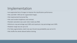
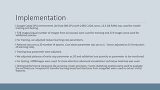
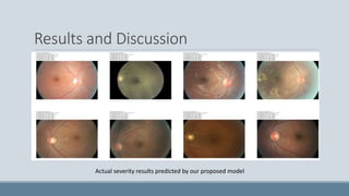
DeepDR: An image guided diabetic retinopathy
detection technique using attention-based deep
learning scheme
RUBINA NAZ, UMAIR SAEED, JAWERIA TANVEER, NOMAN ISLAM, KAMLESH KUMAR, AFTAB
AHMED SHAIKH
S I N D H M A D R AS S AT U L I S L A M U N I V ERS I T Y, K A R AC HI
I Q R A U N I V ERS I T Y, K A RAC HI
Overview
Introduction
Related work
Datastet
Methodology
Implementation
Results and Discussion
Conclusion
References
Q&A
Feedback
Introduction
Diabetes mellitus has reached to an epidemic level globally.
According to some statistics it will reach to 360 million people by 2030.
Diabetic retinopathy (DR) is still the leading causes of visual loss all over the world and account
for 28% of diabetes patients in USA.
It has been observed that an earlier diagnosis of retinopathy can prevent or avoid a significant
proportion of visual loss.
Introduction
Accurate diagnosis of this disease and identifying the stage of the disease is a challenge.
Early detection of disease and treatment is very essential to combat the increasingly large
number of retinopathy patients.
It can be said that a multidisciplinary approach is required for catering to this challenge
Related Work
Nursel Yalçin et al. [1] proposed a deep learning approach for DR disease classification.
Omer Deperlioglu et al. [2] proposed a CNN based deep learning model.
Darshit Doshi et al [3] proposed a CNN model.
Arkadiusz Kwasigroch et al [4] proposed CNN based decision support system for DR disease
classification.
Fully connected convolutional neural network was proposed by Manaswini Jena et al [5].
XiaoliangWang et al [6] used deep learning model with 63.23% validation accuracy. The
proposed model was based on pre-trained model inceptionNetV3.
Related Work
Hai Quan Chen et al [7] obtained validation accuracy up to 80.0%. Deep neural network model
was discussed in his paper.
Abhay Shah et al [8] described a CNN model with 53.57% accuracy.
IgiArdiyanto et al [9] proposed a Deep learning model for assessment DR disease in embedded
system.
Hanung Adi Nugroho [10] discussed the three different approaches. First approach was based
on pathologies. Second approach was based on foveal avascular zone (FAZ) structure. In third
approach, deep learning was proposed with more than 95% validation accuracy.
FengLiYu et al. [11] obtained 95.42% validation accuracy using deep learning model.
Related Work
Bhavani Sambaturu et al [12] achieved 91% validation accuracy via deep learning techniques.
Yashal Shakti Kanungo et al. [13] discussed deep learning model with 88% training accuracy.
Syahidahizza Rufaida et al. [14] achieved 51.05% accuracy using CNN deep learning model.
Ratul Ghosh et al. [15] proposed two deep learning techniques for two DR stages. 95% and 85%
validation accuracy were achieved respectively.
Roye [16] explained a model based on fuzzy C mean based technique to extract the features and
support vector machine to classify the feature.
Dong et al. [17] proposed a wavelet based feature classification techniques with up to 84% validation
accuracy.
S. Choudhury et al. [18] extracted features using Fuzzy C mean based feature extraction technique.
These extracted features were classified using support vector machines.
Models Comparison
S. # First author, Year Model Validation Accuracy (%)
1 NurselYalcin, 2018 CNN 98.5
2 Omer Deperlioglu, 2018 CNN 96.67
3 Arkadiusz Kwasigroch, 2018 CNN 82
4 Manaswini Jena, 208 CNN 91.6
5 Xiaoliang Wang, 2018 CNN, InceptionNetV3 63.3
6 HaiQuan Chen, 2018 CNN 80
7 Abhay Shah, 2018 CNN 53.5
8 IgiArdiyanto, 2017 CNN 73.3
9 FengLi Yu, 2017 CNN 95.4
10 Bhavani Sambaturu, 2017 CNN 91
11 Yashal Shakti Kanungo, 2017 CNN 88
12 SyahidahizzaRufaida, 2017 CNN 50.05
13 Ratul Ghosh, 2017 CNN 95
14 Arisha Roy, 2017 Fuzzy C mean, SVM 96.23
15 Yanyan Dong, 2017 CNN, SVM 94.07
16 S. Choudhury, 2016 Fuzzy C mean, SVM 97.6
17 Darshit Doshi, 2016 CNN 38.6
18 Our Proposed Model CNN 94.3
Dataset
Images of Diabetic retinopathy were used from Kaggle dataset.
This dataset contains 35,000 color images. 5 class labels were defined as “No DR”, “Mild,
Moderate”, “Severe” and “Proliferative DR”.
Retina images are high-resolution taken under a diversity of imaging circumstances.
 A left and right eyes images are provided for every patient.
Proposed Methodology (Pre-processing)
Noise is observed in the images.
Due to the lighting effects, pixel intensity varies and it causes variation dissimilarity to
classification pathology.
Images were normalized by using Gaussian Smoothing Filters.
Unsharp masking techniques were used to enhance the edges in images.
Filtering technique of Contrast Limited Adaptive Histogram Equalization was used to adjust the
contrast in images.
Proposed Methodology (CNN Model)
Several architectures were trained and test with different pre-trained models like DenseNet,
MobileNet, InceptionV3, VGG16 and VGG19.
Optimized results were obtained with InceptionV3 architecture.
Initially we utilized attention mechanism based CNN with pre-trained IncptionV3 model
discussed in Kaggle for this dataset.
This model was proposed by Kevin Mader initially.
We contributed in this model by adding some layers to improve the performance and accuracy.
Initials layers were used to learn deeper features.
Proposed Methodology (CNN Model)
An attention layer was added with liner activation function.
This layer was not being trained during training process (trainable = False) because this layer
was used for attention purpose.
Mask features were calculated with the help of Initial extracted features generated by pre-
trained model and deeper features extracted after adding further Convolutional layers.
To build attention mechanism, Global average pooling was being used.
GAP features and GAP mask were obtained from mask features and attention layers
respectively.
Lambda layer was used to rescale the features.
Description of Proposed CNN Architecture
Layer (type) Output Shape Param # Connected to
input_3 (InputLayer) (None, 512, 512, 3) 0
xception (Model) (None, 16, 16, 2048) 20861480 input_3[0][0]
batch_normalization_10 (BatchNo (None, 16, 16,
2048)
8192 xception[1][0]
dropout_4 (Dropout) (None, 16, 16, 2048) 0 batch_normalization_10[0][0]
conv2d_15 (Conv2D) (None, 16, 16, 64) 131136 dropout_4[0][0]
conv2d_16 (Conv2D) (None, 16, 16, 16) 1040 conv2d_15[0][0]
conv2d_17 (Conv2D) (None, 16, 16, 8) 136 conv2d_16[0][0]
conv2d_18 (Conv2D) (None, 16, 16, 4) 36 conv2d_17[0][0]
conv2d_19 (Conv2D) (None, 16, 16, 1) 5 conv2d_18[0][0]
conv2d_20 (Conv2D) (None, 16, 16, 2048) 2048 conv2d_19[0][0]
multiply_2 (Multiply) (None, 16, 16, 2048) 0 conv2d_20[0][0]
batch_normalization_10[0][0]
global_average_pooling2d_3 (GAP) (None, 2048) 0 multiply_2[0][0]
global_average_pooling2d_4 (GAP) (None, 2048) 0 conv2d_20[0][0]
RescaleGAP (Lambda) (None, 2048) 0 global_average_pooling2d_3[0][0]
global_average_pooling2d_4[0][0]
dropout_5 (Dropout) (None, 2048) 0 RescaleGAP[0][0]
dense_4 (Dense) (None, 128) 262272 dropout_5[0][0]
dropout_6 (Dropout) (None, 128) 0 dense_4[0][0]
dense_5 (Dense) (None, 64) 8256 dropout_6[0][0]
dense_6 (Dense) (None, 5) 325 dense_5[0][0]
Total params: 21,274,926
Trainable params: 407,302
Non-trainable params: 20,867,624
Implementation
we augmented lots of images to improve the classification performance.
We used 640 × 640 size for augmented images.
We implemented horizontal flip.
We used random brightness and contrast.
Random saturation was used. Color mode was RGB.
Minimum crop percentage was 0.001 and maximum crop percentage was 0.005.
Rotation range was set up to 10.
For Data augmentation, batch size was 16 and crop probability was set to 0.5.
We shuffle the whole dataset before training.
Implementation
Google Colab GPU environment (1xTesla K80 GPU with 2496 CUDA cores, 12.6 GB RAM) was used for model
training and testing.
778 images (equal number of images from all classes) were used for training and 274 images were used for
validation process.
For training, we adjusted reduce learning rate parameters.
Patience was set as 20 number of epochs. Cool down parameter was set as 5. Factor adjusted as 0.4 (reduction
of learning rate).
Training stop parameter were adjusted.
We adjusted patience of early stop parameter as 20 and validation lose quantity as parameter to be monitored.
For testing, 1008images were used. To show attention advanced visualization technique heatmap was used.
Testing performance measures like accuracy, recall, precision, f-score statistical analysis were used to evaluate
the architecture. InceptionV3 transfer learning based architectures from imageNet were used to extract initial
features.
Results and Discussion
 More than 94% validation accuracy was achieved.
 On Test dataset, 65% accuracy was obtained. Compared with initial proposed model, up to 5% test
accuracy was improved
Results and Discussion
Confusion matrix of our proposed model ROC Curve of our proposed model
Results and Discussion
Actual severity results predicted by our proposed model
Results and Discussion
Activation heatmap of our proposed learning model
Conclusion
Global weighted average pooling-based Attention mechanism in convolutional neural network
increased the performance and accuracy to detect the diabetic retinopathy in imbalanced and
noisy dataset.
Further pre-processing and balance dataset will increase the performance and accuracy.
Our study shows that CNN-based deep learning model can detect severity level of diabetic
retinopathy at initial stage via retinal fundus medical images.
CNN models are capable enough to understand the training images and learn from raw values
of pixels.
Our Heatmap visualization demonstrates that our model learned the features resides in image
portion correctly.
These features were clearly visible by specialist.
Q & A
You can send us your Questions and concerns on following Email addresses;
Dr. Noman Islam : noman.islam@gmail.com
Dr. Kamlesh Kumar : kamlesh@smiu.edu.pk
Umair Saeed : umairsaeedmixit@gmail.com
Feedback
Please provide your feedback on following email addressee;
Dr. Noman Islam : noman.islam@gmail.com
Dr. Kamlesh Kumar : kamlesh@smiu.edu.pk
Umair Saeed : umairsaeedmixit@gmail.com
References
1. Nurselyalçin NY, Seyfullahalver SA, Neclauluhatun NU. Classification of retinal images with deep learning for early detection of
diabetic retinopathy disease. SIU. 2018;1-4.
2. Omer deperlioglu O, Utkuköse U. Diagnosis of Diabetic Retinopathy by Using Image Processing and Convolutional Neural Network.
ISMSIT. 2018.
3. Darshitdoshi D, Aniket shenoy A, Deep sidhpura S, Prachi gharpure P. Diabetic retinopathy detection using deep convolutional neural
networks. CAST. 2018;261-266.
4. Arkadiusz kwasigroch K, Bartlomiej jarzembinski J, Michal grochowski G. Deep CNN based decision support system for detection and
assessing the stage of diabetic retinopathy. IIPhDW. 2018;111-116.
5. Manaswinijena M, Smitapravamishra S, debahutimishra D. Detection of Diabetic Retinopathy Images Using a Fully Convolutional
Neural Network. ICDSBA. 2018.
6. xiaoliang wang W, yongjinlu L, yujuan wang Y, wei-bang chen C. Diabetic Retinopathy Stage Classification Using Convolutional Neural
Networks. IRI. 2018; 465-471.
7. Haiquanchen C, Xianglongzeng Z, Yuan luo L, Wenbin ye Y. Detection of Diabetic Retinopathy using Deep Neural Network. DSP. 2018;
1-5.
8. Abhay shah A, Stephanie lynch S, Meindertniemeijer M, Ryan amelon R, Warren clarida W, James Folk J, Stephen Russell SR,
Xiaodong Wu X, Michael D. Abràmoff MD. Susceptibility to misdiagnosis of adversarial images by deep learning based retinal image
analysis algorithms. ISBI. 2018; 1454-1457.
References
9. Hanung Adi Nugroho H. Towards development of a computerised system for screening and monitoring of diabetic retinopathy. EECSI. 2017; 1-1.
10. Fengliyu Y, Jing sun S, Annan li L, Jun cheng C, Cheng wan W. Image quality classification for DR screening using deep learning. EMBC. 2017; 664-667.
11. Bhavani sambaturu B, Bhargav srinivasan S, Sahanamuraleedhara prabhu M, Kumar thirunellairajamani T, Thennarasupalanisamy P, Girish Haritz G, Digvijay Singh BS. A
novel deep learning based method for retinal lesion detection. ICACCI. 2017; 33-37
12. Yashal shakti kanungo K, Bhargav srinivasan S, Savita choudhary C. Detecting diabetic retinopathy using deep learning. RTEICT. 2017; 801-804.
13. Syahidahizzarufaida S, Mohamad ivanfanany M. Residual convolutional neural network for diabetic retinopathy. ICACSIS. 2017; 367-374.
14. Ratulghosh R, Kuntalghosh K, Sanjitmaitra S. Automatic detection and classification of diabetic retinopathy stages using CNN. SPIN. 2017; 550-554.
15. Bariqiabdillah B, Alhadibustamam A, Dewisarwinda D. Classification of diabetic retinopathy through texture features analysis. ICACSIS. 2017; 333-338.
16. Arisharoy R, Debasmitadutta D, Pratyushabhattacharya B, Sabarnachoudhury C. Filter and fuzzy c means based feature extraction and classification of diabetic
retinopathy using support vector machines. ICCSP . 2017; 1844-1848.
17. Yanyan dong D, Qinyanzhang Z, Zhiqiangqiao Q, ji-jiang yang Y. Classification of cataract fundus image based on deep learning. IST. 2017;1-5.
18. S Choudhury C, S Bandyopadhyay B, S K latibL, D K Kole K, c giri G. Fuzzy C means based feature extraction and classification of diabetic retinopathy using support vector
machines. ICCSP. 2016; 1520-152.
19. “Diabetic Retinopathy detection”, https://www.kaggle.com/kmader/inceptionv3-for-retinopathy-gpu-hr, 2018

DeepDRImageGuidedDiabeticRetinopathyDetectionUsingAttentionBasedDeepLearningScheme-Research-Article.pptx

  • 1.
    DeepDR: An imageguided diabetic retinopathy detection technique using attention-based deep learning scheme RUBINA NAZ, UMAIR SAEED, JAWERIA TANVEER, NOMAN ISLAM, KAMLESH KUMAR, AFTAB AHMED SHAIKH S I N D H M A D R AS S AT U L I S L A M U N I V ERS I T Y, K A R AC HI I Q R A U N I V ERS I T Y, K A RAC HI
  • 2.
  • 3.
    Introduction Diabetes mellitus hasreached to an epidemic level globally. According to some statistics it will reach to 360 million people by 2030. Diabetic retinopathy (DR) is still the leading causes of visual loss all over the world and account for 28% of diabetes patients in USA. It has been observed that an earlier diagnosis of retinopathy can prevent or avoid a significant proportion of visual loss.
  • 4.
    Introduction Accurate diagnosis ofthis disease and identifying the stage of the disease is a challenge. Early detection of disease and treatment is very essential to combat the increasingly large number of retinopathy patients. It can be said that a multidisciplinary approach is required for catering to this challenge
  • 5.
    Related Work Nursel Yalçinet al. [1] proposed a deep learning approach for DR disease classification. Omer Deperlioglu et al. [2] proposed a CNN based deep learning model. Darshit Doshi et al [3] proposed a CNN model. Arkadiusz Kwasigroch et al [4] proposed CNN based decision support system for DR disease classification. Fully connected convolutional neural network was proposed by Manaswini Jena et al [5]. XiaoliangWang et al [6] used deep learning model with 63.23% validation accuracy. The proposed model was based on pre-trained model inceptionNetV3.
  • 6.
    Related Work Hai QuanChen et al [7] obtained validation accuracy up to 80.0%. Deep neural network model was discussed in his paper. Abhay Shah et al [8] described a CNN model with 53.57% accuracy. IgiArdiyanto et al [9] proposed a Deep learning model for assessment DR disease in embedded system. Hanung Adi Nugroho [10] discussed the three different approaches. First approach was based on pathologies. Second approach was based on foveal avascular zone (FAZ) structure. In third approach, deep learning was proposed with more than 95% validation accuracy. FengLiYu et al. [11] obtained 95.42% validation accuracy using deep learning model.
  • 7.
    Related Work Bhavani Sambaturuet al [12] achieved 91% validation accuracy via deep learning techniques. Yashal Shakti Kanungo et al. [13] discussed deep learning model with 88% training accuracy. Syahidahizza Rufaida et al. [14] achieved 51.05% accuracy using CNN deep learning model. Ratul Ghosh et al. [15] proposed two deep learning techniques for two DR stages. 95% and 85% validation accuracy were achieved respectively. Roye [16] explained a model based on fuzzy C mean based technique to extract the features and support vector machine to classify the feature. Dong et al. [17] proposed a wavelet based feature classification techniques with up to 84% validation accuracy. S. Choudhury et al. [18] extracted features using Fuzzy C mean based feature extraction technique. These extracted features were classified using support vector machines.
  • 8.
    Models Comparison S. #First author, Year Model Validation Accuracy (%) 1 NurselYalcin, 2018 CNN 98.5 2 Omer Deperlioglu, 2018 CNN 96.67 3 Arkadiusz Kwasigroch, 2018 CNN 82 4 Manaswini Jena, 208 CNN 91.6 5 Xiaoliang Wang, 2018 CNN, InceptionNetV3 63.3 6 HaiQuan Chen, 2018 CNN 80 7 Abhay Shah, 2018 CNN 53.5 8 IgiArdiyanto, 2017 CNN 73.3 9 FengLi Yu, 2017 CNN 95.4 10 Bhavani Sambaturu, 2017 CNN 91 11 Yashal Shakti Kanungo, 2017 CNN 88 12 SyahidahizzaRufaida, 2017 CNN 50.05 13 Ratul Ghosh, 2017 CNN 95 14 Arisha Roy, 2017 Fuzzy C mean, SVM 96.23 15 Yanyan Dong, 2017 CNN, SVM 94.07 16 S. Choudhury, 2016 Fuzzy C mean, SVM 97.6 17 Darshit Doshi, 2016 CNN 38.6 18 Our Proposed Model CNN 94.3
  • 9.
    Dataset Images of Diabeticretinopathy were used from Kaggle dataset. This dataset contains 35,000 color images. 5 class labels were defined as “No DR”, “Mild, Moderate”, “Severe” and “Proliferative DR”. Retina images are high-resolution taken under a diversity of imaging circumstances.  A left and right eyes images are provided for every patient.
  • 10.
    Proposed Methodology (Pre-processing) Noiseis observed in the images. Due to the lighting effects, pixel intensity varies and it causes variation dissimilarity to classification pathology. Images were normalized by using Gaussian Smoothing Filters. Unsharp masking techniques were used to enhance the edges in images. Filtering technique of Contrast Limited Adaptive Histogram Equalization was used to adjust the contrast in images.
  • 11.
    Proposed Methodology (CNNModel) Several architectures were trained and test with different pre-trained models like DenseNet, MobileNet, InceptionV3, VGG16 and VGG19. Optimized results were obtained with InceptionV3 architecture. Initially we utilized attention mechanism based CNN with pre-trained IncptionV3 model discussed in Kaggle for this dataset. This model was proposed by Kevin Mader initially. We contributed in this model by adding some layers to improve the performance and accuracy. Initials layers were used to learn deeper features.
  • 12.
    Proposed Methodology (CNNModel) An attention layer was added with liner activation function. This layer was not being trained during training process (trainable = False) because this layer was used for attention purpose. Mask features were calculated with the help of Initial extracted features generated by pre- trained model and deeper features extracted after adding further Convolutional layers. To build attention mechanism, Global average pooling was being used. GAP features and GAP mask were obtained from mask features and attention layers respectively. Lambda layer was used to rescale the features.
  • 13.
    Description of ProposedCNN Architecture Layer (type) Output Shape Param # Connected to input_3 (InputLayer) (None, 512, 512, 3) 0 xception (Model) (None, 16, 16, 2048) 20861480 input_3[0][0] batch_normalization_10 (BatchNo (None, 16, 16, 2048) 8192 xception[1][0] dropout_4 (Dropout) (None, 16, 16, 2048) 0 batch_normalization_10[0][0] conv2d_15 (Conv2D) (None, 16, 16, 64) 131136 dropout_4[0][0] conv2d_16 (Conv2D) (None, 16, 16, 16) 1040 conv2d_15[0][0] conv2d_17 (Conv2D) (None, 16, 16, 8) 136 conv2d_16[0][0] conv2d_18 (Conv2D) (None, 16, 16, 4) 36 conv2d_17[0][0] conv2d_19 (Conv2D) (None, 16, 16, 1) 5 conv2d_18[0][0] conv2d_20 (Conv2D) (None, 16, 16, 2048) 2048 conv2d_19[0][0] multiply_2 (Multiply) (None, 16, 16, 2048) 0 conv2d_20[0][0] batch_normalization_10[0][0] global_average_pooling2d_3 (GAP) (None, 2048) 0 multiply_2[0][0] global_average_pooling2d_4 (GAP) (None, 2048) 0 conv2d_20[0][0] RescaleGAP (Lambda) (None, 2048) 0 global_average_pooling2d_3[0][0] global_average_pooling2d_4[0][0] dropout_5 (Dropout) (None, 2048) 0 RescaleGAP[0][0] dense_4 (Dense) (None, 128) 262272 dropout_5[0][0] dropout_6 (Dropout) (None, 128) 0 dense_4[0][0] dense_5 (Dense) (None, 64) 8256 dropout_6[0][0] dense_6 (Dense) (None, 5) 325 dense_5[0][0] Total params: 21,274,926 Trainable params: 407,302 Non-trainable params: 20,867,624
  • 14.
    Implementation we augmented lotsof images to improve the classification performance. We used 640 × 640 size for augmented images. We implemented horizontal flip. We used random brightness and contrast. Random saturation was used. Color mode was RGB. Minimum crop percentage was 0.001 and maximum crop percentage was 0.005. Rotation range was set up to 10. For Data augmentation, batch size was 16 and crop probability was set to 0.5. We shuffle the whole dataset before training.
  • 15.
    Implementation Google Colab GPUenvironment (1xTesla K80 GPU with 2496 CUDA cores, 12.6 GB RAM) was used for model training and testing. 778 images (equal number of images from all classes) were used for training and 274 images were used for validation process. For training, we adjusted reduce learning rate parameters. Patience was set as 20 number of epochs. Cool down parameter was set as 5. Factor adjusted as 0.4 (reduction of learning rate). Training stop parameter were adjusted. We adjusted patience of early stop parameter as 20 and validation lose quantity as parameter to be monitored. For testing, 1008images were used. To show attention advanced visualization technique heatmap was used. Testing performance measures like accuracy, recall, precision, f-score statistical analysis were used to evaluate the architecture. InceptionV3 transfer learning based architectures from imageNet were used to extract initial features.
  • 16.
  • 17.
    Results and Discussion More than 94% validation accuracy was achieved.  On Test dataset, 65% accuracy was obtained. Compared with initial proposed model, up to 5% test accuracy was improved
  • 18.
    Results and Discussion Confusionmatrix of our proposed model ROC Curve of our proposed model
  • 19.
    Results and Discussion Actualseverity results predicted by our proposed model
  • 20.
    Results and Discussion Activationheatmap of our proposed learning model
  • 21.
    Conclusion Global weighted averagepooling-based Attention mechanism in convolutional neural network increased the performance and accuracy to detect the diabetic retinopathy in imbalanced and noisy dataset. Further pre-processing and balance dataset will increase the performance and accuracy. Our study shows that CNN-based deep learning model can detect severity level of diabetic retinopathy at initial stage via retinal fundus medical images. CNN models are capable enough to understand the training images and learn from raw values of pixels. Our Heatmap visualization demonstrates that our model learned the features resides in image portion correctly. These features were clearly visible by specialist.
  • 22.
    Q & A Youcan send us your Questions and concerns on following Email addresses; Dr. Noman Islam : noman.islam@gmail.com Dr. Kamlesh Kumar : kamlesh@smiu.edu.pk Umair Saeed : umairsaeedmixit@gmail.com
  • 23.
    Feedback Please provide yourfeedback on following email addressee; Dr. Noman Islam : noman.islam@gmail.com Dr. Kamlesh Kumar : kamlesh@smiu.edu.pk Umair Saeed : umairsaeedmixit@gmail.com
  • 24.
    References 1. Nurselyalçin NY,Seyfullahalver SA, Neclauluhatun NU. Classification of retinal images with deep learning for early detection of diabetic retinopathy disease. SIU. 2018;1-4. 2. Omer deperlioglu O, Utkuköse U. Diagnosis of Diabetic Retinopathy by Using Image Processing and Convolutional Neural Network. ISMSIT. 2018. 3. Darshitdoshi D, Aniket shenoy A, Deep sidhpura S, Prachi gharpure P. Diabetic retinopathy detection using deep convolutional neural networks. CAST. 2018;261-266. 4. Arkadiusz kwasigroch K, Bartlomiej jarzembinski J, Michal grochowski G. Deep CNN based decision support system for detection and assessing the stage of diabetic retinopathy. IIPhDW. 2018;111-116. 5. Manaswinijena M, Smitapravamishra S, debahutimishra D. Detection of Diabetic Retinopathy Images Using a Fully Convolutional Neural Network. ICDSBA. 2018. 6. xiaoliang wang W, yongjinlu L, yujuan wang Y, wei-bang chen C. Diabetic Retinopathy Stage Classification Using Convolutional Neural Networks. IRI. 2018; 465-471. 7. Haiquanchen C, Xianglongzeng Z, Yuan luo L, Wenbin ye Y. Detection of Diabetic Retinopathy using Deep Neural Network. DSP. 2018; 1-5. 8. Abhay shah A, Stephanie lynch S, Meindertniemeijer M, Ryan amelon R, Warren clarida W, James Folk J, Stephen Russell SR, Xiaodong Wu X, Michael D. Abràmoff MD. Susceptibility to misdiagnosis of adversarial images by deep learning based retinal image analysis algorithms. ISBI. 2018; 1454-1457.
  • 25.
    References 9. Hanung AdiNugroho H. Towards development of a computerised system for screening and monitoring of diabetic retinopathy. EECSI. 2017; 1-1. 10. Fengliyu Y, Jing sun S, Annan li L, Jun cheng C, Cheng wan W. Image quality classification for DR screening using deep learning. EMBC. 2017; 664-667. 11. Bhavani sambaturu B, Bhargav srinivasan S, Sahanamuraleedhara prabhu M, Kumar thirunellairajamani T, Thennarasupalanisamy P, Girish Haritz G, Digvijay Singh BS. A novel deep learning based method for retinal lesion detection. ICACCI. 2017; 33-37 12. Yashal shakti kanungo K, Bhargav srinivasan S, Savita choudhary C. Detecting diabetic retinopathy using deep learning. RTEICT. 2017; 801-804. 13. Syahidahizzarufaida S, Mohamad ivanfanany M. Residual convolutional neural network for diabetic retinopathy. ICACSIS. 2017; 367-374. 14. Ratulghosh R, Kuntalghosh K, Sanjitmaitra S. Automatic detection and classification of diabetic retinopathy stages using CNN. SPIN. 2017; 550-554. 15. Bariqiabdillah B, Alhadibustamam A, Dewisarwinda D. Classification of diabetic retinopathy through texture features analysis. ICACSIS. 2017; 333-338. 16. Arisharoy R, Debasmitadutta D, Pratyushabhattacharya B, Sabarnachoudhury C. Filter and fuzzy c means based feature extraction and classification of diabetic retinopathy using support vector machines. ICCSP . 2017; 1844-1848. 17. Yanyan dong D, Qinyanzhang Z, Zhiqiangqiao Q, ji-jiang yang Y. Classification of cataract fundus image based on deep learning. IST. 2017;1-5. 18. S Choudhury C, S Bandyopadhyay B, S K latibL, D K Kole K, c giri G. Fuzzy C means based feature extraction and classification of diabetic retinopathy using support vector machines. ICCSP. 2016; 1520-152. 19. “Diabetic Retinopathy detection”, https://www.kaggle.com/kmader/inceptionv3-for-retinopathy-gpu-hr, 2018