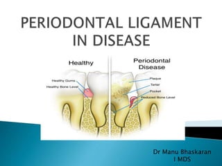
2. Introduction
Fibroblasts and collagen degradation
Fibrosis
Collagen
Pathological alterations in PDL
Connective tissue alterations
MMPs and Reactive oxygen species
Polypeptide mediators in diseased PDL
Invasion of PDL by pathogens
Functional demand to PDL
Stages of tissue response to occlusal forces
Hyalinisation
Conditions an overview.
Conclusion & References
3. Specialized connective tissue situated
between cementum covering the root and the
bone forming the socket wall.
Cell reservoir for tissue homeostasis and
repair/regeneration
PDL consists of cells and an extracellular
compartment comprising collagenous and
non collagenous matrix constituents.
5. Extracellular compartment
consists of well defined collagen
fiber bundles embedded in an
amorphous ground substance
Any injury or inflammation can disturb the collagen
fibres and increase the tissue fluids within the
matrix of the ground substance.
6. Principal cells of PDL
Characterized by their rapid
turnover of the extracellular
compartment in particular,collagen
The collagen fibrils of the bundles are
continuously being remodeled by
the fibroblasts, which are capable of
simultaneously synthesizing and degrading
collagen.
7. Activated fibroblast can be induced by appropriate stimuli from macrophages,
lymphocytes,mechanical force, and bacteria. Activated fibroblasts may secrete
tissue inhibitor of metalloproteinases ,matrix metalloproteinase ,cytokines [p
(PGE-2), plasminogen activator inhibitor (PAI), interleukin-6 .The fibroblast that
is induced to secrete matrix proteins can produce collagens, elastin, and
glycosaminoglycans (GAG'S).
8. in connective tissue remodelling, fibroblasts
are capable of the synthesis and phagocytosis
of collagen and components of extracellular
matrix
also produce cytokines with the capacity to
mediate tissue destruction and to stimulate
osteoclastic bone resorption.
An eg-matrix metalloproteinase-1 is a
fibroblast/macrophage derived enzyme with
the ability to degrade extracellular matrix
collagen at physiological pH and temperature.
9. During normal tissue homeostasis, collagen
degradation can take place within the
lysosomal apparatus of phagocytic cells.
Phagocytosis of collagen starts with
recognition of collagen fibrils through
membrane bound receptors
Partial digestion of fibrils by
proteinases such as gelatinases
(MMP-2,9) ,final degradation by
lysosomal enzymes.
Cells defective in phagocytosis
result in fibrosis and
compromise normal wound
repair and tissue regeneration.
10. After exposure to cytokines like
prostaglandin E2, IL-1 , collagenase
production and in some circumstances
phagocytosis can be upregulated.
EM study in rats show that the PDL fibroblasts
contain more phagocytosed collagen than
gingival fibroblasts (Svoboda ELA 1981)
suggesting site specific differences in
phagocytic capacity.
11. • Cultured PDL fibroblasts appear to
synthesize more collagen and fibronectin
than gingival fibroblasts
• Fibronectin which coats collagen fibrils in
vivo (Pitaru et al 1987) has been proposed
to initiate phagocytosis by acting as a
recognition site for fibroblasts.
Fibroblast-derived proinflammatory mediators and
cytokines such as PGE2, IL-1b, IL- 6, or IL-8 may be
directly or indirectly implicated in periodontal tissue
destruction by promoting fibrosis, granuloma formation
or bone resorption.
12. Thickening and scarring of connective tissue
as a result of injury
results from abnormal activation of the repair
process
Fibrosis may be due to-
1. Abnormal release of mediators during
inflammation.
2. Persistence of changes in the abnormal
growth factor ⁄ cytokine profile
3. Cell phenotypes such as myofibroblasts that
characterize fibrosis become established.
13. Predominant collagens are type I,III and XII.
Collagen undegoes degradation and remodelling
during development, inflammation & wound repair
and during bone resorption.
Enzymes responsible for collagen degradation are
matrix metalloproteinases
Diseases assosc.with collagen alteration include
Ehlers-Danlos syndrome,Crohn’s disease
14. •Ehlers-Danlos VIII (EDS-VIII) is an
autosomal dominant disorder characterized by
severe early-onset periodontal disease
•Pathogenic mutations in the genes encoding
collagen types I, III, and V, and the collagen-
processing enzymes lysyl hydroxylase and
procollagen N-peptidase have been found to
underlie.
•The underlying molecular cause of EDS-VIII is
unknown. A reduction of collagen type III was
reported in a single case (Lapiere and Nusgens
1981)
15. Crohn’s disease (CD) is characterised by
inflammation, muscle layer overgrowth, and
collagenous fibrosis of the intestinal tract,
with no effective therapy against collagen
accumulation(Type-III).
Includes perioral erythema with
scaling,mucogingivitis,aphthous
stomatitis,cobblestoning
16. Because of the exceptionally high rate of turnover of
collagen in the PDL ,any interference with fibroblast
function by disease result in loss of tooth supporting
tissue.
In inflammatory changes ,increased expression
of matrix metalloproteinases(MMPs) are
present that destroy collagen.
17. PDL fibroblasts produce collagenases that are thought to
be involved in normal tissue turnover. Neutrophils and
macrophages produce MMPs, with neutrophils being the
major source of collagenase in periodontitis.
In periodontal diseases, MMPs play key roles in the
degradation of the ECM, basement membrane and
protective serpins as well as in the modification of
cytokine action and activation of osteoclasts.
18. Hallmark of inflammatory PDL diseases is
connective tissue destruction.
During inflammation, PMN and
macrophages(major destructive cells) cause
matrix degradation that occurs through
phagocytosis or by MMP released by these
cells.
Chronic periodontitis leads to destruction of
PDL.
19. Collagen level is reduced ,fibrosis occurs
resulting in scarring of gingiva.
PMNs secretes large quantities of MMP-
2,9,1,8 from granules during any acute
inflammation causing extensive destruction.
Microbial plaque is another source of matrix
degradation.
Black-pigmented bacteroide species
synthesize proteases capable of disrupting
periodontal extracellular matrix
20. P. gingivalis, C. histolyticus and facultative
bacillus species from plaque secrete collagen
degrading enzymes.
These enzymes could also facilitate MMP
activity by activating their inactive precursors
and by damaging MMP- inhibitors.
Degrade proteoglycans and influence the ECM
indirectly through activation of interleukins
and affect fibroblast function.
21. Connective tissue alterations in acquired
diseases are brought about by interaction of
fibroblasts with inflammatory mediators and
cytokines present at the site of injury
These substances are released from damaged
tissue and inflammatory cells.
Growth factors Function
PDGF,TGF beta Cell growth and matrix
synthesis,
IFN-gamma,TNF and PGE2 Suppress collagen synthesis
22. Topographical changes in the distribution of
glycosaminoglycans, proteoglycans, and other
extracellular matrix macromolecules in the PDL
have been associated with periodontitis.
Degradation of the extracellular matrix can occur
through a number of different pathways, including
activation of matrix metalloproteinases (MMPs),
release of reactive oxygen species and phagocytosis
of matrix components.
23. Encodes 24 homologous proteinases such as
collagenases, gelatinases,and other MMPs.
synthesized in a latent, nonactive form and
require activation for enzyme function.
Matrix metalloproteinases play a major role in
connective tissue breakdown.
To date, MMP-1, -2, -3, -8, -9, and -13 have
been identified in inflamed periodontal tissues.
24. MMP activity is controlled in 3 ways-
enzymes are synthesized and secreted as
inactive precursors, and conversion to the
active form requires activation.
interleukin-1 (IL-1) may increase matrix
metalloproteinase synthesis, whereas
transforming growth factor-b (TGF-b) may
decrease matrix metalloproteinase
synthesis in inflamed tissues
25. matrix metalloproteinase activity can be
neutralized by endogenous serum and
tissue inhibitors. The major serum inhibitor
is a2-macroglobulin, which covalently
crosslinks with and inactivates target matrix
metalloproteinases. Tissues also contain
another group of matrix metalloproteinase
inhibitors known as tissue inhibitor of
metalloproteinases (TIMPs-1,2,3,4).
26. Tissues and cells may be exposed to
oxygen-derived free radicals during inflammatory
reactions, particularly where polymorphonuclear
leukocytes and macrophages are in abundance.
Oxygen-derived free radicals are highly reactive
molecular species which can disrupt cellular proteins,
nucleic acids and membrane lipids as well as
cause depolymerization of matrix components such
as collagen, hyaluronan and proteoglycans.
additional role for reactive oxygen species in inflamed
periodontal tissues may involve activation of
neutrophil collagenase .
27. With an advancing disease process, a typical
pattern of cytokine production may be
established
levels of cytokines IL-1a, IL-1b, TNF-a, IL-6, IL-
8, TGF-b, PDGF, keratinocyte growth factor,
VEGF, and prostaglandins are affected in
inflammatory cells, fibroblasts, and epithelial
cells.
TGF-beta is responsible for the increase in the
synthesis and accumulation of extracellular
matrix components and the decrease in matrix
metalloproteinase synthesis
28. Though these molecules are present in
normal connective tissue, their action is
induced when challenged by bacterial
endotoxins during inflammation.
IL-1a is a key regulator of inflammation
where it induces the expression of genes for
cell adhesion molecules, cytokines and MMPs.
IL-6 has been implicated in periodontitis as it
activates osteoblasts and bone resorption.
32. bacterial pathogens constantly interact with epithelial
cells, fibroblasts and inflammatory cells. Pathogens are
recognized by Toll-like receptors (TLR).
TLR function as pattern recognition receptors and play
a crucial role in host defense by activating the innate
host immune system.
TLR-2 recognizes peptidoglycans and lipopeptide from
gram-positive bacteria
TLR-4 recognizes lipopolysaccharide endotoxin from
gram-negative bacteria.
33. The expression of Toll-like receptor mRNA is often
affected by the endotoxins, and their excessive
activation results in exuberant inflammatory injury.
fibroblasts, and inflammatory cells respond to
lipopolysaccharide by increasing the expression of
cytokines, growth factors, matrix components and
matrix metalloproteinases.
Another transcription factor that plays a major
role in periodontal diseases is the activator protein-1
which regulates the expression of matrix
metalloproteinases in fibroblasts.
34. The first critical step of bacterial colonization is
adherence to host tissue.
Periodontopathic bacterial invasion of PDL is seen
as an important component of virulence, which
contributes to periodontal tissue breakdown.
35. A. actinomycetemcomitans and P. gingivalis with
fimbriae have been found to mediate binding of this
organism to different types of epithelial cells ,
fibronectin and fibrinogen and salivary components,
such as proline-rich proteins.
P.gingivalis type II and IV fimA are assosciated with
severe PDL disease. The prevention of host cell
apoptosis may promote P. gingivalis survival within
invaded oral epithelial cells in the human
periodontium.
Nonfimbriated P. gingivalis, i.e. YPF1 strain can
also invade primary oral epithelial cells, albeit less
efficiently.
36. A. actinomycetemcomitans invasion has been
shown to be an attachment and protein synthesis
required, energy consumptive and receptor-
mediated endocytotic process
Invasion-20 min for P. gingivalis , 30 min for A.
actinomycetemcomitans, replicate in epithelial
cells once inside, and appear to be capable of
spreading to surrounding cells.
Other oral bacterial species that have been
shown to invade epithelial cells include T.
forsythia, E. corrodens and F. nucleatum
37.
38. Capacity to adapt to functional changes
When functional demand increases,
1.width of PDL increases up to 50%
2.collagen fibers increase in thickness
When functional demand decreases,
1.narrowing of PDL
2.collagen fibers decrease in thickness
39. Trauma from occlusion refers to tissue injury
resulting from occlusal forces exceeding the adaptive
capacity of the tissues.
The radiographic signs include:
1. Increased width of periodontal space
2. Vertical destruction of interdental septum
3. Radioluscence and condensation of alveolar bone
4. Root resorption
40. Factors that help increase traumatic forces:
(Magnitude, direction & duration)
A) When magnitude of occlusal forces is increased:
1. the periodontium responds with a widening of the
periodontal ligament space.
2. an increase in the number and width of
periodontal ligament fibers.
3. increase in the density of alveolar bone
41. B) Direction of the occlusal forces
• the periodontal ligament fibers are arranged
so that the occlusal forces are
applied along the long axis of the tooth.
• Change in the direction of the occlusal forces
lead to change the
orientation of periodontal ligament fibers.
C) Duration and frequency of occlusal forces.
• Constant pressure on the bone is more injurious than
intermittent forces.
• The more frequent the application of a continuous force,
the more injurious the force to the periodontium
42. occur in three stages:
1. Stage I: Injury
2. Stage II: Repair
3. Stage III: Adaptive Remodeling of
periodontium.
43. Caused by excessive occlusal forces.
Under the forces of occlusion or tooth rotates
around a fulcrum which creates pressure and tension
on opposite sides of fulcrum.
Slightly excessive pressure stimulates resorption of
alveolar bone, with compression of PDL fibres.
Slightly excessive tension causes elongation of PDL
fibers & apposition of alveolar bone.
In areas of increased pressure, the blood vessels are
numerous and reduced in size and in increased
tension they are enlarged.
44. Compression of fibres produces hyalinisation.
Injury to fibroblasts and other connective
tissue cells leads to necrosis.
Vascular changes:
Within 30 min: retardation and stasis of
blood flow
2-3 hrs: blood vessels paused with
erythrocytes which start to fragment
In 1-7 days disintegration of the blood vessel
walls and release of the contents into the
surrounding tissue
45. Increased pressure in a localized region of PDL
exceeds the optimum and inhibit the differentiation of
osteoclasts.
Direct resorption of alveolar bone which would releive
the pressure in the PDL cannot occur
Series of degenerative tissue reactions take place
commencing within a few hours.
Hyalinisation is the term used to describe these tissue
reactions
46. Form of tissue degeneration characterized by formation
of a clear, eosinophilic homogenous substances
Denotes a compressed and locally degenerated PDL.
Reversible process
Occurs in almost all forms of orthodontic tooth
movement but the areas are wider when the force
applied is extreme
47. Changes observed during formation of hyalinized zone are:
Gradual shrinkage of PDL fibres.
Cellular structures become indistinct
Collagenous tissues gradually unite into a more or less cell
free mass
Changes also occur in the ground substance.
Break down of blood vessel walls leading to spilling of
their contents.
Osteoclasts are formed after a period of 20-30 hrs.
48. The presence of hyalinised zone indicates that the
ligament is non-functional and therefore bone
resorption cannot occur.
The tooth is hence not capable of further movement
until the local damaged tissue has been removed and
the adjacent alveolar bone resorbs .
49. Greater the forces wider is the area of hyalinization.
Thus larger areas of the ligament becomes functionless
,thereby showing larger areas of rearward resorption.
If lighter forces are used,the hyalinised zone is smaller
and a larger area of functioning ligament is available
and frontal resorption predominates.
The location and extend of hyalinised tissue largely
depends upon nature of tooth movement
50. Widening of the periodontal ligament.
Thrombosis
Hemorrhage
Tearing of the periodontal ligament
Resorption of the alveolar bone.
51. These return to normal levels after dissipation of the forces.
Injury to the periodontium produces a temporary depression in
mitotic activity and the rate of proliferation of fibroblasts,
collagen and bone formation.
The areas of the periodontium most susceptible to injury from
excessive occlusal forces are the furcations
52. When bone is resorbed by excessive occlusal forces, the body attempts to
reinforce the thinned bone with new bone. This attempt to compensate
for lost bone is called buttressing bone formation and is an important
feature associated with TFO.
The damaged tissues are removed and new connective tissue cells,
fibers, bone, and cementum are formed in an attempt to restore the
injured periodontium.
Repair is constantly occurring in the normal periodontium and
TFO stimulates increased reparative activity.
53. Buttressing bone formation occurs within the jaw
(central buttressing) and on the bone surface
(peripheral buttressing).
In central buttressing the
endosteal cells deposit new
bone, which restores the bony
trabeculae and reduces the
size of the marrow spaces.
Peripheral buttressing occurs on the facial and
lingual surfaces of the alveolar plate.This can
cause bulging of bone contours called as lipping.
54. If the repair process cannot keep pace with the
destruction, the periodontium is remodeled in an
effort to create a structured relationship in which
forces are no longer injurious to the tissues.
This results in a thickened periodontal ligament, which
is funnel- shaped at the crest, and angular defects in
the bone with no pocket formation, increased mobility
and increased vascularisation (Dutto et al 1967).
55. Pathological deepening of gingival sulcus by coronal
movement of the gingiva,apical displacement of
gingival attachment or by combination of the two.
56. Periodontal pocket produces destruction of the
supporting periodontal tissues, thereby leading to the
loosening and exfoliation of the teeth.
2 types -
1. Suprabony (supracrestal or supraalveolar) occurs
when the bottom of the pocket is coronal to the
underlying alveolar bone
Horizontal bone
loss
57. 2.Intrabony pocket (infrabony, subcrestal, or
intraalveolar) occurs when the bottom of the pocket is
apical to the level of the adjacent alveolar bone. With
this second type, the lateral pocket wall lies between
the tooth surface and the alveolar bone
Vertical
bone loss
58. Pocket formation starts as an inflammatory
change in the connective tissue wall of the
gingival sulcus.
The cellular and fluid inflammatory
exudate causes degeneration of the surrounding
connective tissue, including the gingival fibers
Just apical to the junctional epithelium, collagen
fibers are destroyed, and the area is occupied by
inflammatory cells and edema
59. Collagen loss happens in 2 mechanisms-
1. 1. collagenases and other enzymes secreted by
various cells in healthy and inflamed tissue, such as
fibroblasts, PMNs, and macrophages, become
extracellular and destroy collagen
matrix metalloproteinases degrade collagen and
other matrix macromolecules into small peptides.
60. 2.Fibroblasts phagocytize collagen fibers by
extending cytoplasmic processes to the
ligament–cementum interface and degrading
the inserted collagen fibrils and the fibrils of
the cementum matrix
61. After loss of collagen, the apical cells of the
junctional epithelium proliferate along the root
and extend fingerlike projections that are two
or three cells in thickness
62. Periodontal abscess is a localized painful swelling
that affects deeper periodontal structures,
including deep pockets, furcations and vertical
osseous defects, and is usually located beyond the
mucogingival line.
63. invasion of bacteria into the soft tissues
surrounding the periodontal pocket
develop an inflammatory process through the
chemotactic factors released by bacteria that
attract inflammatory cells
destruction of the connective tissues, the
encapsulation of the bacterial infection and the
production of pus.
Depends on growth of bacteria inside the focus,
their virulence and the local pH (an acidic
environment favors the lysosomal enzymes)
64. a normal oral epithelium and lamina propria.
an acute inflammatory infiltrate.
an intense focus of inflammation, with neutrophils and
lymphocytes present in an area of destroyed and
necrotic connective tissue.
a destroyed and ulcerated pocket epithelium.
65. necrosis affects the periodontal ligament and the
alveolar bone, leading to loss of attachment.
there is concomitant necrosis of the soft tissue, the
presence of pockets is not an usual finding
the interdental papilla is divided into a buccal part and
a lingual/ palatal part, with a necrotic area in the
middle, known as the interproximal crater.
66. 1. Dental restorations and appliances
Subgingivally placed onlays,crowns,fillings,orthodontic
bands result in gingival inflammation
Any restoration may impinge on the biologic width
Can result in loss of attachment and bone with apical
migration of junctional epithelium
67. 2.Root fractures
Caused by traumatic forces or by restorative or
endodontic procedures
Lead to periodontal involvement through apical
migration of plaque along the fracture
Fracture originates coronal to clinical attachment,which
results in alveolar ridge defect when exposed to oral
environment
68. 3.Cervical root resorption and cemental tears
Periodontal destruction occurs when the lesion communicates
with the oral cavity and allows bacteria to migrate
subgingivally
Avulsed teeth that are reimplanted results in cervical root
resorption
69. Tooth morphology –
Any developmental variation in morphology
may result in accumulation and retention of
plaque that may result in a localized
periodontal lesion
71. 1. Down’s syndrome
Increase in P .Intermedia in children
Anterior mandibular region had more periodontal
destruction (Cohen and goldman,1960 ;Johnson and
Young,1963)
A study in a group of Mongoloid patients showed
90%of severe periodontitis.
Severity of chronic inflammatory periodontal
disease depend on a combination of genetic and
environmental factors.
72.
73. 2.Papillon –Lefevre syndrome
Characterized by aggressive periodontitis and
hyperkeratosis of palms and soles with premature
destruction of PDL of deciduous and permanent teeth
resulting in their early loss.
Due to rapid bone loss, mobility and pathological
migration occurs resulting in loss of entire dentition
at an early age.
74.
75. Hypophosphatasia
Rare metabolic bone disease characterized by
deficiency of tissue non specific alkaline
phosphatase.
Premature loss of primary teeth caused by lack of
cementum on root surfaces
76.
77. 1. Actinomycosis-PDL lesions secondary to cervicofacial
actinomycosis.
2. Tuberculosis-PDL lesions secondary to pulmonary
TB,painless ulcer orally can rarely extend to PDL and
cause tooth loss.
3. Leprosy-Gingival ulceration in lepromatous leprosy
spread to cause PDL destruction particularly maxillary
incisors
4. Aspergillosis
78. 5.Cytomegalovirus-Morgan (1993) reported a case of
necrotizing periodontitis in an HIV patient having
cytomegalovirus infected endothelial cells lining
blood vessels in inflamed periodontal pocket wall.
6.Histoplasmosis
7.Myiasis
8.Herpes zoster-Wright et al (1983) reported a case of
alveolar bone necrosis
79. True incidence of PDL tumours are unknown.
Secondary involvement of PDL by infitration from
primary tumours arising in adjacent tissues such as
gingival carcinoma or by blood borne metastasis
eg.Leukemia is more common
82. PDL FEATURES IN BLOOD AND
LYMPHORETICUAR DISORDERS
1.Disorders of Hemostasis
Hemophilia
Thrombocytopenia
2.Red blood cell disorders
Anemia
Sickle cell disease
Polycythemia
3.Leukocyte disorders
Neutropenia,Agranulocytosis,Leukemia
83. 4.Disorders of white blood cells
Chediak-Higashi syndrome
Infectious mononucleosis
5.Leukocyte related conditions that affect the PDL
through invasions or depositions
Burkitts lymphoma
Non-Hodgkins Lymphoma
Hodgkins Lymphoma
84. Soft tissue sacs containing fluids ,semi fluid or gas and
may contain pus when secondarily infected
PDL is involved by non-neoplastic cysts
1.Odontogenic cysts
Radicular cyst
Paradental cyst
86. Connective tissue disorders consists of group of
diseases affecting connective tissues
Strongest link that binds them is common
histopathological pattern :
Increased interfibrillar ground substance
Proliferation of fibroblasts
Fibrinoid necrosis
87. Progressive systemic sclerosis
3:1 ratio of females to males
Characterized by diffuse sclerosis of skin, GIT,lungs
Assosciated with CREST syndrome
Radiographically:
Widening of PDL space in less than 30% patients
Teeth is not hypermobile
88. All of these aspects offer potential for
development of agents to modify host
responses to inflammatory stimuli.
89. Human PDL in disease is a sequelae of
gingivitis persistent gingivitis eventually lead
to periodontitis.
Periodontium consists of both fibrous and
mineralized tissues whose homeostasis differs
dramatically due to protein
composition,cellular elements, mineralization,
metabolism and function.
90. In health the primary component of the PDL is
a systematic arrangement.
Disease of PDL and other components of
periodontium accounts for a great deal of total
loss in the adult population.
91. Periodontal tissues in health and disease:introduction
,perio2000,vol 40
Structure of periodontal tissues in health and
disease,perio2000,vol40
Molecular and cell biology of periodontal
tissues,perio2000,vol40.
Role of bacteria in health and disease of periodontal
tissues,perio2000,vol40
Carranzas 10th and 12th edition
Berkovitz –perodontal ligament in health and disease-2nd edition
Shafers –oral pathology-7th edition
Fibroblast Heterogeneity in Periodontium – a Review Dr. A.
Archana et ,IJDSR,2014
Periodontal Ligament Cell Populations: The Central Role of
Fibroblasts in Creating a Unique Tissue P. Lekic and c.A.G.
Mcculloch,1996.