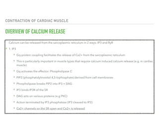
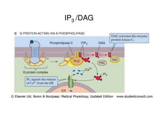
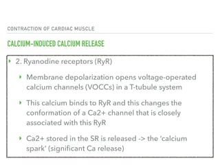
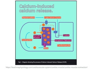
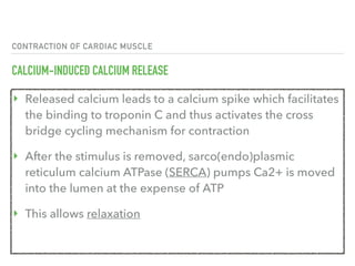
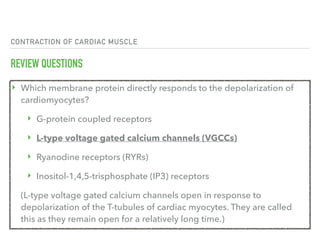
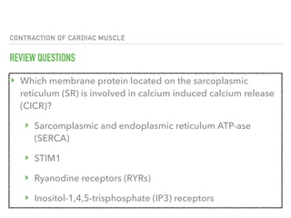
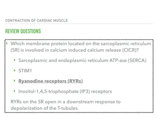
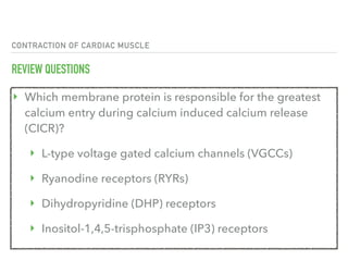
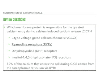
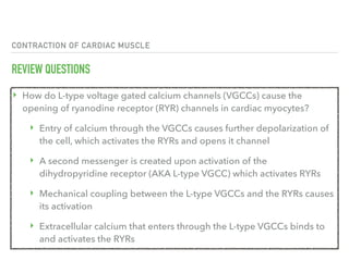
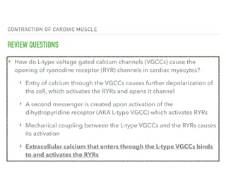
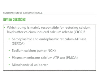
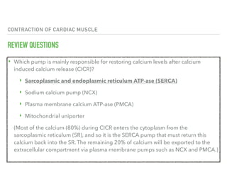
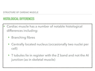
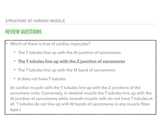
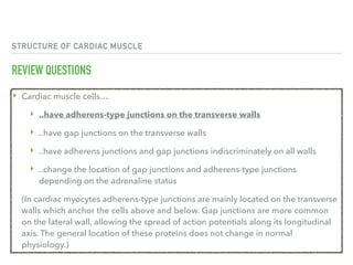
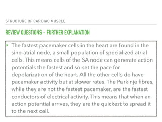
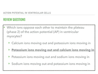
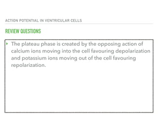
CARDIAC MUSCLE
PHYSIOLOGY: CARDIOVASCULAR
CONTRACTION OF
CARDIAC MUSCLE
CONTRACTION OF CARDIAC MUSCLE
OVERVIEW
‣ As in skeletal muscle, cardiac myocytes contract according
to the sliding filament theory of muscle contraction
Learning Goal
‣ To look at the process of calcium-induced calcium release
and the electrical coupling of cardiac myocytes
CONTRACTION OF CARDIAC MUSCLE
OVERVIEW OF CALCIUM RELEASE
Calcium can be released from the sarcoplasmic reticulum in 2 ways: IP3 and RyR
‣ 1. IP3
‣ Gq-protein coupling facilitates the release of Ca2+ from the sarcoplasmic reticulum
‣ This is particularly important in muscle types that require calcium induced calcium release (e.g. in cardiac
muscle)
‣ Gq activates the effector: Phospholipase C
‣ PIP2 (phosphatidylinositol 4,5-biphosphate) derived from cell membranes
‣ Phospholipase breaks PIP2 into IP3 + DAG
‣ IP3 binds IP3R of the SR
‣ DAG acts on various proteins (e.g PKC)
‣ Action terminated by IP3 phosphatase (IP3 cleaved to IP2)
‣ Ca2+ channels on the SR open and Ca2+ is released
CONTRACTION OF CARDIAC MUSCLE
CALCIUM-INDUCED CALCIUM RELEASE
‣ 2. Ryanodine receptors (RyR)
‣ Membrane depolarization opens voltage-operated
calcium channels (VOCCs) in a T-tubule system
‣ This calcium binds to RyR and this changes the
conformation of a Ca2+ channel that is closely
associated with this RyR
‣ Ca2+ stored in the SR is released -> the ‘calcium
spark’ (significant Ca release)
https://teachmephysiology.com/cardiovascular-system/cardiac-muscle/cardiac-muscle-contraction/
CONTRACTION OF CARDIAC MUSCLE
CALCIUM-INDUCED CALCIUM RELEASE
‣ Released calcium leads to a calcium spike which facilitates
the binding to troponin C and thus activates the cross
bridge cycling mechanism for contraction
‣ After the stimulus is removed, sarco(endo)plasmic
reticulum calcium ATPase (SERCA) pumps Ca2+ is moved
into the lumen at the expense of ATP
‣ This allows relaxation
CONTRACTION OF CARDIAC MUSCLE
PATHWAY OF CONTRACTION
‣ Pacemaker cells in the SA and AV nodes initiate an action
potential which is conducted around the heart via gap junctions
‣ The action potential travels down the T-tubules between
sarcomeres resulting in an influx of calcium ions into the
sarcoplasm through VOCCs
‣ When calcium enters the sarcoplasm (through VOCCs and
ryanodine receptors) it binds to cardiac troponin-C which
moves the tropomyosin away from the actin binding site thus
exposing it and initiating contraction
CONTRACTION OF CARDIAC MUSCLE
SLIDING FILAMENT MODEL OF CONTRACTION
‣ Cardiac muscle contraction occurs via the sliding filament model of contraction,
much like skeletal muscle
‣ Once calcium is bound to troponin-C and the conformational change of
tropomyosin has occurred myosin heads can bind to actin
‣ Following this ADP and inorganic phosphate are released from the myosin head
so the power stroke can occur, in this the myosin pivots and bends, pulling on
the actin and moving it, causing muscle contraction
‣ After this occurs a new molecule of ATP binds to the myosin head, causing it to
detach from the actin
‣ Finally, the ATP is hydrolyzed into ADP and inorganic phosphate, following this
the cycle can begin again and further contraction can occur
https://teachmephysiology.com/cardiovascular-system/cardiac-muscle/cardiac-muscle-contraction/
CONTRACTION OF CARDIAC MUSCLE
REMOVAL OF CALCIUM
‣ After the stimulus is removed intracellular calcium is then reduced by
two main methods:
‣ By entering the sarcoplasmic reticulum for storage via
a SERCA (sarco(endo)plasmic reticulum calcium-ATPase) channel
at the expense of an ATP molecule
‣ Through an NCX (sodium-calcium exchange) channel which
extrudes a calcium ion and admits a sodium ion when membrane
repolarization starts
‣ Calcium is no longer is bound to troponin C and the actin binding site
is covered up ending contraction and relaxing the muscle
https://teachmephysiology.com/cardiovascular-system/cardiac-muscle/cardiac-muscle-contraction/
CONTRACTION OF CARDIAC MUSCLE
REVIEW QUESTIONS
‣ Which membrane protein directly responds to the
depolarization of cardiomyocytes?
‣ G-protein coupled receptors
‣ L-type voltage gated calcium channels (VGCCs)
‣ Ryanodine receptors (RYRs)
‣ Inositol-1,4,5-trisphosphate (IP3) receptors
CONTRACTION OF CARDIAC MUSCLE
REVIEW QUESTIONS
‣ Which membrane protein directly responds to the depolarization of
cardiomyocytes?
‣ G-protein coupled receptors
‣ L-type voltage gated calcium channels (VGCCs)
‣ Ryanodine receptors (RYRs)
‣ Inositol-1,4,5-trisphosphate (IP3) receptors
(L-type voltage gated calcium channels open in response to
depolarization of the T-tubules of cardiac myocytes. They are called
this as they remain open for a relatively long time.)
CONTRACTION OF CARDIAC MUSCLE
REVIEW QUESTIONS
‣ Which membrane protein located on the sarcoplasmic
reticulum (SR) is involved in calcium induced calcium release
(CICR)?
‣ Sarcomplasmic and endoplasmic reticulum ATP-ase
(SERCA)
‣ STIM1
‣ Ryanodine receptors (RYRs)
‣ Inositol-1,4,5-trisphosphate (IP3) receptors
CONTRACTION OF CARDIAC MUSCLE
REVIEW QUESTIONS
‣ Which membrane protein located on the sarcoplasmic reticulum
(SR) is involved in calcium induced calcium release (CICR)?
‣ Sarcoplasmic and endoplasmic reticulum ATP-ase (SERCA)
‣ STIM1
‣ Ryanodine receptors (RYRs)
‣ Inositol-1,4,5-trisphosphate (IP3) receptors
RYRs on the SR open in a downstream response to
depolarization of the T-tubules.
CONTRACTION OF CARDIAC MUSCLE
REVIEW QUESTIONS
‣ Which membrane protein is responsible for the greatest
calcium entry during calcium induced calcium release
(CICR)?
‣ L-type voltage gated calcium channels (VGCCs)
‣ Ryanodine receptors (RYRs)
‣ Dihydropyridine (DHP) receptors
‣ Inositol-1,4,5-trisphosphate (IP3) receptors
CONTRACTION OF CARDIAC MUSCLE
REVIEW QUESTIONS
‣ Which membrane protein is responsible for the greatest
calcium entry during calcium induced calcium release (CICR)?
‣ L-type voltage gated calcium channels (VGCCs)
‣ Ryanodine receptors (RYRs)
‣ Dihydropyridine (DHP) receptors
‣ Inositol-1,4,5-trisphosphate (IP3) receptors
80% of the calcium that enters the cell during CICR comes from
the sarcoplasmic reticulum via RYRs
CONTRACTION OF CARDIAC MUSCLE
REVIEW QUESTIONS
‣ How do L-type voltage gated calcium channels (VGCCs) cause the
opening of ryanodine receptor (RYR) channels in cardiac myocytes?
‣ Entry of calcium through the VGCCs causes further depolarization of
the cell, which activates the RYRs and opens it channel
‣ A second messenger is created upon activation of the
dihydropyridine receptor (AKA L-type VGCC) which activates RYRs
‣ Mechanical coupling between the L-type VGCCs and the RYRs causes
its activation
‣ Extracellular calcium that enters through the L-type VGCCs binds to
and activates the RYRs
CONTRACTION OF CARDIAC MUSCLE
REVIEW QUESTIONS
‣ How do L-type voltage gated calcium channels (VGCCs) cause the
opening of ryanodine receptor (RYR) channels in cardiac myocytes?
‣ Entry of calcium through the VGCCs causes further depolarization of
the cell, which activates the RYRs and opens it channel
‣ A second messenger is created upon activation of the
dihydropyridine receptor (AKA L-type VGCC) which activates RYRs
‣ Mechanical coupling between the L-type VGCCs and the RYRs causes
its activation
‣ Extracellular calcium that enters through the L-type VGCCs binds
to and activates the RYRs
CONTRACTION OF CARDIAC MUSCLE
REVIEW QUESTIONS
‣ Which pump is mainly responsible for restoring calcium
levels after calcium induced calcium release (CICR)?
‣ Sarcoplasmic and endoplasmic reticulum ATP-ase
(SERCA)
‣ Sodium calcium pump (NCX)
‣ Plasma membrane calcium ATP-ase (PMCA)
‣ Mitochondrial uniporter
CONTRACTION OF CARDIAC MUSCLE
REVIEW QUESTIONS
‣ Which pump is mainly responsible for restoring calcium levels after calcium
induced calcium release (CICR)?
‣ Sarcoplasmic and endoplasmic reticulum ATP-ase (SERCA)
‣ Sodium calcium pump (NCX)
‣ Plasma membrane calcium ATP-ase (PMCA)
‣ Mitochondrial uniporter
(Most of the calcium (80%) during CICR enters the cytoplasm from the
sarcoplasmic reticulum (SR), and so it is the SERCA pump that must return this
calcium back into the SR. The remaining 20% of calcium will be exported to the
extracellular compartment via plasma membrane pumps such as NCX and PMCA.)
STRUCTURE OF
CARDIAC MUSCLE
STRUCTURE OF CARDIAC MUSCLE
OVERVIEW
‣ Cardiac muscle is similar to skeletal muscle in that
‣ It is striated 
‣ The sarcomere is the contractile unit
‣ Contraction being achieved by the relationship between
calcium, troponins and the myofilaments
Learning Goal
‣ To consider the structure of cardiac muscle as well as
relevant clinical conditions
STRUCTURE OF CARDIAC MUSCLE
HISTOLOGICAL DIFFERENCES
‣ Cardiac muscle has a number of notable histological
differences including:
‣ Branching fibres
‣ Centrally located nucleus (occasionally two nuclei per
cell)
‣ T tubules lie in register with the Z band and not the AI
junction (as in skeletal muscle)
STRUCTURE OF CARDIAC MUSCLE
COORDINATION
‣ Arguably the most important feature of cardiac muscle is its 
intercalated discs and gap junctions
‣ The disks act as the Z band would in skeletal muscle, demarcating
where a cardiac muscle cell meets the next
‣ The transverse portions form adherens-type junctions (attachments)
and desmosomes, holding the muscle cells together, while the lateral
portions form gap junctions that allow for quick conduction of action
potentials
‣ This is crucial to allow the heart to contract in a coordinated manner
and pump the blood
STRUCTURE OF CARDIAC MUSCLE
CONTRACTILITY
‣ Cardiac muscle is myogenic (creates its own action potentials) but is regulated
by the pacemaker cells – the sino-atrial node and atrioventricular node
‣ These cells spontaneously depolarize to trigger a cardiac action potential
‣ However the cells of the bundle of His and the Purkinje cells are also capable
of spontaneous depolarization
‣ However it should be noted that purkinje fibres are NOT cardiac muscle cells
‣ they are larger cells with far fewer myofilaments and extensive gap
junctions, as well as lots of glycogen
‣ They conduct action potentials much faster allowing for synchronous
contraction of the ventricles
https://teachmephysiology.com/cardiovascular-system/cardiac-muscle/structure-cardiac-muscle/
STRUCTURE OF CARDIAC MUSCLE
REVIEW QUESTIONS
‣ Which of these is true of cardiac myocytes?
‣ The T tubules line up with the AI junction of sarcomeres
‣ The T tubules line up with the Z junction of sarcomeres
‣ The T tubules line up with the M band of sarcomeres
‣ It does not have T tubules
STRUCTURE OF CARDIAC MUSCLE
REVIEW QUESTIONS
‣ Which of these is true of cardiac myocytes?
‣ The T tubules line up with the AI junction of sarcomeres
‣ The T tubules line up with the Z junction of sarcomeres
‣ The T tubules line up with the M band of sarcomeres
‣ It does not have T tubules
(In cardiac muscle cells the T tubules line up with the Z junctions of the
sarcomere units. Conversely, in skeletal muscle the T tubules line up with the
AI junction of sarcomeres while smooth muscle cells do not have T tubules at
all. T tubules do not line up with M bands of sarcomeres in any muscle fibre
type.)
STRUCTURE OF CARDIAC MUSCLE
REVIEW QUESTIONS
‣ Cardiac muscle cells…
‣ ..have adherens-type junctions on the transverse walls
‣ ..have gap junctions on the transverse walls
‣ ..have adherens junctions and gap junctions
indiscriminately on all walls
‣ ..change the location of gap junctions and adherens-
type junctions depending on the adrenaline status
STRUCTURE OF CARDIAC MUSCLE
REVIEW QUESTIONS
‣ Cardiac muscle cells…
‣ ..have adherens-type junctions on the transverse walls
‣ ..have gap junctions on the transverse walls
‣ ..have adherens junctions and gap junctions indiscriminately on all walls
‣ ..change the location of gap junctions and adherens-type junctions
depending on the adrenaline status
(In cardiac myocytes adherens-type junctions are mainly located on the transverse
walls which anchor the cells above and below. Gap junctions are more common
on the lateral wall, allowing the spread of action potentials along its longitudinal
axis. The general location of these proteins does not change in normal
physiology.)
STRUCTURE OF CARDIAC MUSCLE
REVIEW QUESTIONS
‣ Cardiac muscle is myogenic. This means that…
‣ ..it can generate more cardiac muscle cells if some are
destroyed
‣ ..it can generate its own action potential
‣ ..it cannot depolarize and contract if separated from
the brain
‣ ..it contains many cardiac muscle precursor cells
STRUCTURE OF CARDIAC MUSCLE
REVIEW QUESTIONS - FURTHER EXPLANATION
‣ The myogenic property of cardiac muscle means that it can
generate its own action potential due to spontaneous
depolarization without any outside input. Therefore if
separated from the brain (and external electrical influences)
the heart can still depolarize and contract for a while, at
least until it becomes starved of oxygen and nutrients. It
cannot generate more cardiac muscle cells as this tissue is
not capable of regeneration as it is a permanent tissue.
Smooth muscle is the only type of muscle that is capable of
regeneration to replace damaged tissue.
STRUCTURE OF CARDIAC MUSCLE
REVIEW QUESTIONS
‣ Select the single best answer. The fastest pacemaker (rate
not conduction) cells in the heart are found in the…
‣ ..Purkinje fibres
‣ ..atrial fibres
‣ ..atrio-ventricular node
‣ ..sino-atrial node
STRUCTURE OF CARDIAC MUSCLE
REVIEW QUESTIONS
‣ Select the single best answer. The fastest pacemaker (rate
not conduction) cells in the heart are found in the…
‣ ..Purkinje fibres
‣ ..atrial fibres
‣ ..atrio-ventricular node
‣ ..sino-atrial node
STRUCTURE OF CARDIAC MUSCLE
REVIEW QUESTIONS - FURTHER EXPLANATION
‣ The fastest pacemaker cells in the heart are found in the
sino-atrial node, a small population of specialized atrial
cells. This means cells of the SA node can generate action
potentials the fastest and so set the pace for
depolarization of the heart. All the other cells do have
pacemaker activity but at slower rates. The Purkinje fibres,
while they are not the fastest pacemaker, are the fastest
conductors of electrical activity. This means that when an
action potential arrives, they are the quickest to spread it
to the next cell.
STRUCTURE OF CARDIAC MUSCLE
REVIEW QUESTIONS
‣ The main purpose of gap junctions in cardiac myocytes is…
‣ ..to spread out ions between cells so it doesn't reach
too high levels in a single myocyte
‣ ..to allow spread of adrenaline between myocytes
‣ ..to keep myocytes physically connected to each other
‣ ..to allow very quick conduction of action potentials
between myocytes
STRUCTURE OF CARDIAC MUSCLE
REVIEW QUESTIONS
‣ The main purpose of gap junctions in cardiac myocytes is…
‣ ..to spread out ions between cells so it doesn't reach too high levels in a
single myocyte
‣ ..to allow spread of adrenaline between myocytes
‣ ..to keep myocytes physically connected to each other
‣ ..to allow very quick conduction of action potentials between
myocytes
(Gap junctions in cardiac myocytes allow very quick conduction of action
potentials as they allow cells to share a cytosol. Desmosomes are the proteins
whose main function is to keep cells physically connected.
STRUCTURE OF CARDIAC MUSCLE
REVIEW QUESTIONS
‣ An important role of the atrio-ventricular node is…
‣ ..to act as a pacemaker which sets the pace for depolarization of
the ventricles
‣ ..to act as a physical connection between the atria and the
ventricles
‣ ..to introduce a delay between atrial and ventricular
depolarization
‣ ..to enhance depolarization of the ventricles by acting as a
second pacemaker
STRUCTURE OF CARDIAC MUSCLE
REVIEW QUESTIONS
‣ The AV node introduces a delay between atrial and
ventricular depolarization, which is important to ensure
the atria have finished their contraction before the
ventricles depolarize and contract. It is also a pacemaker
but does not set the pace of contraction in health as it is
not normally the fastest pacemaker in the heart.
STRUCTURE OF CARDIAC MUSCLE
REVIEW QUESTIONS
‣ Select the incorrect answer. Purkinje fibres…
‣ ..have lots of glycogen
‣ ..are cardiac muscle cells
‣ ..have extensive gap junctions allowing relatively fast
conduction
‣ ..have relatively few myofilaments
STRUCTURE OF CARDIAC MUSCLE
REVIEW QUESTIONS
‣ Select the incorrect answer. Purkinje fibres…
‣ ..have lots of glycogen
‣ ..are cardiac muscle cells
‣ ..have extensive gap junctions allowing relatively fast
conduction
‣ ..have relatively few myofilaments
STRUCTURE OF CARDIAC MUSCLE
REVIEW QUESTIONS
‣ Purkinje fibres are not cardiac muscle cells even though
they form part of the conduction system of the heart. They
differ from cardiac myocytes in that they do not have T-
tubules and intercalated discs, have few myofilaments that
make up the contractile unit and they have lots of
glycogen and mitochondria.
STRUCTURE OF CARDIAC MUSCLE
REVIEW QUESTIONS
‣ Hypertrophy of cardiac muscle refers to…
‣ ..an increase in the speed of contraction
‣ ..an increase in the rate of contraction
‣ ..an increase in cell number
‣ ..an increase in cell size
STRUCTURE OF CARDIAC MUSCLE
REVIEW QUESTIONS
‣ Hypertrophy of cardiac muscle refers to…
‣ ..an increase in the speed of contraction
‣ ..an increase in the rate of contraction
‣ ..an increase in cell number
‣ ..an increase in cell size
STRUCTURE OF CARDIAC MUSCLE
REVIEW QUESTIONS
‣ Hypertrophy refers to an increase in cell size. This is the
only way cardiac tissue can grow. An increase in cell
number is called hyperplasia, however cardiac tissue is not
capable of this as it is a permanent tissue lacking the
progenitor cells required to replace damaged tissue.
STRUCTURE OF CARDIAC MUSCLE
REVIEW QUESTIONS
‣ With respect to familial cardiomyopathic hypertrophy,
which of the following statements is false?
‣ It can be caused by pressure overload (i.e hypertension)
‣ It is caused by genetic defects
‣ It involves defects in the sarcomere (such as of myosin
and troponin)
‣ It can result in arrhythmias
STRUCTURE OF CARDIAC MUSCLE
REVIEW QUESTIONS
‣ With respect to familial cardiomyopathic hypertrophy, which
of the following statements is false?
‣ It can be caused by pressure overload (i.e
hypertension)
‣ It is caused by genetic defects
‣ It involves defects in the sarcomere (such as of myosin and
troponin)
‣ It can result in arrhythmias
STRUCTURE OF CARDIAC MUSCLE
REVIEW QUESTIONS
‣ While pressure overload can cause cardiac hypertrophy,
the condition has "familial" in its name, indicating it is a
genetic condition and not the kind of hypertrophy caused
by pressure overload. This genetic defect involves defects
in the sarcomere and the hypertrophy that follows can
cause arrhythmias. As it is normally inherited in an
autosomal dominant manner, an affected individual will
normally have at least one affected parent.
THE ACTION POTENTIAL
IN VENTRICULAR CELLS
ACTION POTENTIAL IN VENTRICULAR CELLS
OVERVIEW
‣ Action potentials in ventricular myocytes trigger the Ca2+ entry that
is necessary for their contraction
‣ Their synchronicity, characteristic shape and length safeguards the
heart against abnormal electrical activity
‣ When these safeguards go wrong it can be potentially life
threatening
Learning Goal
‣ To look at how action potentials spread in ventricular cells, their
shape and modulation in disease states
ACTION POTENTIAL IN VENTRICULAR CELLS
GAP JUNCTIONS
‣ Gap junctions are regulated pores that exist between cardiac myocytes
‣ They are made from proteins which form a unit called a connexon
‣ Connexons are embedded in the plasma membrane of adjacent cells
‣ They are mostly located at either end of the cell at the region of the
intercalated disks
‣ When the connexons of two adjacent cells come together they can
form a channel which allows the cytosol of these cells to mix
‣ In this way ions can easily pass from cell to cell and the cells are said to
be electrically coupled
ACTION POTENTIAL IN VENTRICULAR CELLS
GAP JUNCTIONS
‣ Therefore ions that cause an action potential in one cell can spread
to its adjacent cell to initiate an action potential there
‣ Electrical coupling ensures the electrical activity of the heart
is synchronized
‣ As cells share cytosol, ions will quickly spread by passive diffusion
down a large electrochemical gradient
‣ This almost instantaneously evokes an action potential in the next
cell
‣ Gap junctions ensure a unidirectional spread of the action potential
https://teachmephysiology.com/cardiovascular-system/cardiac-muscle/action-potential-ventricular-cells/
ACTION POTENTIAL IN VENTRICULAR CELLS
PHASES OF THE ACTION POTENTIAL
‣ The ventricular action potential is generally split into 5 phases (phases
0-4)
‣ Phase 4 is the baseline from where the membrane potential begins and
ends
‣ Like any action potential, each phase is driven by the opening and closing
of a variety of specific ion channels
‣ This is because opening an ion channel will push the membrane potential
closer to the equilibrium potential of the ions it conducts
‣ This is a great way of controlling the membrane potential in a predictable
manner
ACTION POTENTIAL IN VENTRICULAR CELLS
PHASE 4 - BASELINE
‣ K+ currents are the main determinant of the resting
membrane potential as the membrane is far more
permeable to K+ than any other ion
‣ At rest K+ channels are open, therefore resting membrane
potential tends towards the equilibrium potential for
K+ (EK)
ACTION POTENTIAL IN VENTRICULAR CELLS
PHASE 0 - FAST DEPOLARIZATION
‣ Voltage gated Na+ channels open in response to depolarization that spreads into the cell
through gap junctions
‣ The influx of Na+ ions depolarizes the cell further causing the opening of more
Na+ channels
‣ This continues in a positive feedback mechanism to cause a fast and steep depolarization
‣ The Na+ channels become inactivated almost immediately after opening
‣ It is not possible for them to open while in this inactivated state
‣ These channels can only recover from inactivation to enter the closed state at very
negative membrane potentials
‣ This means that as long as the myocyte is depolarized, these Na+ channels will not be
able to open and cannot induce another action potential in that cell
ACTION POTENTIAL IN VENTRICULAR CELLS
PHASE 1 - NOTCH
‣ These transient opening K+ channels rapidly repolarize 
the cell before the plateau phase
‣ Therefore they set the membrane potential of the plateau
phase
‣ Greater K+ currents during this notch phase allows more
repolarization so that the plateau occurs at lower voltages
‣ Fewer K+ currents means that less repolarization occurs
and the plateau phase occurs at higher voltages
ACTION POTENTIAL IN VENTRICULAR CELLS
PHASE 2 - PLATEAU
‣ L-type Ca2+ channels are located in the T-tubules that penetrate the cell
‣ These channels are in close proximity to the sarcoplasmic reticulum (SR)
‣ Therefore the Ca2+ that enters through them binds to ryanodine receptors located on the SR
‣ This triggers massive Ca2+ release from the SR through a channel in the ryanodine receptor
‣ This phenomenon is called calcium induced calcium release (CICR)
‣ The majority (75%) of Ca2+ that enters the cell comes from the SR
‣ CICR is essential for excitation-contraction coupling within the cell
‣ Calcium ions bind to troponin C and initiate the movement of tropomyosin away from the
myosin head binding site on the actin molecule – permitting contraction
‣ This method of calcium release is different in skeletal muscle
ACTION POTENTIAL IN VENTRICULAR CELLS
PHASE 3 - REPOLARIZATION
‣ As Ca2+ channels close, K+ currents succeed in
repolarizing the cell, driving the membrane potential
toward EK
‣ During this phase Na+ channels will begin to recover from
inactivation as the membrane potential becomes more
negative
‣ This permits the cycle to restart
https://teachmephysiology.com/cardiovascular-system/cardiac-muscle/action-potential-ventricular-cells/
ACTION POTENTIAL IN VENTRICULAR CELLS
CLINICAL RELAVANCE - HYPERKALEMIA
‣ In hyperkalemia, initially the raised extracellular K+ makes
the environment outside the cell more positive. This
increases the driving force for Na+ entry during fast
depolarization as it is repelled by the positive charges on
K+. This makes depolarization happen more quickly. This
may cause tachycardia in the short term.
ACTION POTENTIAL IN VENTRICULAR CELLS
CLINICAL RELAVANCE - HYPERKALEMIA
‣ Eventually the cell will re-equilibriate moving closer to the
new Ek. As hyperkalemia makes the Ek less negative this
moves the membrane potential closer to threshold. At
these depolarized potentials voltage gated Na+ channels
become inactive. This means fewer Na+ channels are
available to participate in action potential generation.
Action potentials are less likely to occur under these
conditions causing bradycardia in the long term.
https://teachmephysiology.com/cardiovascular-system/cardiac-muscle/action-potential-ventricular-cells/
ACTION POTENTIAL IN VENTRICULAR CELLS
CLINICAL RELAVANCE - HYPOKALEMIA
‣ In hypokalemia, initially the lower extracellular K+ makes
the environment outside the cell relatively more negative.
This decreases the driving force for Na+ entry during fast
depolarization. This makes depolarization happen more
slowly and may cause bradycardia in the short term.
ACTION POTENTIAL IN VENTRICULAR CELLS
CLINICAL RELAVANCE - HYPOKALEMIA
‣ Eventually the cell will re-equilibriate moving closer to the
new Ek. As hypokalemia makes the Ek more negative this
moves the membrane potential further from threshold.
‣ At these hyperpolarized potentials fewer voltage gated
Na+ channels are inactive. This means more Na+ channels
are available to participate in action potential generation.
Action potentials are more likely to occur under these
conditions causing tachycardia in the long term.
ACTION POTENTIAL IN VENTRICULAR CELLS
REVIEW QUESTIONS
‣ What is the main role of gap junctions between cardiac
myocytes?
‣ Prevent electrical coupling of myocytes
‣ Ensure synchronized electrical activity of the heart
‣ Inhibit leakage of ions outside the cell
‣ Provide structural stability to myocytes
ACTION POTENTIAL IN VENTRICULAR CELLS
REVIEW QUESTIONS
‣ Which phase represents the baseline membrane potential
in ventricular myocytes?
‣ Phase 1
‣ Phase 2
‣ Phase 3
‣ Phase 4
ACTION POTENTIAL IN VENTRICULAR CELLS
REVIEW QUESTIONS
‣ Which phase represents the baseline membrane potential
in ventricular myocytes?
‣ Phase 1
‣ Phase 2
‣ Phase 3
‣ Phase 4
ACTION POTENTIAL IN VENTRICULAR CELLS
REVIEW QUESTIONS
‣ The resting membrane potential of the cardiac myocyte is
produced by the movement of which ion?
‣ Potassium ions moving through leak potassium channels
‣ Potassium ions moving through voltage gated potassium
channels
‣ Sodium ions moving through leak sodium channels
‣ Sodium ions moving through voltage gated sodium
channels
ACTION POTENTIAL IN VENTRICULAR CELLS
REVIEW QUESTIONS
‣ At rest, the membrane is most permeable to potassium ions.
Since the membrane potential will equilibrate to the
equilibrium potential of whichever ions they are permeable too,
it is potassium that is mostly responsible for setting the
membrane potential at rest. Voltage gated channels are not
generally open at rest as they typically open in response to
depolarization. However, there are usually many leak channels
open at rest. Therefore, it is the movement of potassium ions
into the cell through leak potassium channels that is largely
responsible for setting the resting membrane potential of these
cells.
ACTION POTENTIAL IN VENTRICULAR CELLS
REVIEW QUESTIONS
‣ The movement of which ion is responsible for the notch (phase 1) of
the action potential (AP) in ventricular myocytes?
‣ Chloride ions moving in through voltage gated chloride channels.
‣ Sodium ions moving out through transiently opening voltage
gated sodium channels
‣ Calcium ions moving out through ATP activated calcium channels
‣ Potassium ions moving out through transiently opening voltage
gated potassium channels
ACTION POTENTIAL IN VENTRICULAR CELLS
REVIEW QUESTIONS
‣ The movement of which ion is responsible for the notch (phase 1) of
the action potential (AP) in ventricular myocytes?
‣ Chloride ions moving in through voltage gated chloride channels.
‣ Sodium ions moving out through transiently opening voltage
gated sodium channels
‣ Calcium ions moving out through ATP activated calcium channels
‣ Potassium ions moving out through transiently opening
voltage gated potassium channels
ACTION POTENTIAL IN VENTRICULAR CELLS
REVIEW QUESTIONS
‣ The movement of potassium ions out of the cell through
transiently opening voltage gated potassium channels
causes the slight repolarization that occurs in the notch of
the ventricular AP. As these channels only open transiently
the repolarization does not last very long which creates
this notch.
ACTION POTENTIAL IN VENTRICULAR CELLS
REVIEW QUESTIONS
‣ Which ions oppose each other to maintain the plateau
(phase 2) of the action potential (AP) in ventricular
myocytes?
‣ Calcium ions moving out and potassium ions moving in
‣ Potassium ions moving out and calcium ions moving in
‣ Potassium ions moving out and sodium ions moving in
‣ Sodium ions moving out and potassium ions moving in
ACTION POTENTIAL IN VENTRICULAR CELLS
REVIEW QUESTIONS
‣ The plateau phase is created by the opposing action of
calcium ions moving into the cell favouring depolarization
and potassium ions moving out of the cell favouring
repolarization.
References
These slide reflect a summary of the contents of
TeachMePhysiology.com and are to be used for educational
purposes only in compliance with the terms of use policy.
Specific portions referenced in this summary are as follows:
‣ https://teachmephysiology.com/cardiovascular-system/cardiac-muscle/cardiac-
muscle-contraction/
‣ https://teachmephysiology.com/cardiovascular-system/cardiac-muscle/structure-
cardiac-muscle/
‣ https://teachmephysiology.com/cardiovascular-system/cardiac-muscle/action-
potential-ventricular-cells/
Additional sources are referenced on the slide containing
that specific content.

1. cardiac muscle

  • 1.
  • 2.
  • 3.
    CONTRACTION OF CARDIACMUSCLE OVERVIEW ‣ As in skeletal muscle, cardiac myocytes contract according to the sliding filament theory of muscle contraction Learning Goal ‣ To look at the process of calcium-induced calcium release and the electrical coupling of cardiac myocytes
  • 4.
    CONTRACTION OF CARDIACMUSCLE OVERVIEW OF CALCIUM RELEASE Calcium can be released from the sarcoplasmic reticulum in 2 ways: IP3 and RyR ‣ 1. IP3 ‣ Gq-protein coupling facilitates the release of Ca2+ from the sarcoplasmic reticulum ‣ This is particularly important in muscle types that require calcium induced calcium release (e.g. in cardiac muscle) ‣ Gq activates the effector: Phospholipase C ‣ PIP2 (phosphatidylinositol 4,5-biphosphate) derived from cell membranes ‣ Phospholipase breaks PIP2 into IP3 + DAG ‣ IP3 binds IP3R of the SR ‣ DAG acts on various proteins (e.g PKC) ‣ Action terminated by IP3 phosphatase (IP3 cleaved to IP2) ‣ Ca2+ channels on the SR open and Ca2+ is released
  • 6.
    CONTRACTION OF CARDIACMUSCLE CALCIUM-INDUCED CALCIUM RELEASE ‣ 2. Ryanodine receptors (RyR) ‣ Membrane depolarization opens voltage-operated calcium channels (VOCCs) in a T-tubule system ‣ This calcium binds to RyR and this changes the conformation of a Ca2+ channel that is closely associated with this RyR ‣ Ca2+ stored in the SR is released -> the ‘calcium spark’ (significant Ca release)
  • 7.
  • 8.
    CONTRACTION OF CARDIACMUSCLE CALCIUM-INDUCED CALCIUM RELEASE ‣ Released calcium leads to a calcium spike which facilitates the binding to troponin C and thus activates the cross bridge cycling mechanism for contraction ‣ After the stimulus is removed, sarco(endo)plasmic reticulum calcium ATPase (SERCA) pumps Ca2+ is moved into the lumen at the expense of ATP ‣ This allows relaxation
  • 9.
    CONTRACTION OF CARDIACMUSCLE PATHWAY OF CONTRACTION ‣ Pacemaker cells in the SA and AV nodes initiate an action potential which is conducted around the heart via gap junctions ‣ The action potential travels down the T-tubules between sarcomeres resulting in an influx of calcium ions into the sarcoplasm through VOCCs ‣ When calcium enters the sarcoplasm (through VOCCs and ryanodine receptors) it binds to cardiac troponin-C which moves the tropomyosin away from the actin binding site thus exposing it and initiating contraction
  • 10.
    CONTRACTION OF CARDIACMUSCLE SLIDING FILAMENT MODEL OF CONTRACTION ‣ Cardiac muscle contraction occurs via the sliding filament model of contraction, much like skeletal muscle ‣ Once calcium is bound to troponin-C and the conformational change of tropomyosin has occurred myosin heads can bind to actin ‣ Following this ADP and inorganic phosphate are released from the myosin head so the power stroke can occur, in this the myosin pivots and bends, pulling on the actin and moving it, causing muscle contraction ‣ After this occurs a new molecule of ATP binds to the myosin head, causing it to detach from the actin ‣ Finally, the ATP is hydrolyzed into ADP and inorganic phosphate, following this the cycle can begin again and further contraction can occur
  • 11.
  • 12.
    CONTRACTION OF CARDIACMUSCLE REMOVAL OF CALCIUM ‣ After the stimulus is removed intracellular calcium is then reduced by two main methods: ‣ By entering the sarcoplasmic reticulum for storage via a SERCA (sarco(endo)plasmic reticulum calcium-ATPase) channel at the expense of an ATP molecule ‣ Through an NCX (sodium-calcium exchange) channel which extrudes a calcium ion and admits a sodium ion when membrane repolarization starts ‣ Calcium is no longer is bound to troponin C and the actin binding site is covered up ending contraction and relaxing the muscle
  • 13.
  • 14.
  • 15.
  • 16.
    CONTRACTION OF CARDIACMUSCLE REVIEW QUESTIONS ‣ Which membrane protein directly responds to the depolarization of cardiomyocytes? ‣ G-protein coupled receptors ‣ L-type voltage gated calcium channels (VGCCs) ‣ Ryanodine receptors (RYRs) ‣ Inositol-1,4,5-trisphosphate (IP3) receptors
  • 17.
    CONTRACTION OF CARDIACMUSCLE REVIEW QUESTIONS ‣ Which membrane protein directly responds to the depolarization of cardiomyocytes? ‣ G-protein coupled receptors ‣ L-type voltage gated calcium channels (VGCCs) ‣ Ryanodine receptors (RYRs) ‣ Inositol-1,4,5-trisphosphate (IP3) receptors (L-type voltage gated calcium channels open in response to depolarization of the T-tubules of cardiac myocytes. They are called this as they remain open for a relatively long time.)
  • 18.
    CONTRACTION OF CARDIACMUSCLE REVIEW QUESTIONS ‣ Which membrane protein located on the sarcoplasmic reticulum (SR) is involved in calcium induced calcium release (CICR)? ‣ Sarcomplasmic and endoplasmic reticulum ATP-ase (SERCA) ‣ STIM1 ‣ Ryanodine receptors (RYRs) ‣ Inositol-1,4,5-trisphosphate (IP3) receptors
  • 19.
    CONTRACTION OF CARDIACMUSCLE REVIEW QUESTIONS ‣ Which membrane protein located on the sarcoplasmic reticulum (SR) is involved in calcium induced calcium release (CICR)? ‣ Sarcoplasmic and endoplasmic reticulum ATP-ase (SERCA) ‣ STIM1 ‣ Ryanodine receptors (RYRs) ‣ Inositol-1,4,5-trisphosphate (IP3) receptors RYRs on the SR open in a downstream response to depolarization of the T-tubules.
  • 20.
    CONTRACTION OF CARDIACMUSCLE REVIEW QUESTIONS ‣ Which membrane protein is responsible for the greatest calcium entry during calcium induced calcium release (CICR)? ‣ L-type voltage gated calcium channels (VGCCs) ‣ Ryanodine receptors (RYRs) ‣ Dihydropyridine (DHP) receptors ‣ Inositol-1,4,5-trisphosphate (IP3) receptors
  • 21.
    CONTRACTION OF CARDIACMUSCLE REVIEW QUESTIONS ‣ Which membrane protein is responsible for the greatest calcium entry during calcium induced calcium release (CICR)? ‣ L-type voltage gated calcium channels (VGCCs) ‣ Ryanodine receptors (RYRs) ‣ Dihydropyridine (DHP) receptors ‣ Inositol-1,4,5-trisphosphate (IP3) receptors 80% of the calcium that enters the cell during CICR comes from the sarcoplasmic reticulum via RYRs
  • 22.
    CONTRACTION OF CARDIACMUSCLE REVIEW QUESTIONS ‣ How do L-type voltage gated calcium channels (VGCCs) cause the opening of ryanodine receptor (RYR) channels in cardiac myocytes? ‣ Entry of calcium through the VGCCs causes further depolarization of the cell, which activates the RYRs and opens it channel ‣ A second messenger is created upon activation of the dihydropyridine receptor (AKA L-type VGCC) which activates RYRs ‣ Mechanical coupling between the L-type VGCCs and the RYRs causes its activation ‣ Extracellular calcium that enters through the L-type VGCCs binds to and activates the RYRs
  • 23.
    CONTRACTION OF CARDIACMUSCLE REVIEW QUESTIONS ‣ How do L-type voltage gated calcium channels (VGCCs) cause the opening of ryanodine receptor (RYR) channels in cardiac myocytes? ‣ Entry of calcium through the VGCCs causes further depolarization of the cell, which activates the RYRs and opens it channel ‣ A second messenger is created upon activation of the dihydropyridine receptor (AKA L-type VGCC) which activates RYRs ‣ Mechanical coupling between the L-type VGCCs and the RYRs causes its activation ‣ Extracellular calcium that enters through the L-type VGCCs binds to and activates the RYRs
  • 24.
    CONTRACTION OF CARDIACMUSCLE REVIEW QUESTIONS ‣ Which pump is mainly responsible for restoring calcium levels after calcium induced calcium release (CICR)? ‣ Sarcoplasmic and endoplasmic reticulum ATP-ase (SERCA) ‣ Sodium calcium pump (NCX) ‣ Plasma membrane calcium ATP-ase (PMCA) ‣ Mitochondrial uniporter
  • 25.
    CONTRACTION OF CARDIACMUSCLE REVIEW QUESTIONS ‣ Which pump is mainly responsible for restoring calcium levels after calcium induced calcium release (CICR)? ‣ Sarcoplasmic and endoplasmic reticulum ATP-ase (SERCA) ‣ Sodium calcium pump (NCX) ‣ Plasma membrane calcium ATP-ase (PMCA) ‣ Mitochondrial uniporter (Most of the calcium (80%) during CICR enters the cytoplasm from the sarcoplasmic reticulum (SR), and so it is the SERCA pump that must return this calcium back into the SR. The remaining 20% of calcium will be exported to the extracellular compartment via plasma membrane pumps such as NCX and PMCA.)
  • 26.
  • 27.
    STRUCTURE OF CARDIACMUSCLE OVERVIEW ‣ Cardiac muscle is similar to skeletal muscle in that ‣ It is striated  ‣ The sarcomere is the contractile unit ‣ Contraction being achieved by the relationship between calcium, troponins and the myofilaments Learning Goal ‣ To consider the structure of cardiac muscle as well as relevant clinical conditions
  • 28.
    STRUCTURE OF CARDIACMUSCLE HISTOLOGICAL DIFFERENCES ‣ Cardiac muscle has a number of notable histological differences including: ‣ Branching fibres ‣ Centrally located nucleus (occasionally two nuclei per cell) ‣ T tubules lie in register with the Z band and not the AI junction (as in skeletal muscle)
  • 29.
    STRUCTURE OF CARDIACMUSCLE COORDINATION ‣ Arguably the most important feature of cardiac muscle is its  intercalated discs and gap junctions ‣ The disks act as the Z band would in skeletal muscle, demarcating where a cardiac muscle cell meets the next ‣ The transverse portions form adherens-type junctions (attachments) and desmosomes, holding the muscle cells together, while the lateral portions form gap junctions that allow for quick conduction of action potentials ‣ This is crucial to allow the heart to contract in a coordinated manner and pump the blood
  • 30.
    STRUCTURE OF CARDIACMUSCLE CONTRACTILITY ‣ Cardiac muscle is myogenic (creates its own action potentials) but is regulated by the pacemaker cells – the sino-atrial node and atrioventricular node ‣ These cells spontaneously depolarize to trigger a cardiac action potential ‣ However the cells of the bundle of His and the Purkinje cells are also capable of spontaneous depolarization ‣ However it should be noted that purkinje fibres are NOT cardiac muscle cells ‣ they are larger cells with far fewer myofilaments and extensive gap junctions, as well as lots of glycogen ‣ They conduct action potentials much faster allowing for synchronous contraction of the ventricles
  • 31.
  • 32.
  • 33.
  • 34.
    STRUCTURE OF CARDIACMUSCLE REVIEW QUESTIONS ‣ Which of these is true of cardiac myocytes? ‣ The T tubules line up with the AI junction of sarcomeres ‣ The T tubules line up with the Z junction of sarcomeres ‣ The T tubules line up with the M band of sarcomeres ‣ It does not have T tubules
  • 35.
    STRUCTURE OF CARDIACMUSCLE REVIEW QUESTIONS ‣ Which of these is true of cardiac myocytes? ‣ The T tubules line up with the AI junction of sarcomeres ‣ The T tubules line up with the Z junction of sarcomeres ‣ The T tubules line up with the M band of sarcomeres ‣ It does not have T tubules (In cardiac muscle cells the T tubules line up with the Z junctions of the sarcomere units. Conversely, in skeletal muscle the T tubules line up with the AI junction of sarcomeres while smooth muscle cells do not have T tubules at all. T tubules do not line up with M bands of sarcomeres in any muscle fibre type.)
  • 36.
    STRUCTURE OF CARDIACMUSCLE REVIEW QUESTIONS ‣ Cardiac muscle cells… ‣ ..have adherens-type junctions on the transverse walls ‣ ..have gap junctions on the transverse walls ‣ ..have adherens junctions and gap junctions indiscriminately on all walls ‣ ..change the location of gap junctions and adherens- type junctions depending on the adrenaline status
  • 37.
    STRUCTURE OF CARDIACMUSCLE REVIEW QUESTIONS ‣ Cardiac muscle cells… ‣ ..have adherens-type junctions on the transverse walls ‣ ..have gap junctions on the transverse walls ‣ ..have adherens junctions and gap junctions indiscriminately on all walls ‣ ..change the location of gap junctions and adherens-type junctions depending on the adrenaline status (In cardiac myocytes adherens-type junctions are mainly located on the transverse walls which anchor the cells above and below. Gap junctions are more common on the lateral wall, allowing the spread of action potentials along its longitudinal axis. The general location of these proteins does not change in normal physiology.)
  • 38.
    STRUCTURE OF CARDIACMUSCLE REVIEW QUESTIONS ‣ Cardiac muscle is myogenic. This means that… ‣ ..it can generate more cardiac muscle cells if some are destroyed ‣ ..it can generate its own action potential ‣ ..it cannot depolarize and contract if separated from the brain ‣ ..it contains many cardiac muscle precursor cells
  • 39.
    STRUCTURE OF CARDIACMUSCLE REVIEW QUESTIONS ‣ Cardiac muscle is myogenic. This means that… ‣ ..it can generate more cardiac muscle cells if some are destroyed ‣ ..it can generate its own action potential ‣ ..it cannot depolarize and contract if separated from the brain ‣ ..it contains many cardiac muscle precursor cells
  • 40.
    STRUCTURE OF CARDIACMUSCLE REVIEW QUESTIONS - FURTHER EXPLANATION ‣ The myogenic property of cardiac muscle means that it can generate its own action potential due to spontaneous depolarization without any outside input. Therefore if separated from the brain (and external electrical influences) the heart can still depolarize and contract for a while, at least until it becomes starved of oxygen and nutrients. It cannot generate more cardiac muscle cells as this tissue is not capable of regeneration as it is a permanent tissue. Smooth muscle is the only type of muscle that is capable of regeneration to replace damaged tissue.
  • 41.
    STRUCTURE OF CARDIACMUSCLE REVIEW QUESTIONS ‣ Select the single best answer. The fastest pacemaker (rate not conduction) cells in the heart are found in the… ‣ ..Purkinje fibres ‣ ..atrial fibres ‣ ..atrio-ventricular node ‣ ..sino-atrial node
  • 42.
    STRUCTURE OF CARDIACMUSCLE REVIEW QUESTIONS ‣ Select the single best answer. The fastest pacemaker (rate not conduction) cells in the heart are found in the… ‣ ..Purkinje fibres ‣ ..atrial fibres ‣ ..atrio-ventricular node ‣ ..sino-atrial node
  • 43.
    STRUCTURE OF CARDIACMUSCLE REVIEW QUESTIONS - FURTHER EXPLANATION ‣ The fastest pacemaker cells in the heart are found in the sino-atrial node, a small population of specialized atrial cells. This means cells of the SA node can generate action potentials the fastest and so set the pace for depolarization of the heart. All the other cells do have pacemaker activity but at slower rates. The Purkinje fibres, while they are not the fastest pacemaker, are the fastest conductors of electrical activity. This means that when an action potential arrives, they are the quickest to spread it to the next cell.
  • 44.
    STRUCTURE OF CARDIACMUSCLE REVIEW QUESTIONS ‣ The main purpose of gap junctions in cardiac myocytes is… ‣ ..to spread out ions between cells so it doesn't reach too high levels in a single myocyte ‣ ..to allow spread of adrenaline between myocytes ‣ ..to keep myocytes physically connected to each other ‣ ..to allow very quick conduction of action potentials between myocytes
  • 45.
    STRUCTURE OF CARDIACMUSCLE REVIEW QUESTIONS ‣ The main purpose of gap junctions in cardiac myocytes is… ‣ ..to spread out ions between cells so it doesn't reach too high levels in a single myocyte ‣ ..to allow spread of adrenaline between myocytes ‣ ..to keep myocytes physically connected to each other ‣ ..to allow very quick conduction of action potentials between myocytes (Gap junctions in cardiac myocytes allow very quick conduction of action potentials as they allow cells to share a cytosol. Desmosomes are the proteins whose main function is to keep cells physically connected.
  • 46.
    STRUCTURE OF CARDIACMUSCLE REVIEW QUESTIONS ‣ An important role of the atrio-ventricular node is… ‣ ..to act as a pacemaker which sets the pace for depolarization of the ventricles ‣ ..to act as a physical connection between the atria and the ventricles ‣ ..to introduce a delay between atrial and ventricular depolarization ‣ ..to enhance depolarization of the ventricles by acting as a second pacemaker
  • 47.
    STRUCTURE OF CARDIACMUSCLE REVIEW QUESTIONS ‣ An important role of the atrio-ventricular node is… ‣ ..to act as a pacemaker which sets the pace for depolarization of the ventricles ‣ ..to act as a physical connection between the atria and the ventricles ‣ ..to introduce a delay between atrial and ventricular depolarization ‣ ..to enhance depolarization of the ventricles by acting as a second pacemaker
  • 48.
    STRUCTURE OF CARDIACMUSCLE REVIEW QUESTIONS ‣ The AV node introduces a delay between atrial and ventricular depolarization, which is important to ensure the atria have finished their contraction before the ventricles depolarize and contract. It is also a pacemaker but does not set the pace of contraction in health as it is not normally the fastest pacemaker in the heart.
  • 49.
    STRUCTURE OF CARDIACMUSCLE REVIEW QUESTIONS ‣ Select the incorrect answer. Purkinje fibres… ‣ ..have lots of glycogen ‣ ..are cardiac muscle cells ‣ ..have extensive gap junctions allowing relatively fast conduction ‣ ..have relatively few myofilaments
  • 50.
    STRUCTURE OF CARDIACMUSCLE REVIEW QUESTIONS ‣ Select the incorrect answer. Purkinje fibres… ‣ ..have lots of glycogen ‣ ..are cardiac muscle cells ‣ ..have extensive gap junctions allowing relatively fast conduction ‣ ..have relatively few myofilaments
  • 51.
    STRUCTURE OF CARDIACMUSCLE REVIEW QUESTIONS ‣ Purkinje fibres are not cardiac muscle cells even though they form part of the conduction system of the heart. They differ from cardiac myocytes in that they do not have T- tubules and intercalated discs, have few myofilaments that make up the contractile unit and they have lots of glycogen and mitochondria.
  • 52.
    STRUCTURE OF CARDIACMUSCLE REVIEW QUESTIONS ‣ Hypertrophy of cardiac muscle refers to… ‣ ..an increase in the speed of contraction ‣ ..an increase in the rate of contraction ‣ ..an increase in cell number ‣ ..an increase in cell size
  • 53.
    STRUCTURE OF CARDIACMUSCLE REVIEW QUESTIONS ‣ Hypertrophy of cardiac muscle refers to… ‣ ..an increase in the speed of contraction ‣ ..an increase in the rate of contraction ‣ ..an increase in cell number ‣ ..an increase in cell size
  • 54.
    STRUCTURE OF CARDIACMUSCLE REVIEW QUESTIONS ‣ Hypertrophy refers to an increase in cell size. This is the only way cardiac tissue can grow. An increase in cell number is called hyperplasia, however cardiac tissue is not capable of this as it is a permanent tissue lacking the progenitor cells required to replace damaged tissue.
  • 55.
    STRUCTURE OF CARDIACMUSCLE REVIEW QUESTIONS ‣ With respect to familial cardiomyopathic hypertrophy, which of the following statements is false? ‣ It can be caused by pressure overload (i.e hypertension) ‣ It is caused by genetic defects ‣ It involves defects in the sarcomere (such as of myosin and troponin) ‣ It can result in arrhythmias
  • 56.
    STRUCTURE OF CARDIACMUSCLE REVIEW QUESTIONS ‣ With respect to familial cardiomyopathic hypertrophy, which of the following statements is false? ‣ It can be caused by pressure overload (i.e hypertension) ‣ It is caused by genetic defects ‣ It involves defects in the sarcomere (such as of myosin and troponin) ‣ It can result in arrhythmias
  • 57.
    STRUCTURE OF CARDIACMUSCLE REVIEW QUESTIONS ‣ While pressure overload can cause cardiac hypertrophy, the condition has "familial" in its name, indicating it is a genetic condition and not the kind of hypertrophy caused by pressure overload. This genetic defect involves defects in the sarcomere and the hypertrophy that follows can cause arrhythmias. As it is normally inherited in an autosomal dominant manner, an affected individual will normally have at least one affected parent.
  • 58.
    THE ACTION POTENTIAL INVENTRICULAR CELLS
  • 59.
    ACTION POTENTIAL INVENTRICULAR CELLS OVERVIEW ‣ Action potentials in ventricular myocytes trigger the Ca2+ entry that is necessary for their contraction ‣ Their synchronicity, characteristic shape and length safeguards the heart against abnormal electrical activity ‣ When these safeguards go wrong it can be potentially life threatening Learning Goal ‣ To look at how action potentials spread in ventricular cells, their shape and modulation in disease states
  • 60.
    ACTION POTENTIAL INVENTRICULAR CELLS GAP JUNCTIONS ‣ Gap junctions are regulated pores that exist between cardiac myocytes ‣ They are made from proteins which form a unit called a connexon ‣ Connexons are embedded in the plasma membrane of adjacent cells ‣ They are mostly located at either end of the cell at the region of the intercalated disks ‣ When the connexons of two adjacent cells come together they can form a channel which allows the cytosol of these cells to mix ‣ In this way ions can easily pass from cell to cell and the cells are said to be electrically coupled
  • 61.
    ACTION POTENTIAL INVENTRICULAR CELLS GAP JUNCTIONS ‣ Therefore ions that cause an action potential in one cell can spread to its adjacent cell to initiate an action potential there ‣ Electrical coupling ensures the electrical activity of the heart is synchronized ‣ As cells share cytosol, ions will quickly spread by passive diffusion down a large electrochemical gradient ‣ This almost instantaneously evokes an action potential in the next cell ‣ Gap junctions ensure a unidirectional spread of the action potential
  • 62.
  • 63.
    ACTION POTENTIAL INVENTRICULAR CELLS PHASES OF THE ACTION POTENTIAL ‣ The ventricular action potential is generally split into 5 phases (phases 0-4) ‣ Phase 4 is the baseline from where the membrane potential begins and ends ‣ Like any action potential, each phase is driven by the opening and closing of a variety of specific ion channels ‣ This is because opening an ion channel will push the membrane potential closer to the equilibrium potential of the ions it conducts ‣ This is a great way of controlling the membrane potential in a predictable manner
  • 64.
    ACTION POTENTIAL INVENTRICULAR CELLS PHASE 4 - BASELINE ‣ K+ currents are the main determinant of the resting membrane potential as the membrane is far more permeable to K+ than any other ion ‣ At rest K+ channels are open, therefore resting membrane potential tends towards the equilibrium potential for K+ (EK)
  • 65.
    ACTION POTENTIAL INVENTRICULAR CELLS PHASE 0 - FAST DEPOLARIZATION ‣ Voltage gated Na+ channels open in response to depolarization that spreads into the cell through gap junctions ‣ The influx of Na+ ions depolarizes the cell further causing the opening of more Na+ channels ‣ This continues in a positive feedback mechanism to cause a fast and steep depolarization ‣ The Na+ channels become inactivated almost immediately after opening ‣ It is not possible for them to open while in this inactivated state ‣ These channels can only recover from inactivation to enter the closed state at very negative membrane potentials ‣ This means that as long as the myocyte is depolarized, these Na+ channels will not be able to open and cannot induce another action potential in that cell
  • 66.
    ACTION POTENTIAL INVENTRICULAR CELLS PHASE 1 - NOTCH ‣ These transient opening K+ channels rapidly repolarize  the cell before the plateau phase ‣ Therefore they set the membrane potential of the plateau phase ‣ Greater K+ currents during this notch phase allows more repolarization so that the plateau occurs at lower voltages ‣ Fewer K+ currents means that less repolarization occurs and the plateau phase occurs at higher voltages
  • 67.
    ACTION POTENTIAL INVENTRICULAR CELLS PHASE 2 - PLATEAU ‣ L-type Ca2+ channels are located in the T-tubules that penetrate the cell ‣ These channels are in close proximity to the sarcoplasmic reticulum (SR) ‣ Therefore the Ca2+ that enters through them binds to ryanodine receptors located on the SR ‣ This triggers massive Ca2+ release from the SR through a channel in the ryanodine receptor ‣ This phenomenon is called calcium induced calcium release (CICR) ‣ The majority (75%) of Ca2+ that enters the cell comes from the SR ‣ CICR is essential for excitation-contraction coupling within the cell ‣ Calcium ions bind to troponin C and initiate the movement of tropomyosin away from the myosin head binding site on the actin molecule – permitting contraction ‣ This method of calcium release is different in skeletal muscle
  • 68.
    ACTION POTENTIAL INVENTRICULAR CELLS PHASE 3 - REPOLARIZATION ‣ As Ca2+ channels close, K+ currents succeed in repolarizing the cell, driving the membrane potential toward EK ‣ During this phase Na+ channels will begin to recover from inactivation as the membrane potential becomes more negative ‣ This permits the cycle to restart
  • 69.
  • 70.
    ACTION POTENTIAL INVENTRICULAR CELLS CLINICAL RELAVANCE - HYPERKALEMIA ‣ In hyperkalemia, initially the raised extracellular K+ makes the environment outside the cell more positive. This increases the driving force for Na+ entry during fast depolarization as it is repelled by the positive charges on K+. This makes depolarization happen more quickly. This may cause tachycardia in the short term.
  • 71.
    ACTION POTENTIAL INVENTRICULAR CELLS CLINICAL RELAVANCE - HYPERKALEMIA ‣ Eventually the cell will re-equilibriate moving closer to the new Ek. As hyperkalemia makes the Ek less negative this moves the membrane potential closer to threshold. At these depolarized potentials voltage gated Na+ channels become inactive. This means fewer Na+ channels are available to participate in action potential generation. Action potentials are less likely to occur under these conditions causing bradycardia in the long term.
  • 72.
  • 73.
    ACTION POTENTIAL INVENTRICULAR CELLS CLINICAL RELAVANCE - HYPOKALEMIA ‣ In hypokalemia, initially the lower extracellular K+ makes the environment outside the cell relatively more negative. This decreases the driving force for Na+ entry during fast depolarization. This makes depolarization happen more slowly and may cause bradycardia in the short term.
  • 74.
    ACTION POTENTIAL INVENTRICULAR CELLS CLINICAL RELAVANCE - HYPOKALEMIA ‣ Eventually the cell will re-equilibriate moving closer to the new Ek. As hypokalemia makes the Ek more negative this moves the membrane potential further from threshold. ‣ At these hyperpolarized potentials fewer voltage gated Na+ channels are inactive. This means more Na+ channels are available to participate in action potential generation. Action potentials are more likely to occur under these conditions causing tachycardia in the long term.
  • 75.
    ACTION POTENTIAL INVENTRICULAR CELLS REVIEW QUESTIONS ‣ What is the main role of gap junctions between cardiac myocytes? ‣ Prevent electrical coupling of myocytes ‣ Ensure synchronized electrical activity of the heart ‣ Inhibit leakage of ions outside the cell ‣ Provide structural stability to myocytes
  • 76.
    ACTION POTENTIAL INVENTRICULAR CELLS REVIEW QUESTIONS ‣ What is the main role of gap junctions between cardiac myocytes? ‣ Prevent electrical coupling of myocytes ‣ Ensure synchronized electrical activity of the heart ‣ Inhibit leakage of ions outside the cell ‣ Provide structural stability to myocytes
  • 77.
    ACTION POTENTIAL INVENTRICULAR CELLS REVIEW QUESTIONS ‣ Which phase represents the baseline membrane potential in ventricular myocytes? ‣ Phase 1 ‣ Phase 2 ‣ Phase 3 ‣ Phase 4
  • 78.
    ACTION POTENTIAL INVENTRICULAR CELLS REVIEW QUESTIONS ‣ Which phase represents the baseline membrane potential in ventricular myocytes? ‣ Phase 1 ‣ Phase 2 ‣ Phase 3 ‣ Phase 4
  • 79.
    ACTION POTENTIAL INVENTRICULAR CELLS REVIEW QUESTIONS ‣ The resting membrane potential of the cardiac myocyte is produced by the movement of which ion? ‣ Potassium ions moving through leak potassium channels ‣ Potassium ions moving through voltage gated potassium channels ‣ Sodium ions moving through leak sodium channels ‣ Sodium ions moving through voltage gated sodium channels
  • 80.
    ACTION POTENTIAL INVENTRICULAR CELLS REVIEW QUESTIONS ‣ The resting membrane potential of the cardiac myocyte is produced by the movement of which ion? ‣ Potassium ions moving through leak potassium channels ‣ Potassium ions moving through voltage gated potassium channels ‣ Sodium ions moving through leak sodium channels ‣ Sodium ions moving through voltage gated sodium channels
  • 81.
    ACTION POTENTIAL INVENTRICULAR CELLS REVIEW QUESTIONS ‣ At rest, the membrane is most permeable to potassium ions. Since the membrane potential will equilibrate to the equilibrium potential of whichever ions they are permeable too, it is potassium that is mostly responsible for setting the membrane potential at rest. Voltage gated channels are not generally open at rest as they typically open in response to depolarization. However, there are usually many leak channels open at rest. Therefore, it is the movement of potassium ions into the cell through leak potassium channels that is largely responsible for setting the resting membrane potential of these cells.
  • 82.
    ACTION POTENTIAL INVENTRICULAR CELLS REVIEW QUESTIONS ‣ The movement of which ion is responsible for the notch (phase 1) of the action potential (AP) in ventricular myocytes? ‣ Chloride ions moving in through voltage gated chloride channels. ‣ Sodium ions moving out through transiently opening voltage gated sodium channels ‣ Calcium ions moving out through ATP activated calcium channels ‣ Potassium ions moving out through transiently opening voltage gated potassium channels
  • 83.
    ACTION POTENTIAL INVENTRICULAR CELLS REVIEW QUESTIONS ‣ The movement of which ion is responsible for the notch (phase 1) of the action potential (AP) in ventricular myocytes? ‣ Chloride ions moving in through voltage gated chloride channels. ‣ Sodium ions moving out through transiently opening voltage gated sodium channels ‣ Calcium ions moving out through ATP activated calcium channels ‣ Potassium ions moving out through transiently opening voltage gated potassium channels
  • 84.
    ACTION POTENTIAL INVENTRICULAR CELLS REVIEW QUESTIONS ‣ The movement of potassium ions out of the cell through transiently opening voltage gated potassium channels causes the slight repolarization that occurs in the notch of the ventricular AP. As these channels only open transiently the repolarization does not last very long which creates this notch.
  • 85.
    ACTION POTENTIAL INVENTRICULAR CELLS REVIEW QUESTIONS ‣ Which ions oppose each other to maintain the plateau (phase 2) of the action potential (AP) in ventricular myocytes? ‣ Calcium ions moving out and potassium ions moving in ‣ Potassium ions moving out and calcium ions moving in ‣ Potassium ions moving out and sodium ions moving in ‣ Sodium ions moving out and potassium ions moving in
  • 86.
    ACTION POTENTIAL INVENTRICULAR CELLS REVIEW QUESTIONS ‣ Which ions oppose each other to maintain the plateau (phase 2) of the action potential (AP) in ventricular myocytes? ‣ Calcium ions moving out and potassium ions moving in ‣ Potassium ions moving out and calcium ions moving in ‣ Potassium ions moving out and sodium ions moving in ‣ Sodium ions moving out and potassium ions moving in
  • 87.
    ACTION POTENTIAL INVENTRICULAR CELLS REVIEW QUESTIONS ‣ The plateau phase is created by the opposing action of calcium ions moving into the cell favouring depolarization and potassium ions moving out of the cell favouring repolarization.
  • 88.
    References These slide reflecta summary of the contents of TeachMePhysiology.com and are to be used for educational purposes only in compliance with the terms of use policy. Specific portions referenced in this summary are as follows: ‣ https://teachmephysiology.com/cardiovascular-system/cardiac-muscle/cardiac- muscle-contraction/ ‣ https://teachmephysiology.com/cardiovascular-system/cardiac-muscle/structure- cardiac-muscle/ ‣ https://teachmephysiology.com/cardiovascular-system/cardiac-muscle/action- potential-ventricular-cells/ Additional sources are referenced on the slide containing that specific content.