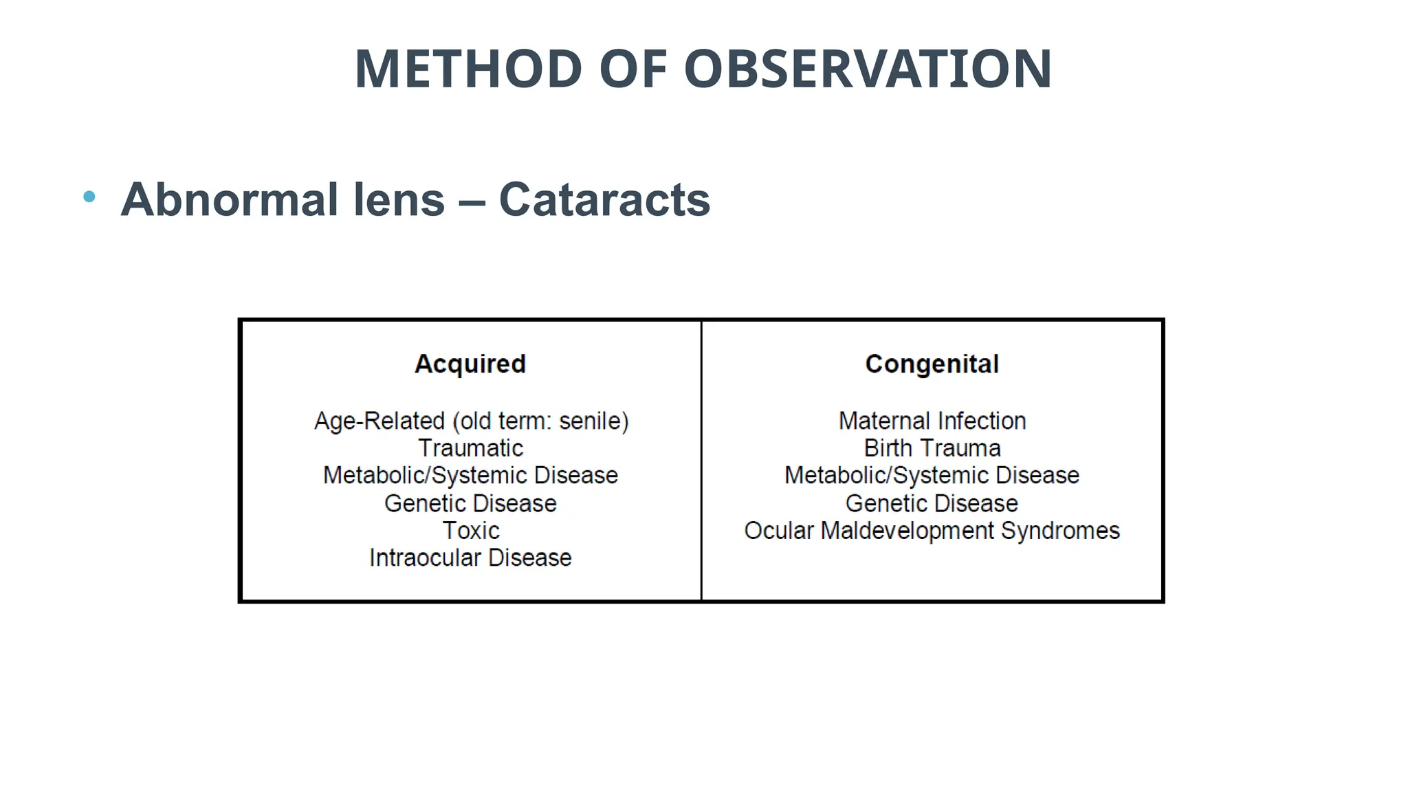
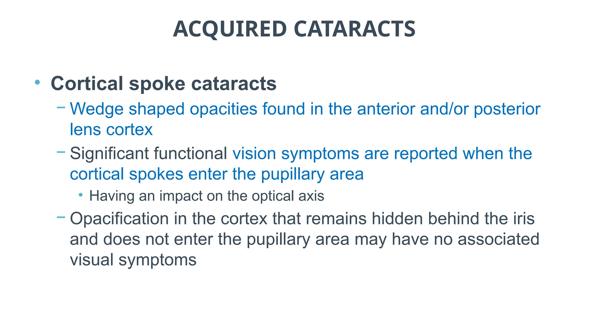
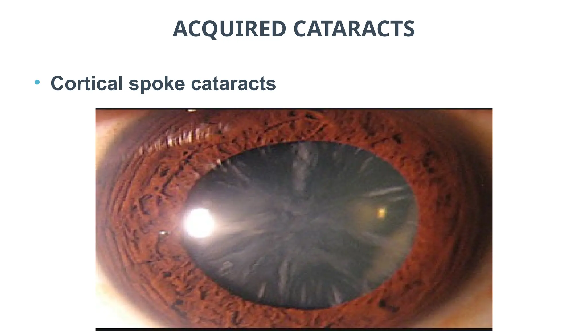
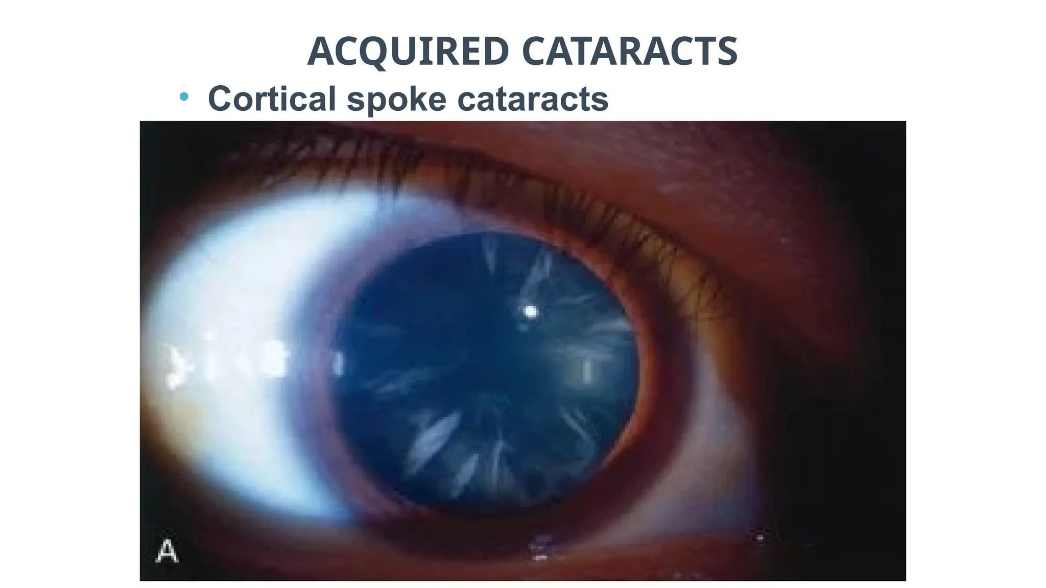
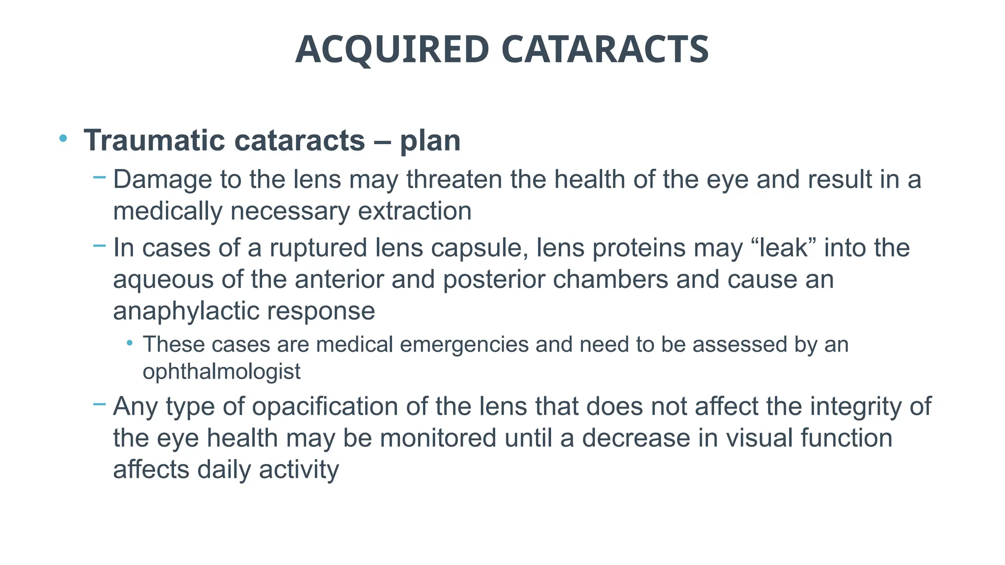
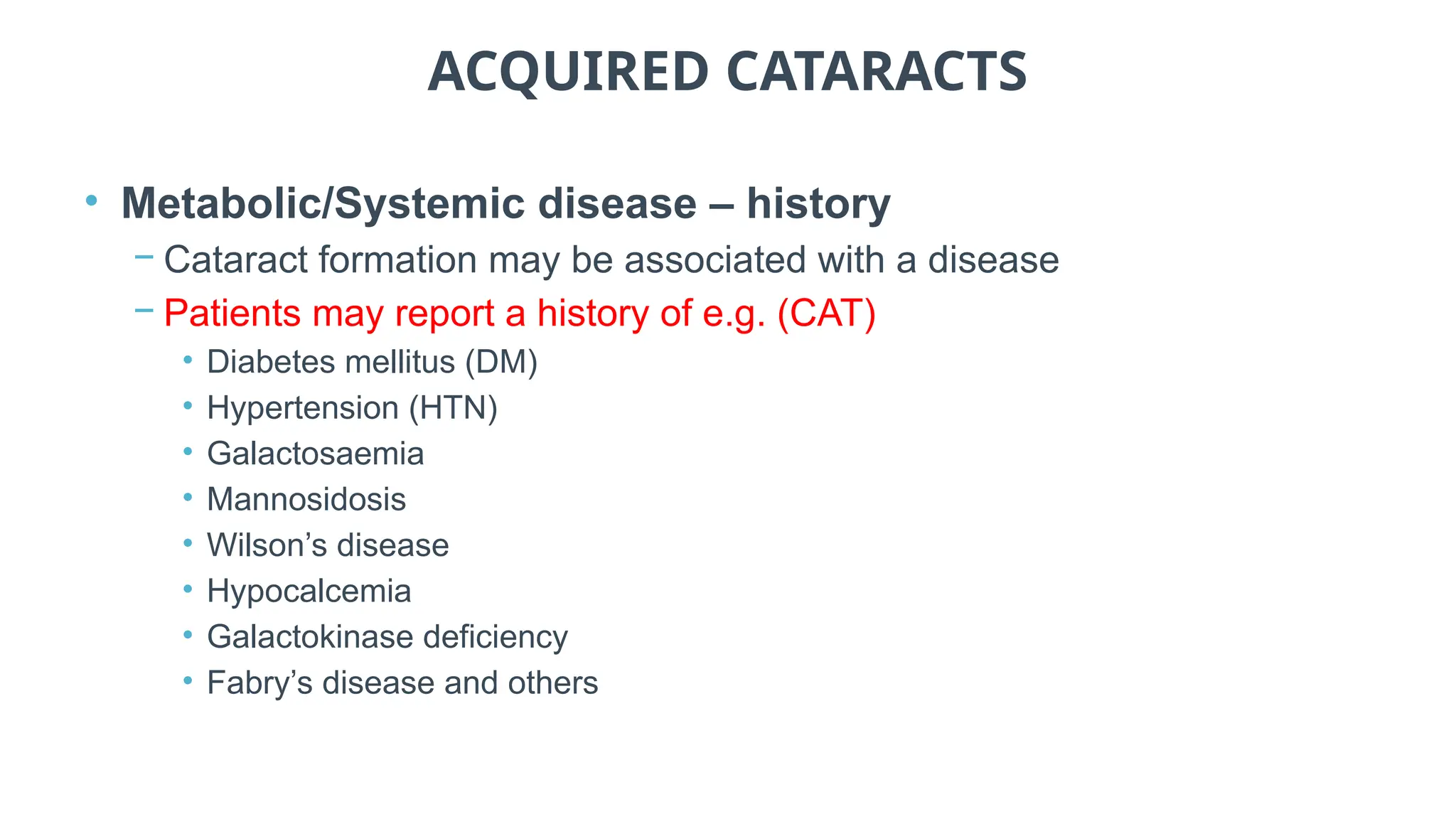
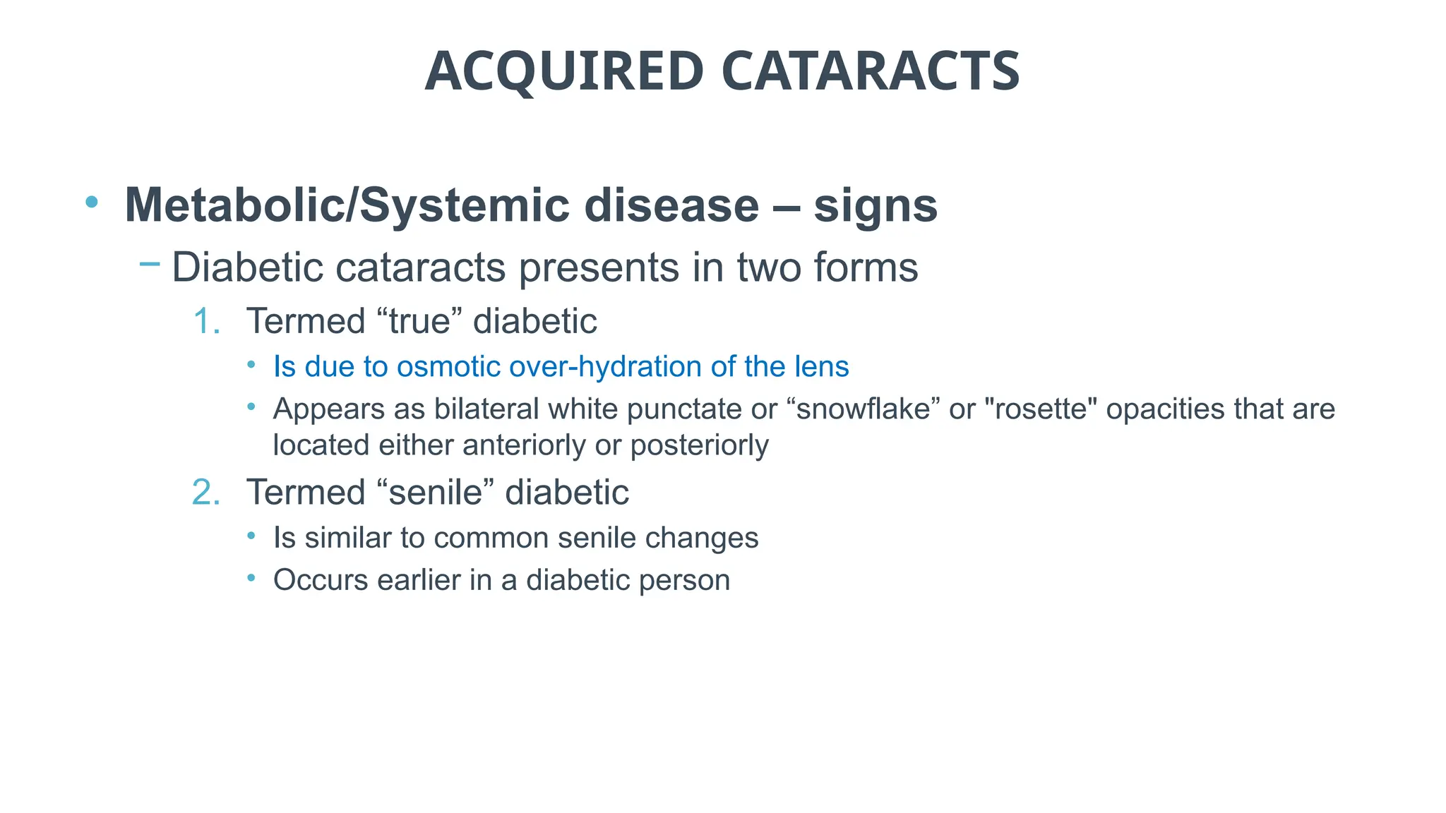
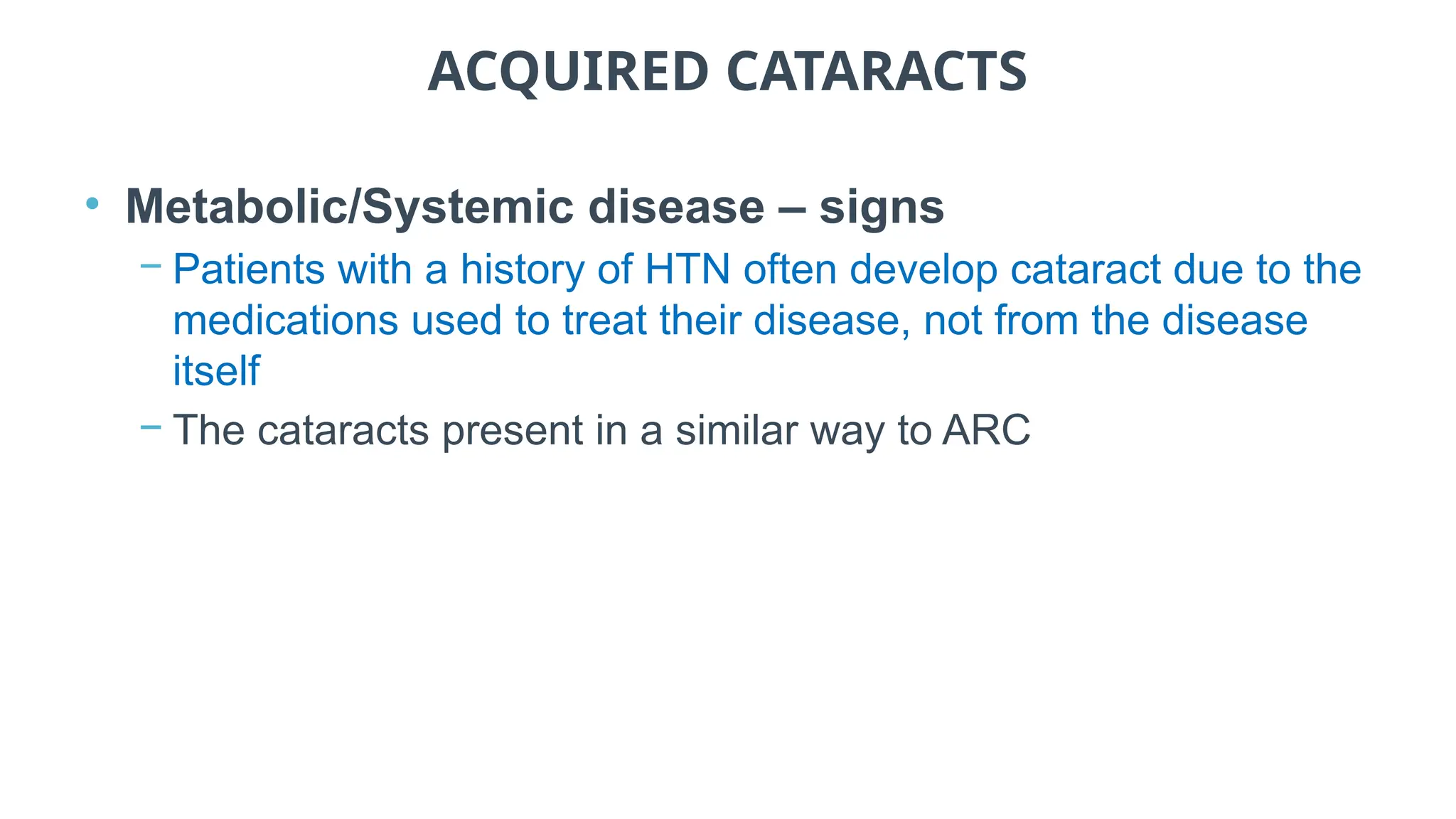
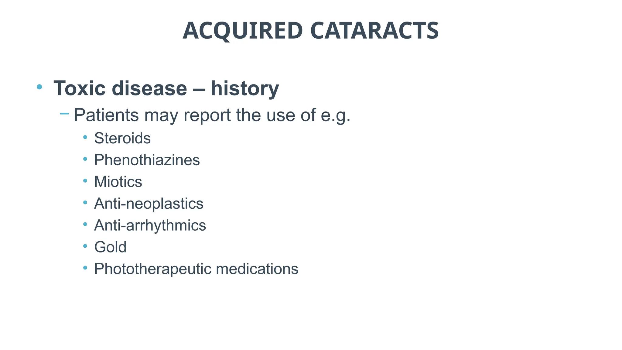
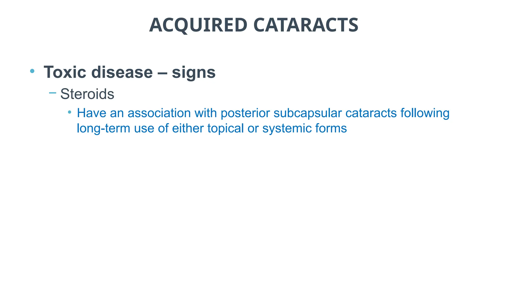
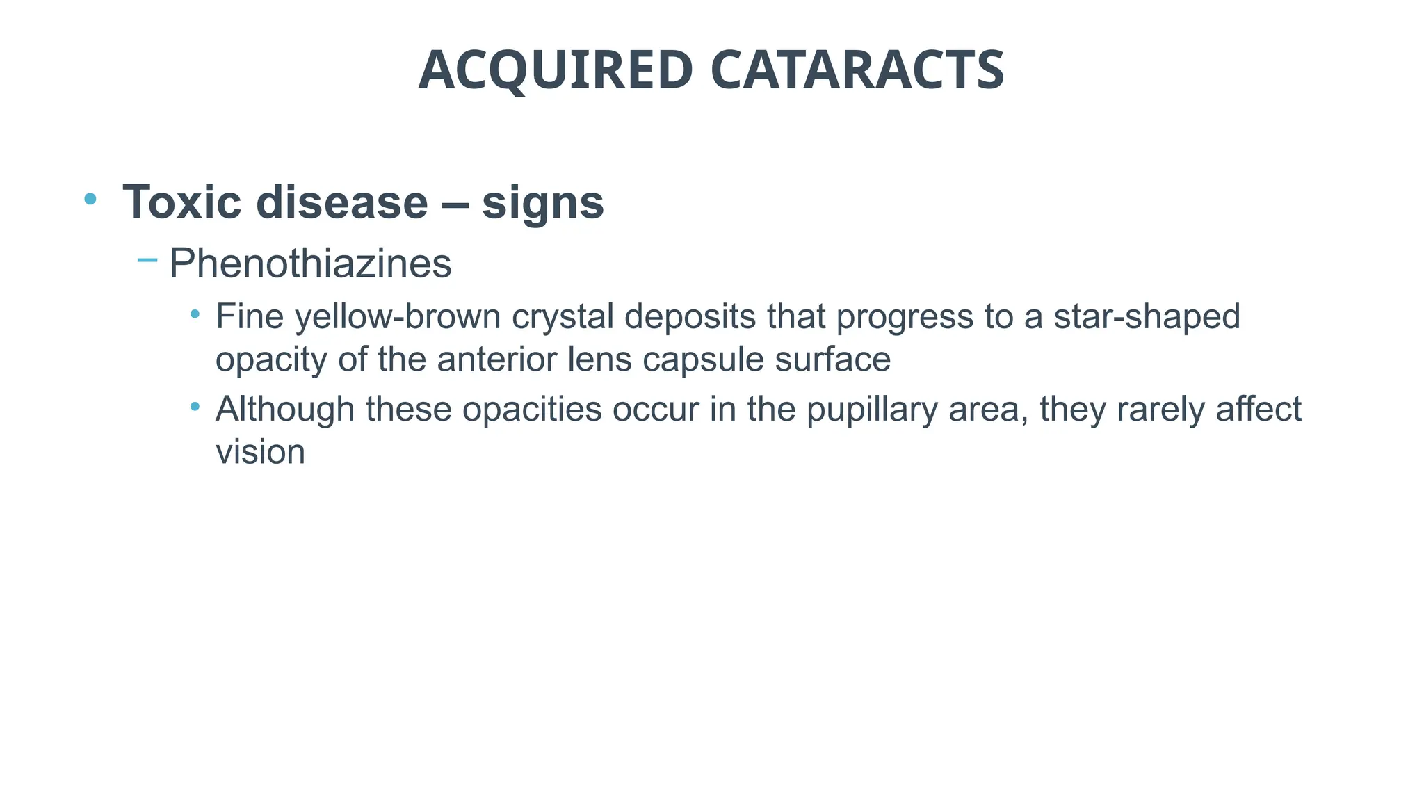
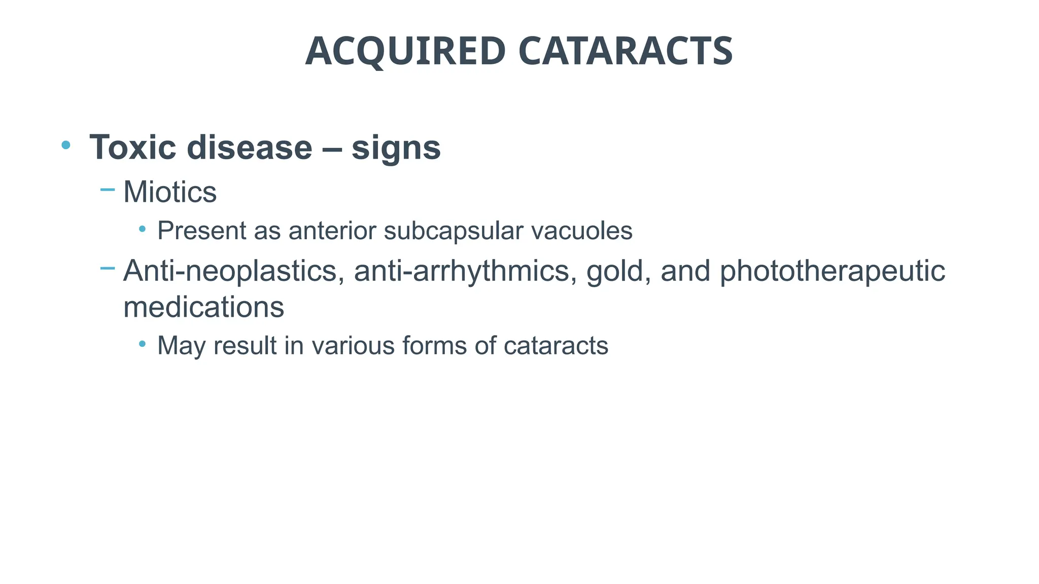
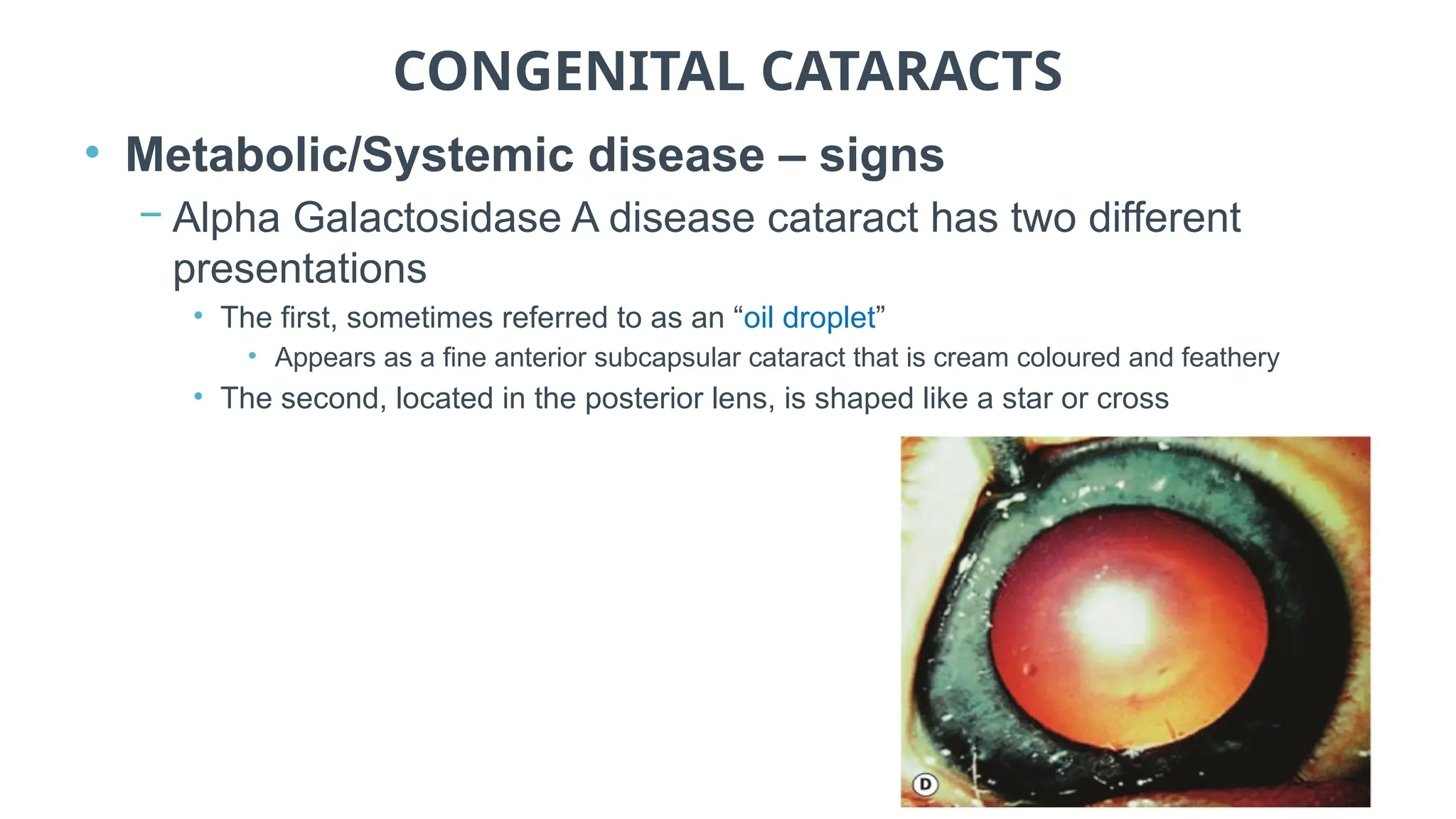
COMMON EYE DISORDERS
Normal and abnormal lens
CHAPTER CONTENTS
1. Method of observation
2. Acquired cataracts
3. Congenital cataracts
4. Abnormalities in lens shape and position
METHOD OF OBSERVATION
• Examine the lens with the slit-lamp using:
− Direct illumination
− Parallelepiped : Narrowing the beam to 1-2mm in width, Microscope is placed directly in front of
patients cornea and Light source is approximately 45 degree.
− Optic section: is a very thin parallelepiped “0.1mm” and optically cuts a very thin slice of
the cornea
− Retro-illumination: A vertical slit beam 1-4mm wide can be used
• Retro-illumination may also be performed using the direct
ophthalmoscope
METHOD OF OBSERVATION
• Normal lens
− The examiner can view the layers of the lens using a parallelepiped
and optic section
− Layers, from anterior to posterior, are:
• Anterior capsule
• Anterior sub-capsular space
• Anterior cortex
• Nucleus
• Posterior cortex
• Posterior sub-capsular space
• Posterior capsule
METHOD OF OBSERVATION
• Abnormal lens – Cataracts
ACQUIRED CATARACTS
• Age-related cataracts (ARC)
− The most common form of cataract
• Nuclear sclerotic (NS), cortical spoke (CS) and posterior subcapsular
cataract are forms of ARC
• Other, less common aetiologies of cataract do exist and may have
significant impact on functional vision
− Aetiology of cataractogenesis should be identified where
possible
• Through clinical experience, history taking, and access to other
resources, etc.
ACQUIRED CATARACTS
• Age-related cataracts – symptoms
− Include decreasing vision, glare and difficulties with night
driving
• Patients may commonly report that “my glasses are dirty, but cleaning
them doesn’t help”
− Other symptoms may include monocular diplopia
• Often associated with cortical cataracts
ACQUIRED CATARACTS
• Age-related cataracts – risk factors
− Case history may uncover other risk factors for ARC
• May be a family history of cataract as age-related nuclear and cortical
cataracts have an association with hereditary factors
• There may be a history of cigarette smoking
• An identified risk factor associated with nuclear cataracts
ACQUIRED CATARACTS
• Age-related cataracts – risk factors
− PSC are the least common form of ARC and are more often
associated with non age-related risk factors(CAT)
• These risks include diabetes, history of systemic steroid use
• Secondary to ocular disease such as retinitis pigmentosa (RP), uveitis,
or eye trauma, etc.
ACQUIRED CATARACTS
• Age-related cataracts – signs
− Three main morphological types of ARC include:
• Nuclear sclerotic (NS)
• Cortical spoke (CS)
• Posterior sub-capsular (PSC)
ACQUIRED CATARACTS
• Nuclear sclerotic cataracts
− Presents as a homogeneous increase in light scatter in the lens
nucleus
− Can be associated with increased yellowing or brunescence
• Indicative of blue wavelength-dependent light absorption
− These changes also occur to a lesser degree with normal ageing
− Use of a slit-lamp optic section technique is an accurate way of
detecting and assessing nuclear cataract and it is best performed
with a dilated pupil
• It is possible through an undilated pupil
ACQUIRED CATARACTS
• Nuclear sclerotic cataract
ACQUIRED CATARACTS
• Nuclear sclerotic cataracts
• Often associated with myopia due to an increase in the
refractive index of the nucleus
− Nuclear yellowing and opacification can be graded on a scale
of 0 to 4+
• 0 being the absence of opacity
• 4+ being the most severe form of opacity
− Several standardized systems of grading cataracts exist
− Digital imaging is useful for comparison over time
ACQUIRED CATARACTS
• Cortical spoke cataracts
− Wedge shaped opacities found in the anterior and/or posterior
lens cortex
− Significant functional vision symptoms are reported when the
cortical spokes enter the pupillary area
• Having an impact on the optical axis
− Opacification in the cortex that remains hidden behind the iris
and does not enter the pupillary area may have no associated
visual symptoms
ACQUIRED CATARACTS
• Cortical spoke cataracts
ACQUIRED CATARACTS
• Cortical spoke cataracts
ACQUIRED CATARACTS
• Cortical spoke cataracts
− Opacities are most often found in the inferior-nasal part of the
lens
• Suggests the association of ultra-violet B radiation in their
aetiology(CAT)
− Opacification is due to the scattering of light when it meets
irregular interfaces between regions of different refractive
indices
ACQUIRED CATARACTS
• Cortical spoke cataracts
− Opacities are best seen using retro-illumination from the fundus
• Opacities appear black against the red fundal glow
− The brightest reflection from the fundus is obtained when the
illuminating beam strikes the optic nerve head
• To achieve this the illumination system is placed on the temporal side
of the biomicroscope
ACQUIRED CATARACTS
• Posterior subcapsular cataracts (PSC)
− Presents at the posterior portion of the lens just in front of the
posterior capsule
− In age-related vacuolar type of PSC cataract, localized
reductions in refractive index and vacuoles are found in the early
stages
− In the later stages of development there is a posterior migration
of epithelial cells from the lens equator
• These epithelial cells converge on the posterior pole, forming the
balloon or bladder cells of Wedl
ACQUIRED CATARACTS
• Posterior subcapsular cataracts (PSC)
ACQUIRED CATARACTS
• Posterior subcapsular cataracts (PSC)
− A membranous appearance occurs in the later stages of PSC
development
• Due in part to the many organelles in the epithelial cells and the
membrane formed by the migrating cells
− Although an optic section may aid in determining the depth of
any opacity of the lens a PSC is best viewed through a dilated
pupil using retro-illumination
ACQUIRED CATARACTS
• Posterior subcapsular cataracts (PSC)
− PSC are the least common form of ARC and are more often
associated with non age-related risk factors
• These risks include diabetes, history of systemic steroid use
• Secondary to ocular disease such as retinitis pigmentosa (RP), uveitis, or eye
trauma, etc.
− Documentation of the extent of a PSC may be completed by using
digital imaging or by simply drawing the cataract on your medical
record
• Recording the dimension of the undilated pupil as a dashed line on the
diagram will aid in locating the PSC with respect to the optical axis
ACQUIRED CATARACTS
• Clinical assessment
− Objective assessments should include refraction
• Nuclear and cortical cataracts can cause significant changes in refractive
error
− Retinoscopy is often difficult with cataracts due to minimal light
returning to the examiner
• This can sometimes be overcome by:
• Using a larger sight hole
• Using as few trial lenses as possible
• To reduce light loss due to reflections from the lenses
• Working closer to the patient than the traditional 50 or 67cm
ACQUIRED CATARACTS
• Clinical assessment
− Assessment of visual function should includeCAT)
• Contrast sensitivity
• Disability glare testing
• Visual acuity
• Preferably using a logMAR chart
− The former two tests provide a better measure of the patient’s
functional vision compared to the high contrast with high (or
low) luminance test provided by a clinical visual acuity
assessment
ACQUIRED CATARACTS
• Clinical assessment
− It is important to differentially diagnose the morphological
type(s) of cataracts as they have different risk factors and
management plans
• A significant myopic shift in refractive error in an older patient strongly
suggests a nuclear cataract
• A significant shift in the power or axis of astigmatism in an older patient
can also suggest cortical cataract
ACQUIRED CATARACTS
• Clinical assessment
− A PSC may be obstructed by the corneal and lens reflexes
when viewed through an undilated pupil
− Care should be given to conduct a thorough, dilated
examination of the lens to identify the extent of cataract
development
ACQUIRED CATARACTS
• Clinical assessment
− Cataracts are monitored until the decrease in functional visual
acuity affects daily activity
− Ultimately, surgery is required to remove the cataract and
replace it with an artificial lens
• Intraocular lens (IOL)
− Surgical referral is dependent on individual patient symptoms
ACQUIRED CATARACTS
• Clinical assessment
− Prior to referral, there are several options that should be considered
to improve the patient’s visual function(CAT)
1. Provide updated refractive correction
• Particularly in nuclear cataracts (myopic shifts) and cortical cataracts (astigmatic
changes)
2. Suggest anti-reflection (AR) coating
• Particularly useful if they read with their back to a window, limiting reflections from the
back surface of the spectacle lenses
3. Suggest ultra-violet (UV) coating
• Some cataracts contain fluorescent pigments that can scatter light and reduce vision
further
ACQUIRED CATARACTS
• Clinical assessment
4. Suggest wearing a broad brimmed hat
• Can be better than suggesting tinted spectacles as the tint reduces the light from
the object they are looking at as well as the sun’s light
5. Suggest a tint for glasses
• A dilated pupil may allow for light to pass around a PSC allowing for reduced light
scatter and less distorted visual acuity
• Providing a tint for any patient that complains of discomfort due to glare may be
beneficial
• Use caution in cases of CS cataracts as tints can dilate the pupil slightly and
expose more of the spokes, potentially reducing vision
ACQUIRED CATARACTS
• Clinical assessment
6. Suggest that the patients read or do their near activities
with the window/sun behind their back and/or with a
lamp shining over their shoulder
• Light should fall on the near task and not into their eyes
• This can be further improved using a typoscope
• A black card with a wide slit in it
• The card stops light reflecting from the white pages of the
book/newspaper into their eye
• The patients must move the slit in the card as they read the
page
ACQUIRED CATARACTS
• Traumatic cataracts - history
− Patients may report a history of e.g.
• Penetrating injuries display opacification local to the area of involvement
• Blunt trauma may cause a characteristic flower-shaped opacity
• Electric shock is a rare cause of cataract, patterns including diffuse milky-
white opacification and multiple snowflake-like opacities
• Infrared radiation, if intense as in glassblowers, may rarely cause true
exfoliation of the anterior lens capsule
• Ionizing radiation exposure such as for ocular tumour treatment may cause
posterior subcapsular opacities
TRAUMATIC CATARACTS
ACQUIRED CATARACTS
• Traumatic cataracts – signs
− Penetrating injuries display opacification local to the area of
involvement
− Concussive injury may lead to a Vossius ring
• Viewed as an imprinting of iris pigment onto the anterior lens capsule
− A more diffuse opacification of the lens may be seen in cases of
cataracts resulting from electric shock or exposure to radiation
− Exfoliation of the anterior lens capsule may result from intense
UV exposure
ACQUIRED CATARACTS
• Traumatic cataracts – plan
− Damage to the lens may threaten the health of the eye and result in a
medically necessary extraction
− In cases of a ruptured lens capsule, lens proteins may “leak” into the
aqueous of the anterior and posterior chambers and cause an
anaphylactic response
• These cases are medical emergencies and need to be assessed by an
ophthalmologist
− Any type of opacification of the lens that does not affect the integrity of
the eye health may be monitored until a decrease in visual function
affects daily activity
ACQUIRED CATARACTS
• Metabolic/Systemic disease – history
− Cataract formation may be associated with a disease
− Patients may report a history of e.g. (CAT)
• Diabetes mellitus (DM)
• Hypertension (HTN)
• Galactosaemia
• Mannosidosis
• Wilson’s disease
• Hypocalcemia
• Galactokinase deficiency
• Fabry’s disease and others
ACQUIRED CATARACTS
• Metabolic/Systemic disease – signs
− Diabetic cataracts presents in two forms
1. Termed “true” diabetic
• Is due to osmotic over-hydration of the lens
• Appears as bilateral white punctate or “snowflake” or "rosette" opacities that are
located either anteriorly or posteriorly
2. Termed “senile” diabetic
• Is similar to common senile changes
• Occurs earlier in a diabetic person
ACQUIRED CATARACTS
• Metabolic/Systemic disease – signs
− Patients with a history of HTN often develop cataract due to the
medications used to treat their disease, not from the disease
itself
− The cataracts present in a similar way to ARC
ACQUIRED CATARACTS
• Metabolic/Systemic disease – signs
− Alpha Galactosidase A or Fabry's disease cataract has two
different presentations
1. Sometimes referred to as an “oil droplet”
• Appears as a fine anterior sub-capsular cataract that is cream coloured and
feathery
2. Located in the posterior lens
• Is shaped like a star or cross
ACQUIRED CATARACTS
• Metabolic/Systemic disease – signs
− Wilson’s syndrome cataract, called green “sunflower”, consists
of central pigmented opacities with tapering extensions
• Because copper is deposited anteriorly and posteriorly within the lens,
it takes on a yellow-green appearance
ACQUIRED CATARACTS
• Metabolic/Systemic disease – signs
− Hypocalcemia cataract consists of multi-colored crystals or
flecks
• They are small, white or polychromatic crystals located in the anterior
and posterior cortex just beneath the capsule
• A clear zone separates this zonular cortical cataract from the nucleus
ACQUIRED CATARACTS
• Metabolic/Systemic disease – signs
− Mannosidosis cataract varies in presentation
− Galactokinase deficiency is an autosomal recessive disease
that presents with lamellar cataract
ACQUIRED CATARACTS
• Metabolic/Systemic disease – plans
− The cataract is monitored until the decrease in visual acuity
affects daily function
− Referral is based on an individual patient basis
ACQUIRED CATARACTS
• Genetic disease – history
− Patients may report a history of e.g.
• Myotonic dystrophy: an inherited disease in which thw muscles
contracts but have decreasing power to relax (prolonged contraction of
muscles)
• Down’s syndrome
• Other genetic syndromes
ACQUIRED CATARACTS
• Genetic disease – signs
− Myotonic dystrophy causes a “Christmas tree” cataract
• A stellate figure in the posterior subcapsular area with or without multi-
colored dust-like opacities present in the cortex
− Down’s syndrome cataracts vary in appearance but generally
have the potential to decrease VA
• The more common presentations consist of cortical flake opacities or
arcuate opacities
CHRISTMAS TREE CATARACT
ACQUIRED CATARACTS
• Genetic disease – plan
− The cataract is monitored until the decrease in visual function
affects daily activity
− Referral for surgery is based on an individual patient basis
ACQUIRED CATARACTS
• Toxic disease – history
− Patients may report the use of e.g.
• Steroids
• Phenothiazines
• Miotics
• Anti-neoplastics
• Anti-arrhythmics
• Gold
• Phototherapeutic medications
ACQUIRED CATARACTS
• Toxic disease – signs
− Steroids
• Have an association with posterior subcapsular cataracts following
long-term use of either topical or systemic forms
ACQUIRED CATARACTS
• Toxic disease – signs
− Phenothiazines
• Fine yellow-brown crystal deposits that progress to a star-shaped
opacity of the anterior lens capsule surface
• Although these opacities occur in the pupillary area, they rarely affect
vision
ACQUIRED CATARACTS
• Toxic disease – signs
− Miotics
• Present as anterior subcapsular vacuoles
− Anti-neoplastics, anti-arrhythmics, gold, and phototherapeutic
medications
• May result in various forms of cataracts
ACQUIRED CATARACTS
• Toxic disease – signs
− Phospholine iodide
• Is an anticholinesterase
• Causes cataract in the form of anterior sub-capsular vacuoles
− Amiodarone
• Causes anterior subcapsular lens opacities
ACQUIRED CATARACTS
• Toxic disease – plans
− The cataract is monitored until the decrease in visual function
affects daily activity
− Referral for surgery is based on an individual patient basis
ACQUIRED CATARACTS
• Intraocular disease – history
− Patients may have a history of e.g.
• Chronic intraocular inflammation or infection
• Retinal disease
• Vitreo-retinal disease
ACQUIRED CATARACTS
• Intraocular disease – signs
− Uveitic cataracts appear as a colorful lustre at the posterior
pole of the lens
• As well as anterior and posterior subcapsular opacities
− In cases of recurrent or persistent inflammation, a fibro-
vascular membrane may develop on the anterior lens surface
affecting the optical axis
ACQUIRED CATARACTS
• Intraocular disease – signs
− Retinal disease such as retinitis pigmentosa (RP), Leber’s
hereditary optic neuropathy, gyrate atrophy or vitreo-retinal
diseases such as Stickler’s syndrome or Wagner’s syndrome
produce posterior subcapsular lens opacities
ACQUIRED CATARACTS
• Intraocular disease – signs
− Glaukomflecken present as fine whitish-gray anterior
subcapsular opacities in the area of the pupil
• Result from an acute angle-closure attack
ACQUIRED CATARACTS
• Intraocular disease – plans
− The cataract is monitored until the decrease in visual function
affects daily activity
− Referral for surgery is based on an individual patient basis
CONGENITAL CATARACTS
• Maternal infection/Drug ingestion – history
− Patient report e.g. that their biological mother had a history of
gestational viral infection, rubella (German measles) or
ingested medications such as steroids or thalidomide during
pregnancy
CONGENITAL CATARACTS
• Maternal infection/Drug ingestion – signs
− Rubella infection during pregnancy produces congenital
cataracts, which are present unilaterally or bilaterally
• They appear as either a diffuse opacity throughout the lens or
a dense, pearly, nuclear cataract that is surrounded by a less
dense cortical opacity
• Certain cases of rubella may exhibit microspherophakia
CONGENITAL CATARACTS
• Maternal infection/Drug ingestion –
signs
− Ingestion of medications during pregnancy
produces cataracts, which vary in appearance
according to the medication taken
− Long-term use of steroids may result in
posterior subcapsular cataracts
CONGENITAL CATARACTS
• Maternal infection/Drug ingestion –
plans
− The cataract is monitored until the decrease
in visual function affects daily activity
− Referral for surgery is based on an individual
patient basis
CONGENITAL CATARACTS
• Birth trauma – history
− Patients report a history of trauma at birth
− Cataract appearance secondary to birth trauma depends on the
type of trauma involved
CONGENITAL CATARACTS
• Birth trauma – plans
− The cataract is monitored until the decrease in visual function
affects daily activity
− Referral for surgery is based on an individual patient basis
CONGENITAL CATARACTS
• Metabolic/Systemic disease – history
− Patients report a history of Alpha Galactosidase A
− Or no known related history of disease
CONGENITAL CATARACTS
• Metabolic/Systemic disease – signs
− Alpha Galactosidase A disease cataract has two different
presentations
• The first, sometimes referred to as an “oil droplet”
• Appears as a fine anterior subcapsular cataract that is cream coloured and feathery
• The second, located in the posterior lens, is shaped like a star or cross
CONGENITAL CATARACTS
• Metabolic/Systemic disease – plans
− The cataract is monitored until the decrease in visual function
affects daily activity
− Referral for surgery is based on an individual patient basis
CONGENITAL CATARACTS
• Genetic disease – history
− Patient reports a history of Lowe’s oculocerebrorenal syndrome
− Or Down’s syndrome
− Or no known systemic disease
CONGENITAL CATARACTS
• Genetic disease – signs
− Lowe’s syndrome is associated with congenital cataracts
• Lens appears thin and small and is referred to as microphakic
• Opacities may be present in the capsule, cortex or nucleus
CONGENITAL CATARACTS
• Genetic disease – signs
− Down’s syndrome is associated with various forms of cataracts
• The more common presentations consist of cortical flake opacities or
arcuate opacities
• The cataracts usually have the potential of causing decreased vision
CONGENITAL CATARACTS
• Genetic disease – signs
− Congenital nuclear cataracts without systemic association may
present in two forms
1. Is termed the embryonal or nuclear cataract (cataracta centralis
pulverulenta)
• Contains small star-shaped opacities that are located in the embryonal nucleus
• The foetal nucleus is not affected
• This type of cataract has a dominant inheritance pattern, is usually bilateral and does
not affect vision adversely
2. Is termed the total nuclear cataract
• This affects both the embryonal and foetal nucleus and therefore may affect vision
adversely
CONGENITAL CATARACTS
• Genetic disease – plans
− The cataract is monitored until the decrease in visual function
affects daily activity
− Referral for surgery is based on an individual patient basis
CONGENITAL CATARACTS
• Ocular maldevelopment – history
− Patients report a history of Peter’s anomaly
− Or no awareness of a related anomaly
CONGENITAL CATARACTS
• Ocular maldevelopment – signs
− A cataract associated with Peter's anomaly appears in
conjunction with a central corneal defect which is in apposition
to the lens
− The anomaly is referred to as keratolenticular strands
CONGENITAL CATARACTS
• Ocular maldevelopment – plans
− The cataract is monitored until the decrease in visual acuity
affects daily function
− Referral is based on an individual patient basis
ABNORMALITIES IN LENS SHAPE
AND POSITION
• History
− Patients may complain of blurry and/or monocular double vision
− May be history of e.g. Lowe’s syndrome, Weill-Marchesani
syndrome, Marfan’s syndrome, Alport syndrome,
homocystinuria, hyperlysinaemia, or Ehlers-Danlos’ and others
ABNORMALITIES IN LENS SHAPE
AND POSITIONING
• History
− Also, incomplete closure of the choroidal fissure may result in
coloboma of the lens as well as other ocular structures (e.g.
optic nerve, retina, iris, eyelid)
ABNORMALITIES IN LENS SHAPE
AND POSITIONING
• Signs
− Lowe’s syndrome
• May display posterior lenticonus in which the
posterior pole of the lens is conical
• Spherophakia (small lens) may be present
which is defined as an abnormally short lens
radius of curvature
ABNORMALITIES IN LENS SHAPE
AND POSITIONING
• Signs
− Weill-Marchesani syndrome
• May display spherophakia (small lens),
subluxation (partial dislocation) of the lens due
to defective lens zonules
• After subluxation, the lens may dislocate
anteriorly due to spherophakia
ABNORMALITIES IN LENS SHAPE
AND POSITIONING
• Signs
− Marfan’s syndrome
• May present with microspherophakia
• Due to defective lens zonules, patients may
present with lens subluxation
• The lens is usually displaced upward and this is
referred to as ectopia lentis due to defective
lens zonules
ABNORMALITIES IN LENS SHAPE
AND POSITIONING
• Signs
− Alport syndrome
• May present with an anterior lenticonus where
the anterior pole of the lens is conical
• Anterior lenticonus produces irregular astigmatism
− Homocystinuria
• May present with subluxation of the lens due to
defective zonules
• Lens is usually displaced downward
ABNORMALITIES IN LENS SHAPE
AND POSITIONING
• Signs
− Hyperlysinaemia
• May present with microspherophakia
− Ehlers-Danlos
• May present with subluxation of the lens
ABNORMALITIES IN LENS SHAPE
AND POSITIONING
• Signs
− Anomalous, incomplete closure of the
choroidal fissure may result in a lens
coloboma
− This variation presents as a notch at the
equator of the lens with associated defect
of the local zonules
ABNORMALITIES IN LENS SHAPE
AND POSITIONING
• Plans
− Treatment is dependent on anomaly and
presentation
POLAR CATARACT

03 Normal and abnormal lens_1.pp( lens asone of most part used in refraction in eye)

  • 1.
    COMMON EYE DISORDERS Normaland abnormal lens
  • 2.
    CHAPTER CONTENTS 1. Methodof observation 2. Acquired cataracts 3. Congenital cataracts 4. Abnormalities in lens shape and position
  • 3.
    METHOD OF OBSERVATION •Examine the lens with the slit-lamp using: − Direct illumination − Parallelepiped : Narrowing the beam to 1-2mm in width, Microscope is placed directly in front of patients cornea and Light source is approximately 45 degree. − Optic section: is a very thin parallelepiped “0.1mm” and optically cuts a very thin slice of the cornea − Retro-illumination: A vertical slit beam 1-4mm wide can be used • Retro-illumination may also be performed using the direct ophthalmoscope
  • 4.
    METHOD OF OBSERVATION •Normal lens − The examiner can view the layers of the lens using a parallelepiped and optic section − Layers, from anterior to posterior, are: • Anterior capsule • Anterior sub-capsular space • Anterior cortex • Nucleus • Posterior cortex • Posterior sub-capsular space • Posterior capsule
  • 5.
    METHOD OF OBSERVATION •Abnormal lens – Cataracts
  • 6.
    ACQUIRED CATARACTS • Age-relatedcataracts (ARC) − The most common form of cataract • Nuclear sclerotic (NS), cortical spoke (CS) and posterior subcapsular cataract are forms of ARC • Other, less common aetiologies of cataract do exist and may have significant impact on functional vision − Aetiology of cataractogenesis should be identified where possible • Through clinical experience, history taking, and access to other resources, etc.
  • 7.
    ACQUIRED CATARACTS • Age-relatedcataracts – symptoms − Include decreasing vision, glare and difficulties with night driving • Patients may commonly report that “my glasses are dirty, but cleaning them doesn’t help” − Other symptoms may include monocular diplopia • Often associated with cortical cataracts
  • 8.
    ACQUIRED CATARACTS • Age-relatedcataracts – risk factors − Case history may uncover other risk factors for ARC • May be a family history of cataract as age-related nuclear and cortical cataracts have an association with hereditary factors • There may be a history of cigarette smoking • An identified risk factor associated with nuclear cataracts
  • 9.
    ACQUIRED CATARACTS • Age-relatedcataracts – risk factors − PSC are the least common form of ARC and are more often associated with non age-related risk factors(CAT) • These risks include diabetes, history of systemic steroid use • Secondary to ocular disease such as retinitis pigmentosa (RP), uveitis, or eye trauma, etc.
  • 10.
    ACQUIRED CATARACTS • Age-relatedcataracts – signs − Three main morphological types of ARC include: • Nuclear sclerotic (NS) • Cortical spoke (CS) • Posterior sub-capsular (PSC)
  • 11.
    ACQUIRED CATARACTS • Nuclearsclerotic cataracts − Presents as a homogeneous increase in light scatter in the lens nucleus − Can be associated with increased yellowing or brunescence • Indicative of blue wavelength-dependent light absorption − These changes also occur to a lesser degree with normal ageing − Use of a slit-lamp optic section technique is an accurate way of detecting and assessing nuclear cataract and it is best performed with a dilated pupil • It is possible through an undilated pupil
  • 12.
  • 13.
    ACQUIRED CATARACTS • Nuclearsclerotic cataracts • Often associated with myopia due to an increase in the refractive index of the nucleus − Nuclear yellowing and opacification can be graded on a scale of 0 to 4+ • 0 being the absence of opacity • 4+ being the most severe form of opacity − Several standardized systems of grading cataracts exist − Digital imaging is useful for comparison over time
  • 14.
    ACQUIRED CATARACTS • Corticalspoke cataracts − Wedge shaped opacities found in the anterior and/or posterior lens cortex − Significant functional vision symptoms are reported when the cortical spokes enter the pupillary area • Having an impact on the optical axis − Opacification in the cortex that remains hidden behind the iris and does not enter the pupillary area may have no associated visual symptoms
  • 15.
  • 16.
  • 17.
    ACQUIRED CATARACTS • Corticalspoke cataracts − Opacities are most often found in the inferior-nasal part of the lens • Suggests the association of ultra-violet B radiation in their aetiology(CAT) − Opacification is due to the scattering of light when it meets irregular interfaces between regions of different refractive indices
  • 18.
    ACQUIRED CATARACTS • Corticalspoke cataracts − Opacities are best seen using retro-illumination from the fundus • Opacities appear black against the red fundal glow − The brightest reflection from the fundus is obtained when the illuminating beam strikes the optic nerve head • To achieve this the illumination system is placed on the temporal side of the biomicroscope
  • 19.
    ACQUIRED CATARACTS • Posteriorsubcapsular cataracts (PSC) − Presents at the posterior portion of the lens just in front of the posterior capsule − In age-related vacuolar type of PSC cataract, localized reductions in refractive index and vacuoles are found in the early stages − In the later stages of development there is a posterior migration of epithelial cells from the lens equator • These epithelial cells converge on the posterior pole, forming the balloon or bladder cells of Wedl
  • 20.
    ACQUIRED CATARACTS • Posteriorsubcapsular cataracts (PSC)
  • 21.
    ACQUIRED CATARACTS • Posteriorsubcapsular cataracts (PSC) − A membranous appearance occurs in the later stages of PSC development • Due in part to the many organelles in the epithelial cells and the membrane formed by the migrating cells − Although an optic section may aid in determining the depth of any opacity of the lens a PSC is best viewed through a dilated pupil using retro-illumination
  • 22.
    ACQUIRED CATARACTS • Posteriorsubcapsular cataracts (PSC) − PSC are the least common form of ARC and are more often associated with non age-related risk factors • These risks include diabetes, history of systemic steroid use • Secondary to ocular disease such as retinitis pigmentosa (RP), uveitis, or eye trauma, etc. − Documentation of the extent of a PSC may be completed by using digital imaging or by simply drawing the cataract on your medical record • Recording the dimension of the undilated pupil as a dashed line on the diagram will aid in locating the PSC with respect to the optical axis
  • 23.
    ACQUIRED CATARACTS • Clinicalassessment − Objective assessments should include refraction • Nuclear and cortical cataracts can cause significant changes in refractive error − Retinoscopy is often difficult with cataracts due to minimal light returning to the examiner • This can sometimes be overcome by: • Using a larger sight hole • Using as few trial lenses as possible • To reduce light loss due to reflections from the lenses • Working closer to the patient than the traditional 50 or 67cm
  • 24.
    ACQUIRED CATARACTS • Clinicalassessment − Assessment of visual function should includeCAT) • Contrast sensitivity • Disability glare testing • Visual acuity • Preferably using a logMAR chart − The former two tests provide a better measure of the patient’s functional vision compared to the high contrast with high (or low) luminance test provided by a clinical visual acuity assessment
  • 25.
    ACQUIRED CATARACTS • Clinicalassessment − It is important to differentially diagnose the morphological type(s) of cataracts as they have different risk factors and management plans • A significant myopic shift in refractive error in an older patient strongly suggests a nuclear cataract • A significant shift in the power or axis of astigmatism in an older patient can also suggest cortical cataract
  • 26.
    ACQUIRED CATARACTS • Clinicalassessment − A PSC may be obstructed by the corneal and lens reflexes when viewed through an undilated pupil − Care should be given to conduct a thorough, dilated examination of the lens to identify the extent of cataract development
  • 27.
    ACQUIRED CATARACTS • Clinicalassessment − Cataracts are monitored until the decrease in functional visual acuity affects daily activity − Ultimately, surgery is required to remove the cataract and replace it with an artificial lens • Intraocular lens (IOL) − Surgical referral is dependent on individual patient symptoms
  • 28.
    ACQUIRED CATARACTS • Clinicalassessment − Prior to referral, there are several options that should be considered to improve the patient’s visual function(CAT) 1. Provide updated refractive correction • Particularly in nuclear cataracts (myopic shifts) and cortical cataracts (astigmatic changes) 2. Suggest anti-reflection (AR) coating • Particularly useful if they read with their back to a window, limiting reflections from the back surface of the spectacle lenses 3. Suggest ultra-violet (UV) coating • Some cataracts contain fluorescent pigments that can scatter light and reduce vision further
  • 29.
    ACQUIRED CATARACTS • Clinicalassessment 4. Suggest wearing a broad brimmed hat • Can be better than suggesting tinted spectacles as the tint reduces the light from the object they are looking at as well as the sun’s light 5. Suggest a tint for glasses • A dilated pupil may allow for light to pass around a PSC allowing for reduced light scatter and less distorted visual acuity • Providing a tint for any patient that complains of discomfort due to glare may be beneficial • Use caution in cases of CS cataracts as tints can dilate the pupil slightly and expose more of the spokes, potentially reducing vision
  • 30.
    ACQUIRED CATARACTS • Clinicalassessment 6. Suggest that the patients read or do their near activities with the window/sun behind their back and/or with a lamp shining over their shoulder • Light should fall on the near task and not into their eyes • This can be further improved using a typoscope • A black card with a wide slit in it • The card stops light reflecting from the white pages of the book/newspaper into their eye • The patients must move the slit in the card as they read the page
  • 31.
    ACQUIRED CATARACTS • Traumaticcataracts - history − Patients may report a history of e.g. • Penetrating injuries display opacification local to the area of involvement • Blunt trauma may cause a characteristic flower-shaped opacity • Electric shock is a rare cause of cataract, patterns including diffuse milky- white opacification and multiple snowflake-like opacities • Infrared radiation, if intense as in glassblowers, may rarely cause true exfoliation of the anterior lens capsule • Ionizing radiation exposure such as for ocular tumour treatment may cause posterior subcapsular opacities
  • 32.
  • 33.
    ACQUIRED CATARACTS • Traumaticcataracts – signs − Penetrating injuries display opacification local to the area of involvement − Concussive injury may lead to a Vossius ring • Viewed as an imprinting of iris pigment onto the anterior lens capsule − A more diffuse opacification of the lens may be seen in cases of cataracts resulting from electric shock or exposure to radiation − Exfoliation of the anterior lens capsule may result from intense UV exposure
  • 34.
    ACQUIRED CATARACTS • Traumaticcataracts – plan − Damage to the lens may threaten the health of the eye and result in a medically necessary extraction − In cases of a ruptured lens capsule, lens proteins may “leak” into the aqueous of the anterior and posterior chambers and cause an anaphylactic response • These cases are medical emergencies and need to be assessed by an ophthalmologist − Any type of opacification of the lens that does not affect the integrity of the eye health may be monitored until a decrease in visual function affects daily activity
  • 35.
    ACQUIRED CATARACTS • Metabolic/Systemicdisease – history − Cataract formation may be associated with a disease − Patients may report a history of e.g. (CAT) • Diabetes mellitus (DM) • Hypertension (HTN) • Galactosaemia • Mannosidosis • Wilson’s disease • Hypocalcemia • Galactokinase deficiency • Fabry’s disease and others
  • 36.
    ACQUIRED CATARACTS • Metabolic/Systemicdisease – signs − Diabetic cataracts presents in two forms 1. Termed “true” diabetic • Is due to osmotic over-hydration of the lens • Appears as bilateral white punctate or “snowflake” or "rosette" opacities that are located either anteriorly or posteriorly 2. Termed “senile” diabetic • Is similar to common senile changes • Occurs earlier in a diabetic person
  • 37.
    ACQUIRED CATARACTS • Metabolic/Systemicdisease – signs − Patients with a history of HTN often develop cataract due to the medications used to treat their disease, not from the disease itself − The cataracts present in a similar way to ARC
  • 38.
    ACQUIRED CATARACTS • Metabolic/Systemicdisease – signs − Alpha Galactosidase A or Fabry's disease cataract has two different presentations 1. Sometimes referred to as an “oil droplet” • Appears as a fine anterior sub-capsular cataract that is cream coloured and feathery 2. Located in the posterior lens • Is shaped like a star or cross
  • 39.
    ACQUIRED CATARACTS • Metabolic/Systemicdisease – signs − Wilson’s syndrome cataract, called green “sunflower”, consists of central pigmented opacities with tapering extensions • Because copper is deposited anteriorly and posteriorly within the lens, it takes on a yellow-green appearance
  • 40.
    ACQUIRED CATARACTS • Metabolic/Systemicdisease – signs − Hypocalcemia cataract consists of multi-colored crystals or flecks • They are small, white or polychromatic crystals located in the anterior and posterior cortex just beneath the capsule • A clear zone separates this zonular cortical cataract from the nucleus
  • 41.
    ACQUIRED CATARACTS • Metabolic/Systemicdisease – signs − Mannosidosis cataract varies in presentation − Galactokinase deficiency is an autosomal recessive disease that presents with lamellar cataract
  • 42.
    ACQUIRED CATARACTS • Metabolic/Systemicdisease – plans − The cataract is monitored until the decrease in visual acuity affects daily function − Referral is based on an individual patient basis
  • 43.
    ACQUIRED CATARACTS • Geneticdisease – history − Patients may report a history of e.g. • Myotonic dystrophy: an inherited disease in which thw muscles contracts but have decreasing power to relax (prolonged contraction of muscles) • Down’s syndrome • Other genetic syndromes
  • 44.
    ACQUIRED CATARACTS • Geneticdisease – signs − Myotonic dystrophy causes a “Christmas tree” cataract • A stellate figure in the posterior subcapsular area with or without multi- colored dust-like opacities present in the cortex − Down’s syndrome cataracts vary in appearance but generally have the potential to decrease VA • The more common presentations consist of cortical flake opacities or arcuate opacities
  • 45.
  • 46.
    ACQUIRED CATARACTS • Geneticdisease – plan − The cataract is monitored until the decrease in visual function affects daily activity − Referral for surgery is based on an individual patient basis
  • 47.
    ACQUIRED CATARACTS • Toxicdisease – history − Patients may report the use of e.g. • Steroids • Phenothiazines • Miotics • Anti-neoplastics • Anti-arrhythmics • Gold • Phototherapeutic medications
  • 48.
    ACQUIRED CATARACTS • Toxicdisease – signs − Steroids • Have an association with posterior subcapsular cataracts following long-term use of either topical or systemic forms
  • 49.
    ACQUIRED CATARACTS • Toxicdisease – signs − Phenothiazines • Fine yellow-brown crystal deposits that progress to a star-shaped opacity of the anterior lens capsule surface • Although these opacities occur in the pupillary area, they rarely affect vision
  • 50.
    ACQUIRED CATARACTS • Toxicdisease – signs − Miotics • Present as anterior subcapsular vacuoles − Anti-neoplastics, anti-arrhythmics, gold, and phototherapeutic medications • May result in various forms of cataracts
  • 51.
    ACQUIRED CATARACTS • Toxicdisease – signs − Phospholine iodide • Is an anticholinesterase • Causes cataract in the form of anterior sub-capsular vacuoles − Amiodarone • Causes anterior subcapsular lens opacities
  • 52.
    ACQUIRED CATARACTS • Toxicdisease – plans − The cataract is monitored until the decrease in visual function affects daily activity − Referral for surgery is based on an individual patient basis
  • 53.
    ACQUIRED CATARACTS • Intraoculardisease – history − Patients may have a history of e.g. • Chronic intraocular inflammation or infection • Retinal disease • Vitreo-retinal disease
  • 54.
    ACQUIRED CATARACTS • Intraoculardisease – signs − Uveitic cataracts appear as a colorful lustre at the posterior pole of the lens • As well as anterior and posterior subcapsular opacities − In cases of recurrent or persistent inflammation, a fibro- vascular membrane may develop on the anterior lens surface affecting the optical axis
  • 55.
    ACQUIRED CATARACTS • Intraoculardisease – signs − Retinal disease such as retinitis pigmentosa (RP), Leber’s hereditary optic neuropathy, gyrate atrophy or vitreo-retinal diseases such as Stickler’s syndrome or Wagner’s syndrome produce posterior subcapsular lens opacities
  • 56.
    ACQUIRED CATARACTS • Intraoculardisease – signs − Glaukomflecken present as fine whitish-gray anterior subcapsular opacities in the area of the pupil • Result from an acute angle-closure attack
  • 57.
    ACQUIRED CATARACTS • Intraoculardisease – plans − The cataract is monitored until the decrease in visual function affects daily activity − Referral for surgery is based on an individual patient basis
  • 58.
    CONGENITAL CATARACTS • Maternalinfection/Drug ingestion – history − Patient report e.g. that their biological mother had a history of gestational viral infection, rubella (German measles) or ingested medications such as steroids or thalidomide during pregnancy
  • 59.
    CONGENITAL CATARACTS • Maternalinfection/Drug ingestion – signs − Rubella infection during pregnancy produces congenital cataracts, which are present unilaterally or bilaterally • They appear as either a diffuse opacity throughout the lens or a dense, pearly, nuclear cataract that is surrounded by a less dense cortical opacity • Certain cases of rubella may exhibit microspherophakia
  • 60.
    CONGENITAL CATARACTS • Maternalinfection/Drug ingestion – signs − Ingestion of medications during pregnancy produces cataracts, which vary in appearance according to the medication taken − Long-term use of steroids may result in posterior subcapsular cataracts
  • 61.
    CONGENITAL CATARACTS • Maternalinfection/Drug ingestion – plans − The cataract is monitored until the decrease in visual function affects daily activity − Referral for surgery is based on an individual patient basis
  • 62.
    CONGENITAL CATARACTS • Birthtrauma – history − Patients report a history of trauma at birth − Cataract appearance secondary to birth trauma depends on the type of trauma involved
  • 63.
    CONGENITAL CATARACTS • Birthtrauma – plans − The cataract is monitored until the decrease in visual function affects daily activity − Referral for surgery is based on an individual patient basis
  • 64.
    CONGENITAL CATARACTS • Metabolic/Systemicdisease – history − Patients report a history of Alpha Galactosidase A − Or no known related history of disease
  • 65.
    CONGENITAL CATARACTS • Metabolic/Systemicdisease – signs − Alpha Galactosidase A disease cataract has two different presentations • The first, sometimes referred to as an “oil droplet” • Appears as a fine anterior subcapsular cataract that is cream coloured and feathery • The second, located in the posterior lens, is shaped like a star or cross
  • 66.
    CONGENITAL CATARACTS • Metabolic/Systemicdisease – plans − The cataract is monitored until the decrease in visual function affects daily activity − Referral for surgery is based on an individual patient basis
  • 67.
    CONGENITAL CATARACTS • Geneticdisease – history − Patient reports a history of Lowe’s oculocerebrorenal syndrome − Or Down’s syndrome − Or no known systemic disease
  • 68.
    CONGENITAL CATARACTS • Geneticdisease – signs − Lowe’s syndrome is associated with congenital cataracts • Lens appears thin and small and is referred to as microphakic • Opacities may be present in the capsule, cortex or nucleus
  • 69.
    CONGENITAL CATARACTS • Geneticdisease – signs − Down’s syndrome is associated with various forms of cataracts • The more common presentations consist of cortical flake opacities or arcuate opacities • The cataracts usually have the potential of causing decreased vision
  • 70.
    CONGENITAL CATARACTS • Geneticdisease – signs − Congenital nuclear cataracts without systemic association may present in two forms 1. Is termed the embryonal or nuclear cataract (cataracta centralis pulverulenta) • Contains small star-shaped opacities that are located in the embryonal nucleus • The foetal nucleus is not affected • This type of cataract has a dominant inheritance pattern, is usually bilateral and does not affect vision adversely 2. Is termed the total nuclear cataract • This affects both the embryonal and foetal nucleus and therefore may affect vision adversely
  • 71.
    CONGENITAL CATARACTS • Geneticdisease – plans − The cataract is monitored until the decrease in visual function affects daily activity − Referral for surgery is based on an individual patient basis
  • 72.
    CONGENITAL CATARACTS • Ocularmaldevelopment – history − Patients report a history of Peter’s anomaly − Or no awareness of a related anomaly
  • 73.
    CONGENITAL CATARACTS • Ocularmaldevelopment – signs − A cataract associated with Peter's anomaly appears in conjunction with a central corneal defect which is in apposition to the lens − The anomaly is referred to as keratolenticular strands
  • 74.
    CONGENITAL CATARACTS • Ocularmaldevelopment – plans − The cataract is monitored until the decrease in visual acuity affects daily function − Referral is based on an individual patient basis
  • 75.
    ABNORMALITIES IN LENSSHAPE AND POSITION • History − Patients may complain of blurry and/or monocular double vision − May be history of e.g. Lowe’s syndrome, Weill-Marchesani syndrome, Marfan’s syndrome, Alport syndrome, homocystinuria, hyperlysinaemia, or Ehlers-Danlos’ and others
  • 76.
    ABNORMALITIES IN LENSSHAPE AND POSITIONING • History − Also, incomplete closure of the choroidal fissure may result in coloboma of the lens as well as other ocular structures (e.g. optic nerve, retina, iris, eyelid)
  • 77.
    ABNORMALITIES IN LENSSHAPE AND POSITIONING • Signs − Lowe’s syndrome • May display posterior lenticonus in which the posterior pole of the lens is conical • Spherophakia (small lens) may be present which is defined as an abnormally short lens radius of curvature
  • 78.
    ABNORMALITIES IN LENSSHAPE AND POSITIONING • Signs − Weill-Marchesani syndrome • May display spherophakia (small lens), subluxation (partial dislocation) of the lens due to defective lens zonules • After subluxation, the lens may dislocate anteriorly due to spherophakia
  • 79.
    ABNORMALITIES IN LENSSHAPE AND POSITIONING • Signs − Marfan’s syndrome • May present with microspherophakia • Due to defective lens zonules, patients may present with lens subluxation • The lens is usually displaced upward and this is referred to as ectopia lentis due to defective lens zonules
  • 80.
    ABNORMALITIES IN LENSSHAPE AND POSITIONING • Signs − Alport syndrome • May present with an anterior lenticonus where the anterior pole of the lens is conical • Anterior lenticonus produces irregular astigmatism − Homocystinuria • May present with subluxation of the lens due to defective zonules • Lens is usually displaced downward
  • 81.
    ABNORMALITIES IN LENSSHAPE AND POSITIONING • Signs − Hyperlysinaemia • May present with microspherophakia − Ehlers-Danlos • May present with subluxation of the lens
  • 82.
    ABNORMALITIES IN LENSSHAPE AND POSITIONING • Signs − Anomalous, incomplete closure of the choroidal fissure may result in a lens coloboma − This variation presents as a notch at the equator of the lens with associated defect of the local zonules
  • 83.
    ABNORMALITIES IN LENSSHAPE AND POSITIONING • Plans − Treatment is dependent on anomaly and presentation
  • 84.

Editor's Notes

  • #32 Causes of traumatic cataract. (A) Penetrating trauma; (B) blunt trauma; (C) electric shock and lightning strike;  (D) infrared radiation (glassblower’s cataract); (E) ionizing radiation 
  • #33 Vossius ring : Is due to blunt trauma to the eye. When the eye injured a circular ring of fainted or stippled opacity is seen on the anterior surface of the lens due to brown amorphous granules of pigment lying on the capsule.