Orthopaedic aspects of Poliomyelitis for MBBS
•
0 likes•310 views

This presentation is a basic overview of the orthpaedic aspects of poliomyelitis, its clinical features and management for undergraduate teaching (MBBS)

Recommended
More Related Content
What's hot
What's hot (20)
Similar to Orthopaedic aspects of Poliomyelitis for MBBS
Similar to Orthopaedic aspects of Poliomyelitis for MBBS (20)
More from Siddhartha Sinha
More from Siddhartha Sinha (20)
Recently uploaded
Recently uploaded (20)
Orthopaedic aspects of Poliomyelitis for MBBS
- 1. Poliomyelitis Dr Siddhartha Sinha Assistant Professor, Orthopaedics Hamdard Institute of Medical Sciences and Research, New Delhi, India
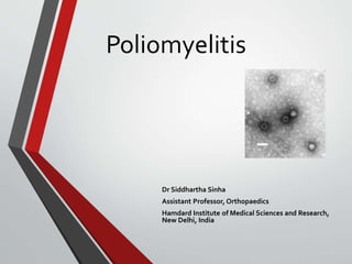
- 2. • small RNA viruses -Enterovirus genus - Picornaviridae family • Targets anterior horn cells and certain brain stem motor nuclei
- 3. Pathogenesis 3 Strains Transmission- Faeco Oral/ Droplets Multiply in GI musoca and secreted in stool After infection Reaches Anterior Horn cell through blood, perineural lymphatics or neurons (lumbar and cervical segments) Anterior Horn cells : • Unaffected • Recovers • Death Mechanism of damage • Direct • Ischaemia • Edema • Haemorrhage
- 4. Stages of poliomyelitis a) Incubation period (6-20 days) b) Pre-paralysis stage c) Stage of Maximum Paralysis d) Stage of recovery e) Post polio residual paralysis
- 5. Acute stage Clinical findings • Lasts 7-10 days. • Wide range of symptoms • Malaise • Encephalomyelitis • Widespread paralysis—diaphragm paralysis • Bulbar polio (medulla affected) • Examination • Fever • Flushing of the skin • Apprehension; muscular pain • Superficial reflexes lost first • Deep tendon reflexes disappear when the muscle group is paralysed. Treatment • Bed rest • Analgesics • Hot packs • Anatomical positioning of limbs to prevent contractures • Gentle passive ROM exercises
- 6. Most commonly affected muscle Quadriceps Muscle undergoing complete paralysis Tibialis anterior Hand muscle most commonly affected Opponens pollicis
- 7. Convalescent stage Clinical features • Recovery phase • 2 days after fever decreases up to 2 yrs • Varying degree of spontaneous recovery in muscle power takes place • > 80% return of strength - recovered muscles • < 30% of normal strength - paralysed muscle • Assess power frequently Treatment • Vigorous passive stretching exercises • Muscle training and gait training • Wedging casts and orthoses- prevent and correct deformities
- 8. Chronic stage • Usually begins 24 months after the acute illness • Aim of management: Achieve the maximal functional activity by management of long term consequences of muscle imbalances. • Goal: 1. Correcting any significant muscle imbalances and preventing or correcting soft tissue or bony deformities. 2. Static joint instability usually can be controlled indefinitely by orthoses. 3. Dynamic joint instability eventually results in a fixed deformity that cannot be controlled with orthoses
- 9. Causes of deformity in Polio • 1. Muscle imbalance • 2. Posture and gravity effect • 3. Dynamics of activity • 4. Dynamics of growth
- 10. Deformities in joints Joint Deformity Hip Flex-Abd- ER Knee • Flexion deformities • Genu Recurvatum • Tibia External rotation • Valgus deformity Foot and ankle • Equino-varus • Equino-valgus • Calcaneo-valgus
- 11. Surgery is indicated for: • Deformity correction when conservative treatment fails • Power loss substitution/ compensation incase paralysis is localized. • Length restoration.
- 12. Surgical options 1.Tenotomy and soft tissue releases a)Tendoachilles lengthening for Equinus foot b) Adductor tenotomy for adduction deformities at hip c) Ober/Yount release for flexion deformity at hip 2.TendonTransfers a) EHL to neck of 1st MT-Tibialis ant weakness b) Peronei to dorsum of foot– Dorsiflexion weakness
- 13. 3. Osteotomy a) Extension osteotomy – flexion deformity of knee b) Supracondylar osteotomy - genu varum 4. Arthrodesis a)Triple arthrodesis – ankle b) Stabilization of other flail joints 5. Limb lengthening a) Illizarov b) LRS
- 14. Other walking aids • AFO • Callipers (KAFO)
- 15. Polio CP Infective Mutifactorial pathogenesis Manifest in any age Usually in children No predisposing factors Many predisposing factors Flaccid pure motor paralysis w/o UMN signs Can be spastic or flaccid with UMN signs Patchy/ irregular pattern Regular patterns No movement in affected limb Uncontrolled movements may be seen
- 16. THANKYOU