
Recommended
More Related Content
What's hot
What's hot (20)
Similar to Knee rthrodesis
Similar to Knee rthrodesis (20)
Recently uploaded
Recently uploaded (20)
Knee rthrodesis
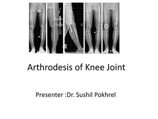
- 1. Arthrodesis of Knee Joint Presenter :Dr. Sushil Pokhrel
- 2. Objectives: • History • Introduction • Indications and contra-indications • Principle of management • Complications • Rehabilitation
- 3. History • Albert of Vienna in 1878 - first arthrodesis - instability of poliomyelitis • Hibbs in 1911 -tuberculous knee • Key (1932) - use external fixation to obtain fusion • Nelson and Evarts (1971)- first described knee arthrodesis as a treatment option for failed knee arthroplasty
- 4. Introduction Arthrodesis: Fusion of joint Knee Arthrodesis: fusion of knee joint Despite the loss of a functioning , knee fusion can provide a functional symptom-free extremity
- 5. INDICATIONS 1.Salvage of failed total knee arthroplasty a.Secondary to infection: 1. presence of resistant organisms 2. immune compromised patient 3. gross instability 4. inadequate skin and soft tissue coverage 5. Deficient extensor mechanism b. Patient is unwilling: revision arthroplasty
- 6. INDICATIONS 2.Relative indication: 1.painful ankylosis after infection 2.loss of the extensor mechanism 3.tuberculosis 4.trauma: severe destruction in young who desire to continue vigorus activity 5.severe deformity in paralytic conditions: -neuropathic arthropathy: DM - malignant or potentially malignant
- 7. CONTRA-INDICATIONS • Contra -lateral knee amputation • Advanced degenerative changes in ipsilateral hip or ankle • Arthrodesis of the contralateral hip or knee
- 8. This study demonstrates the presence pathologic advancement of degenerative osteoarthritis in the contralateral knee following arthrodesis in a select patient cohort. Arthrodesis of the knee creates adverse compensatory stresses on the contralateral knee which lead to symptoms in 58% of patients and the need for surgery in 42%.
- 9. Biomechanics Conway et al. -Increased pelvic inclination -Increased ipsilateral coxal abduction -Increased ipsilateral dorsi-flection of the ankle -Increased energy required for walking (plus 25 to 30%) while amputation knee 25% extra then arthrodesis of knee *Conway JD et al. Arthrodesis of the knee.J Bone Joint Surg Am. 2004 Apr; 86(4):835-48
- 10. Arthrodesis in TB Knee Indications: • Advanced tubercular arthritis- - gross limitation of movements - marked diminution of the joint space -destruction of the apposing joint surfaces • Tubercular arthritis with triple deformity -gross instability -painful ankylosis *SM Tuli Tuberculosis of the Skeletal System(Bones, Joints, Spine and Bursal Sheaths)4 e Triple deformity
- 11. Arthrodesis in TB Knee • Charnley (1953) - recommended compression arthrodesis in tuberculous knee joint in children • compression pins are removed around 4 weeks • Plaster till groin should be used in the best possible functional position for 8 to16 weeks till osseous fusion is demonstrable in the x-rays SM Tuli Tuberculosis of the Skeletal System(Bones, Joints, Spine and Bursal Sheaths)4 e
- 12. Arthrodesis in TB Knee • Patient is encouraged to walk 3 to 4 weeks after operation. • Weight bearing in the plaster cast is commenced 5 to 6 weeks after the operation • walking plaster is retained for 3 to 6 months
- 13. Arthrodesis in charcot arthropathy • Indications: 1.severe instability 2. soft-tissue laxity 3. bony destruction • surgical intervention : destructive phase – ceased radiologically bone reconstruction - started Sina Babazadeh et al. arthroplasty of knee joint ,Orthopedic Reviews 2010; 2:e17
- 14. Arthrodesis in charcot arthropathy • Successful arthrodesis depends on : -carefull removal of all cartilage and debris -removal of sclerotic bone down to bleeding -careful fashinoning of congurent surface for bone apposition - Firm fixatation of bone by intra medullary nail or other methods - Carefull debridement of all synovial tissue and scarred capsule *Drennan DB et .al,Important factors in achieving arthrodesis of the Charcot knee., J Bone Joint Surg Am. 1971 Sep; 53(6):1180-93.
- 15. Patients concerns after knee fusion -attention attract in public -difficulty riding public transportation -difficulty sitting in theaters and stadiums - difficulty getting up after a fall • acceptability test: simulating the functional restrictions imposed by arthrodesis of the knee joint with use of cast of brace
- 16. Pre-Operative Considerations 1. Adress: - systemic primary disease such as DM, CVD, endocrinological diseases ,RA 2. tumour resection: - chemotherapy and radiotherapy-completed to avoid the associated bone- and wound-healing - early rehabilitation 3. examination of the limb: -neurological and vascular status
- 17. Pre-Operative Considerations 4.soft-tissue situation: - consider interdisciplinary treatment with the aid of plastic surgery 5.radiological examinations: -available bone & any bone defects -whole-leg radiographs- the leg axis -LLD 6. considerable bone loss: - autologous as well as allografts. -iliac crest cancellous bone, -pedicled vascularised fibula grafts
- 18. Principles in arthrodesis of knee 1.Use of compression : for rigid fixation - external fixatation -internal fixatation - both 2. Load bearing when possible : which increases the stability and area of bone contact : 3. 1. contact arthrodesis (“tibio-femoral kissing”): -direct contact between femur and tibia – bone fusion occur 3.2. non-contact arthrodesis: -loss of bone stock is significant- need graft 4. Preserve vascularity : soft tissue
- 19. General principles Aim: overall limb alignment with the knee: valgus :5-7 degree flexion:10-15 degree external rotation :10 degree position -extension- minimises the loss of leg length -slight flexion- improves comfort when seated and improves the gait pattern
- 21. General principles –infected TKR cont. • first stage consists – surgical debridement removal of components insertion of an antibiotic impregnated cement spacer followed by 6– 8 weeks of antibiotic treatment based c/s from the tissues serial estimation of inflammatory markers Antibiotics are discontinued - two weeks and inflammatory markers are rechecked • Final step -Arthodesis
- 22. Klinger et al classified bone loss 1. Mild—full bony contact possible 2. Moderate—incomplete bony contact 3. Severe—minimal or no bony contact • severe bone loss-additional bone grafting procedures including vascularised fibular graft or allograft techniques
- 23. Various techniques 1. Long nail Modular nail Non-modular nail 2. External fixation Monoplanar fixators Biplanar fixators Circular frames 3.Hybrid systems intramedular and external techniques 4.Compression plating
- 24. Decision tree for specific knee arthrodesis technique with different clinicl indications Indications for specific knee arthrodesis technique infection Noninfection Severe Soft tissue compromise With or without bony defect Severe bony defect with or without soft tissue compromise No severe soft tissue compromise & No severe bone defect Severe Soft tissue Compromise with or without bony defect Severe bony defect with or without soft tissue compromise No severe soft tissue compromise No severe bone defect External fixatation Consider IMN only with concomitant flap coverage (Consider antibiotic cement coated IMN ) Long or shot IMN Consider IMN coated with antibiotic cement; Circular external fixator External fixatation : Long or short IMN : (Consider antibiotic coated IMN) External fixatation Consider IMN only with concomitant flap coverage External fixatation : Long or short IMN Compression plate External fixatation : Long or short IMN Compression plate Source: Kim K, Snir N, Schwarzkopf R. Modern Techniques in Knee Arthrodesis. International Journal of Orthopaedics 2016; 3(1): 487-496
- 25. • End point of arthrodesis: successful when bony trabeculae traverse from tibia to femur in at least two radiographic projections proper alignment :as per pre-operative assessment
- 26. TECHNIQUES Selection of arthrodesis technique - based on the individual patient and the surgeon’s experience
- 27. COMPRESSION ARTHRODESIS WITH EXTERNAL FIXATION Popularized by charnley –initial compression followed by plaster cast Indication: infected knee minimal bone loss with broad cancellous surfaces with adequate cortical bone to allow good bony apposition and compression
- 28. COMPRESSION ARTHRODESIS WITH EXTERNAL FIXATION cont. Disadvantage : reduced fusion rates compared with intramedullary nailing Advantages: - application of good, stable compression across fusion site - placement of fixation at site remote from the infected or neuropathic joint
- 29. COMPRESSION ARTHRODESIS WITH EXTERNAL FIXATION cont. Procedure : -with long parapatellar incision reflect patella laterally , expose joint and through debridement -cut distal femur and proximal tibia using TKR guide system -Apply biplanner external fixator to compress two surface Denude patellofemoral surface and fix to anterior femoral surface
- 31. COMPRESSION ARTHRODESIS WITH EXTERNAL FIXATION cont. • Post operative care : walk immediately after surgery with assistive devices bearing weight of leg Pin site care once wound healed – follow up 6 weekly till fusion Radiological and clinical union -apply long leg orthosis /cylinder cast and full weight bearing for 4 weeks
- 32. Conclusion: Knee arthrodesis using a monolateral external fixator for failed septic TKR achieved high fusion and infection eradication rates, despite the extended time needed. When fusion is achieved, patients had good pain relief and satisfaction.
- 33. ARTHRODESIS WITH INTRAMEDULLARY ROD FIXATION Extensive bone loss does not allow compression - after tumor resection - failed total knee arthroplasty • Advantages: Immediate weight bearing Easier Rehabilitation Absence of pin track complications High fusion rate
- 34. • Disadvantages: -fat embolism -intramedullary dissemination of infection -potential impediment to obtaining correct alignment - prolong duration of operation time
- 35. • Procedure : periprosthetic TKA infection : 2-stage procedure is recommended Refreshing the distal femur and proximal tibia surfaces IM nail is inserted in an anterograde fashion through the piriformis fossa while the distal aspect of the nail should sit close to the tibial plafond Severe bony defects : allograft and autograft bone grafts or a metal or polyethylene spacer
- 37. • Post operative care: Mobilized as soon as possible post operatively with assistive device Follow up 6 weekly Radiological and clinical union : ambulatory aid dis continued
- 38. ARTHRODESIS WITH PLATE FIXATION indications : difficult salvage cases with severe bone loss with segmental vascularised allogarft advantages : - pin track infection and pin loosening are avoided - earlier weight bearing possible - easier for patient in post operative period
- 39. • Disadvantage: not recommended for even to low garde infection Procedure : Use 12-16 hole plate Secure the plates to femur and tibia with at least five bicortical screw through each plate in each fragment using compression technique
- 41. • Post operative care : apply cylindrical cast Allow touch down weight bearing with crutches till 12 weeks Full weight bearing after radiological and clinical healing
- 42. IM nailing appears to have a higher rate of successful union but a higher risk of recurrent infection when compared with external fixation knee arthrodesis.
- 43. Management of complications following knee arthrodesis 1. Nonunion after knee arthrodesis:- until 6-9 mo after arthrodesis procedure range from 17% to 80% multiple factors : patient comorbidities presence of active knee infection choice of implant Treatment options: -bone grafting the nonunion site plus either exchange intramedullary nailing (EIN) or supplemental plate fixation (SPF)
- 44. • 2. LIMB LENGTH DISCREPANCY AFTER KNEE ARTHRODESIS goal : 2 cm LLD fused side being shorter : allow easier foot clearance when walking, assist in dressing ,relives tenson hamstring tendon and sciatic nerve treatment: Nonoperative :shoe lift balance issues occur more when a lift height of 5 cm or more is required surgical intervention: lengthening over a nail exchange nailing with an internal lengthening device
- 45. 3. THE WELL FUSED BUT INFECTED KNEE ARTHRODESIS: -nail must be removed - long, antibiotic cement-coated IM nail can be placed if a large amount of bone is debrided or if the fusion is disrupted
- 46. TEMPORARY KNEE FUSION FOR TREATMENT OF INFECTED TOTAL KNEE ARTHROPLASTY goals: - eradication of infection - stabilization of the knee. articulating antibiotic cement-coated spacer -may not provide adequate stability - postoperative knee dislocation - inability to bear weight Wood JH et al. Advanced concepts in knee arthrodesis.World J Orthop 2015
- 47. • temporary knee fusion is accomplished by inserting both an antibiotic cement-coated IM knee fusion nail and a static antibiotic cement-coated spacer • Indications: morbidly obese lack an extensor mechanism, have significant soft tissue defects have extensive distal femoral or proximal tibial bone loss *Wood JH et al. Advanced concepts in knee arthrodesis.World J Orthop 2015
- 50. Physiotherapy after knee fusion surgery • Early stages (1-12 weeks): • Modalities to control pain and swelling • Crutch training • Non weight bearing exercises • Strengthening exercises for muscles in unaffected leg (quadriceps, hamstrings, calf muscles) • Ankle exercises in affected leg • Functional, non weight bearing activities using crutches (climbing stairs, sit to stand, moving certain distances etc)
- 51. 3-6 months: • Partial to full weight bearing exercises as tolerated • Strengthening of muscles in hip, knee and ankle in affected and non affected leg • Stretching of quadriceps, hamstrings, hip flexor, and calf muscles in both legs • Range of movement exercises in hip and ankle of affected leg • Range of movement exercises in unaffected leg and back • Gait re-education • Proprioception ( balance) training
- 52. 6 months onwards: • Continuation of strengthening exercises for both lower limbs, upper limbs, core muscles and back • Continuation of range of movement exercises for both lower limbs, upper limbs, core muscles and back • Gait re-education • Proprioception (balance) training
- 53. Quadriceps Knee Strengthening Exercises Calf Strengthening Exercises
- 54. Summary Major indication of arthdrodesis-salvage of infected TKR while other are tb,neuropathic joint , malignant or potentially malignant Patients concerns after knee fusion should be preoperatively counselled Pre-operative considerations are most for successful arthrodesis eg co- morbidities ,soft tissue coverage ,bone loss status , malignant condition , garft asessment Position of limb in valgus :5-7 degree, flexion:10-15 degree,external rotation :10 degree and LLD should be adressed For infected TKR two stage procedure recommended IMN has better union but have higher chances of infection Illozarov’s has advantage of mechanical bone induction along with stabilaization and correction of LLD Post operative physiotherapy ranges from Strengthening Exercises to weight bearing and gait re education should be well educated
- 55. Refrences • Cambell operative orthopedics 13 e • Chapmans orthopaedic surgery 3e • Tuberculosis of skeletal system 4 • Internet