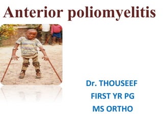
2. Definition
• Poliomyelitis (polio) is a highly infectious viral disease
caused by any of three serotypes of human enteric
poliovirus, which mainly affects young children. The
virus is transmitted through contaminated food and
water, and multiplies in the intestine, from where it can
invade the nervous system.
3. Michael Underwood described poliomyelitis as a debility of the lower
extremities in the second edition of his book Treatise on the Diseases
of Children, 1789.
In 1840, Jacob von Heine described anterior acute poliomyelitis and
the differences with other types of paralysis.
Lesions in the spinal medula were demonstrated in 1870 by Jean-
Martin Charcot & Alex Joffroy.
4. When was it reported?
Early cases
• Poliomyelitis was recorded in
the late 1700’s with the first
epidemic in the late 1800’s.
• The cases that were reported in
1979 where mild and self-
limited and do not result in
paralysis.
5. • The Bavarian neurologist Wilhelm Heinrich Erb
coined the term “anterior acuta poliomielitis” for
clinical adult cases
• In Greek, polios means grey and myelos medula. Of
course, the ending –itis means inflammation of.
6. • Polio= gray matter
• Myelitis= inflammation of the spinal cord
7. Polio An EnterovirusPolio An Enterovirus
• Poliovirus, the causative agent of
poliomyelitis
• A human enterovirus and member of
the family of Picornaviridae.
• Composed of a RNA genome and a
protein capsid. The genome is
single-stranded positive-sense RNA 7
8. Serotypes
• Specificity to receptor restricts mutation rate; slow
genetic drift
• Three serotypes with no cross immunity
– Type 1 polio 90%
Weakest, only 1% causes neuroparalysis
– Type 2 polio 9% (Eliminated)
– Type 3 polio 1%
Greater temperature stability
• Requires trivalent polio vaccine
• Polioviruses can also vary in phenotype of
virulence, host cell lysis, and ability to raise host
defense triggers
9. Polio Infection.
• Incubation 3 – 21 days
• On average 14 days
Predisposing factors.
Severe muscular acitivity can lead to paralysis, as it increases the
blood flow
May produce paralysis in the limb or bulbar region
Injecting vaccines with adjuvant can predispose to paralysis
Patients who underwent tonsillectomy have higher incidence as Ig G
secretion is reduced
Rarely oral Polio vaccine produces poliomyelitis.
10. How is polio transmitted?
• Poliovirus is transmitted through both oral and fecal routes .
• Implantation and replication occurring in either the
orapgaryngeal and or in the intestine of mucosa.
• Most infected for 7-10 days before and after clinical
symptoms begin.
11. Pathogenesis and pathology.
• Enter through Mouth,
• Multiplies in Oropharynx tonsils and Intestines,
• Excreted in Stool.
• Enters the CNS from Blood.
• Spread along the Axons of peripheral nerves to CNS.
• Progress along the fibers of the lower motor neurons spinal
cord or brain.
12. Pathology and Pathogenesis.
• Destroy the Anterior horn cells of the Spinal Cord
• Do not Multiply in Muscles only muscles manifest with
weakness and flaccid paralysis result is secondary.
• Occasionally produce
Myocarditis,
Lymphatic hyperplasia.
13. It cause paralysis?
• Paralytic disease occurs 0.1% to 1% of those who become
infected with the polio virus.
• Paralysis of the respiratory muscles or from cardiac arrest if
the neurons in the medulla oblongata are destroyed.
• Patients have some or full recovery from paralysis usually
apparent with proximally 6 months.
14. Clinical features
• Often child around the age of 9months
• Gives history of mild pyrexia associated with
diarrhoea
• Inability to move a part or whole of the limb.
• Paralysis of varying severity and
assymmetrical
16. Spinal polio
• Spinal polio is the most common
form of paralytic poliomyelitis;
• Results from viral invasion of the
motor neurons of the anterior
horn cells, or the ventral (front)
gray matter section in the spinal
column,
• Virus invasion causes
inflammation of the nerve cells,
leading to damage or destruction
of motor neuron ganglia.
17. Bulbar polio
• Making up about 2% of cases
of paralytic polio,
• Bulbar polio occurs when
poliovirus invades and
destroys nerves within the
bulbar region of the brain
stem.
• Nerves weakens the muscles
supplied by the cranial nerves,
producing symptoms of
encephalitis.
17
18.
19. • Virus mainly localized in anterior horn cells and
certain brain stem motor nuclei
Clinical manifestations:
1. Asymptomatic infection (90-95%)
2. Abortive poliomyelitis
3. Non paralytic polio myelitis
4. Paralytic polio myelitis (1%)
Clinical course
• Three stages - Acute stage
- Convalescent stage
- Chronic stage
20. Acute stage
• 7-10 days
• Superficial reflexes absent
• Deep tendon reflexes disappear when the muscle group is
paralysed
Treatment-
- Bed rest
- Analgesics
- Hot packs
- Anatomical positioning of limbs to prevent flexion
contracture
- Gentle passive ROM exercises
21. Distribution
• Lower limbs 92 %
• Trunk + LL 4 %
• LL + UL 1.33 %
• Bilateral UL 0.67 %
• Trunk + UL + LL 2 %
22. Convalescent stage
• Recovery phase
• Varying degree of spontaneous recovery in
muscle power takes place
• > 80% return of strength - recovered muscles
• < 30% of normal strength - paralysed muscle
23. Treatment:
• Vigorous passive stretching exercises
• Wedging casts for mild –mod contractures
• Surgical release of tight fascia & aponeurosis
• Lengthening of tendons may be necessary for
contractures persisting longer than 6months
• Orthoses used until further no recovery is anticipated
24. Chronic stage
•Usually begins 24 months after the acute illness
•This is the time for orthopaedic intervention
•Most Severely Paralysed Muscle
- Tibialis Anterior
• Most common muscle Paralysed
- Quadriceps femoris
•Most commonly involved muscles in Upper Limb
- Deltoid and Opponens
25. Causes of deformity in Polio
•1. Muscle imbalance
•2. Posture and gravity effect
•3. Dynamics of activity
•4. Dynamics of growth
26. Laboratory Diagnosis.
• Viral isolation from
Throat swabs,
Rectal swabs.
Stool specimens,
• Transported in frozen containers.
• Produce cytopathic effect on
Human and Monkey cells
• Produce cytopathic effects.
26
27. Viral Isolation
• From feces - present in 80% of cases in 1st
week
• In 50 % till 3rd
week
• In 25 % till several weeks
• Collect the fecal sample at the earliest.
• Primary monkey kidney is the ideal cell line for
isolation of virus
• Viral isolation must be interpreted with
caution and clinical presentation
27
29. Goals of treatment
• To achieve maximal functional activity
• Correction of significant muscle imbalances
• Preventing or correcting of limb deformties
• Static joint instability can be controlled by orthoses
• Dynamic joint instability cannot be controlled by
orthoses, that results in fixed deformities
• Soft tissue surgeries such as tendon transfer should be
done before the developement of fixed bony changes
36. What surgeries are done in Polio?
Balancing of power
Stabilization procedures
Correction of deformities
Limb lengthening
37. TENDON TRANSFER
• Tendon transfers are indicated when dynamic
muscle imbalance results in a deformity
• Surgery should be delayed until the maximal
returns of the expected muscle strength has
been achieved
• Objectives of tendon transfer
• To provide active motor power
• To eliminate the deforming effect of a muscle
• To improve stability by improving muscle
balance
38. Criteria and selecting the tendon for
transfer
• Muscle to be transferred must be strong
enough
• Free end of transferred tendon should be
attached as close as possible to the insertion
of paralised tendon
• A transferred tendon should be retained in
its own sheath or should inserted in the
sheath of another tendon or it should be pass
through the subcutaneous fat
39. • Nerve supply and blood supply of transferred
muscle must not be impaired
• Joint must be in satisfactory position
• Contracture must be released before tendon
transfer
• Transferred tendon must be securely attached
to bone under tension slightly greater than
normal
• Agonists muscles are preferable to antagonists
40. • Phasic muscle transfer is preferable to
nonphasic transfer
• A nonphasic muscle should be trained by
extensive physiotherapy before tranfer
• the ideal muscle for tendon transfer wouldthe ideal muscle for tendon transfer would
have the same phasic activity as thehave the same phasic activity as the
paralysed muscle , same size in cross sectionparalysed muscle , same size in cross section
and of equal strength and could be placed inand of equal strength and could be placed in
the proper relationship to the axis of the jointthe proper relationship to the axis of the joint
• Child with dynamic deformity an apropriate
tendon transfer
41. ARTHRODESIS
• Most efficient method for permanent
stabilization of a joint
• When the control of one or more joints
• Bony procedures can be delayed until
skeletal growth is complete
• When the tendon transfer and arthrodesis is
combined in the same operation the
arthrodesis is performed first
42. • Most dependent parts of the body sujected to
significant amount of deforming forces
• M.c deformities includes-
- equinus
- equino varus
- equino valgus
- calcaneous
- cavovarus
- claw toes
- dorsal bunion
PPRP OF FOOT AND ANKLE
44. 1. LIMITED EXTENSOR INVERTOR
INSUFFICIENCY
- tibialis anterior paralysis
- equinus and cavus
- plano valgus
•Transfer of EHL to base od 1st
MT
•If valgus deformity is fixed talonavicular
arthrodesis is combined
45. 2. GROSS EXTENSOR INVERTOR
INSUFFICIENCY
TYPE A
-paralysis of extensors of toes and tibialis anterior
-equinus
-equino valgus
•Transfer of peroneus longus to dorsum of
1st
cunieform bone
•Talonavicular arthrodesis is combined if
deformity is fixed
46. • TYPE B
– paralysis of both tibialis anterior & tibialis
posterior and toe extensors
• Transfer of both peroneals to dorsum of
foot
• Hoke arthrodesis is combined in severe
deformity
47. 3.EVERTOR INSUFFICIENCY
paralysis of peroneal muscles
- varus foot
•Slight-mod impairement:
EHL to base of 5th
MT
•Severe:-tibialis anterior to cuboid
EHL to base of 5th
MT
48. • 4.TRICEPS SURAE INSUFFICIENCY
• Calcaneovarus deformity- tibialis
posterior,FHL
• calcaneovalgus deformity- both peroneals
attached to calcaneum
• calcaneocavus - transfer of
peroneals,tibialis posterior
49. when to operate
1. wait for atleast 1 1/2 years after paralytic attack
2. tendon transfers done in skeletally immature
3. extra articular arthrodesis 3-8 years
4. tendon transfer around ankle and foot after 10yr of age
can be supplimented by arthrodesis to correct the
deformity
4. triple arthrodesis >10-11 years
5. ankle arthrodesis >18 years
50. CLAW TOE
• Hyperextension of MTP and flexion of IP
• Seen when long toe extensors
are used to substitute dorsiflexion of ankle
Treatment:
For lateral toesdivision of extensor tendon by z-plasty
incision,dorsal capsulotomy of MTP
For great toeFHL transferred to prox.phalanx,IP joint
arthrodesis (or)
division of EHL ,proximal slip attached to
neck of 1st
MT,distal slip to soft tissues+ IP arthrodesis
51. Dorsal bunion
• Shaft of 1st
MT is dorsiflexed and graet toe is
plantar flexed
• Seen in muscle imbalance, between anterior
tibial and peroneus longus muscle
52. Lapidus operation
• remove abnormal bone from MT head
• If anterior tibial is overactive- detach its
tendon And transfer it to 2nd
or 3rd
cuneiform
bone
• remove the inferior wedge of bone from 1st
metatarso cuneiform joint
• bring the end of the FHL through the tunnel in
1st
MT and anchor to the capsule over dorsum
of MTP joint
56. • Treatment:
• Young children4-8 yrs:
• Stretching of plantar fascia and posterior ankle structure
with wedging casting
• TA lengthening
• Posterior capsulotomy
• Anterior transfer of tibialis posterior or
• Split transfer of tibialis anterior to insertion of p.brevis (if
tibialis posterior is weak)
• Children >8yrs:
• Triple arthrodesis
• Anterior transfer of tibialis posterior
• Modified jones procedure
57. EQUINO VALGUS DEFORMITY
• Anterior and posterior
muscle weakness with
strong peroneals and
gastroconemius-soleus
muscle
58. • Treatment:
• Skeletally immature:
• Repeated stretching and wedging cast
• TA lengthening
• Anterior transfer of peroneals
• Subtalar arthrodesis and anterior transfer of peroneals
(Grice and green arthrodesis)
• Skeletally mature :
• TA lengthening
• Triple arthrodesis followed by anterior transfer of
peroneals
59. CAVOVARUS DEFORMITY
• Seen due to imbalance of extrinsic muscles or by
unopposed short toe flexors and other intrinsic muscle
•
• Plantar fasciotomy , Release of intrinsic muscles and
resecting motor branch of medial and lateral plantar
nerves before tendon surgery
• Peroneus longus is transferred to the base of the second
MT
• Extensor hallucis longus is transferred
to the neck ofneck of 1st
MT
61. Keeping in slight equinus position during acute
stage of poliomyelitis
•Plantar fasciotomy ,intrinsic muscle release
before tendon transfer
•Depends on residual strength of GS muscle
•Transfer of peroneus brevis and tibialis posterior
to the heel
•Both peroneals trasfered for calcaneo valgus
deformity
•Posterior tibial and FHL can be transfered for
cavovarus deformity
•Anterior tibial tendon can be transferred
posteriorly-DRENNAN TECHNIQUE
62. • For mild deformity –braces used
• Tenodesis of achilles tendon to fibula
• There is progressive equinous deformity with
subsequent growth in pt with achilles
tenodesis
63. Flail foot
• All muscles paralised distal to the knee
• Equinus deformity results because passive
plantar flexion and
• cavoequinus deformity because – intrinsic
muscle may retain some function
• Radical plantar release
• tenodesis
• In older pt mid foot wedge resection may be
required
• ANKLE ARTHRODESIS