Atherosclerosis (2)
•Download as PPSX, PDF•
7 likes•561 views

Clopidogrel, Prasugrel, Ticagrelor

Recommended
More Related Content
What's hot
What's hot (20)
Similar to Atherosclerosis (2)
Similar to Atherosclerosis (2) (20)
More from Shany Thomas
Recently uploaded
Recently uploaded (20)
Atherosclerosis (2)
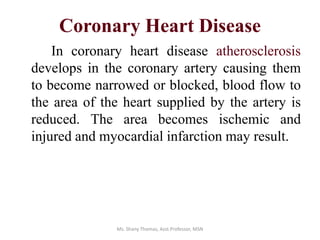
- 1. Coronary Heart Disease In coronary heart disease atherosclerosis develops in the coronary artery causing them to become narrowed or blocked, blood flow to the area of the heart supplied by the artery is reduced. The area becomes ischemic and injured and myocardial infarction may result. Ms. Shany Thomas, Asst.Professor, MSN
- 2. Etiology The primary cause is inflammation and lipid deposition in the wall of the artery
- 3. Risk Factors Non modifiable •Gender •Heredity •Age Modifiable •High blood pressure •High blood cholesterol •Smoking •Physical inactivity •Obesity •Diabetes Contributing factors •Response to stress •Homocysteine levels •Inflammatory responses •Menopause
- 4. PATHOPHYSIOLOGY CAD is a progressive disease. It is initiated by the edothelial injury caused by inflammatory response in the intimal layer and deposition of lipid in to the wall. The process has been shown to occur in 5 phases that include 6 progressive type of lesions.
- 8. Phase I • Present in 30 yrs of age • Clinically silent lesions of type I to type III Type I: Microscopic changes of smooth muscles Type II: Development of fatty streaks Type III: preatheroma- deposition of more lipids
- 9. Phase II • Development of vulnerable lesions Type IV: Atheroma- accumulation of large amount of extracellular lipids forms lipid core Type V : fibrous connective tissue forms a thin cap over the atheroma
- 10. Phase III • Development of complicated lesions Type VI: Fissure in the wall, formation of hematoma and thrombus
- 11. Phase IV • Thrombus blocks significantly Phase V • Plaque calcifies
- 13. Clinical features Signs & Symptoms None Chest Pain Shortness Of Breath Heart Attack • None: This is referred to as silent ischemia. Blood to the heart may be restricted due to CAD, but doesn’t produce symptoms. • Chest pain: If the coronary arteries can’t supply enough blood to meet the oxygen demands of the heart, the result may be chest pain called angina. • Shortness of breath: Some people may not be aware they have CAD until they develop symptoms of congestive heart failure- extreme fatigue with exertion, shortness of breath and swelling in feet and ankles. • Heart attack: Results when an artery to the heart muscle becomes completely blocked.
- 14. Diagnostic studies • Electrocardiography • Echo cardiography • Stress testing • Cardiac catheterization
- 15. ECG Changes
- 16. Exercise stress testing • A stress test monitors the patient's heart rhythms, blood pressure, and clinical status. • It can tell how well the heart handles work and if parts of the heart have decreased blood supply. A typical stress test involves: • The patient walks on a treadmill or rides a stationary bicycle. Exercise continues until the heart is beating at least 85% of its maximum rate, until symptoms of heart trouble occur, or the patient simply wants to stop. • An ECG is used to monitor heart rhythms during a stress test. An echocardiogram or more advanced imaging technique may also be used to visualize the actions of the heart and blood flow.
- 17. Interpretation • Exercise capacity. • ST waves on the ECG • Heart rate • Changes in systolic blood pressure. • Oxygen levels may also be measured
- 20. Cardiac catheterization • Cardiac catheterization is the insertion of a catheter into a chamber or vessel of the heart. This is done both for diagnostic and interventional purposes. • Subsets of this technique are mainly coronary catheterization, involving the catheterization of the coronary arteries, and catheterization of cardiac chambers and valves of the cardiac system.
- 22. Management 1. Reduce the risk factors: 2. Restore blood supply: Angioplasty (PTCA) Coronary atherectomy Stenting Bypass surgery Laser Heart Surgery or Trans myocardial revascularization
- 23. Reducing risk factors •Regular medical checkups. •Control of blood pressure. •Control of blood cholesterol. •Smoking cessation. •Exercise regularly. •Maintain a healthy weight. •Eat a heart-healthy diet. •Manage stress. •Life style modification
- 24. PTCA • A balloon catheter is passed through the guiding catheter to the area near the narrowing. A guide wire inside the balloon catheter is then advanced through the artery until the tip is beyond the narrowing. • The angioplasty catheter is moved over the guide wire until the balloon is within the narrowed segment. • Balloon is inflated, compressing the plaque against the artery wall • once plaque has been compressed and the artery has been sufficiently opened, the balloon catheter will be deflated and removed.
- 27. Stenting • a stent is introduced into a blood vessel on a balloon catheter and advanced into the blocked area of the artery • the balloon is then inflated and causes the stent to expand until it fits the inner wall of the vessel, conforming to contours as needed • the balloon is then deflated and drawn back • The stent stays in place permanently, holding the vessel open and improving the flow of blood.
- 30. Coronary Artery Bypass Graft • Construction of new conduits. GRAFTS: Saphenous vein Internal mammary artery Radial artery Gastroepiploic artery Inferior epigastric artery
- 32. Laser Heart Surgery or Trans myocardial revascularization • High powered laser is used to open up channels in the heart through a relatively small chest incision by punching holes in the beating heart which is believed to promote angiogenesis.
- 34. Medical management • Antiplatelet therapy: Aspirin, Heparin