Live Doppler FLow Velocity System Demo April 14th
•Download as PPTX, PDF•
0 likes•41 views

Enjoy our hands-on demonstration of the Doppler Flow Velocity System, a non-invasive, real-time pulsed Doppler measurement device for studying cardiovascular function in a variety of small animal models, such as mice and rats.

Recommended
Recommended
More Related Content
More from Scintica Instrumentation
More from Scintica Instrumentation (20)
Recently uploaded
Recently uploaded (20)
Live Doppler FLow Velocity System Demo April 14th
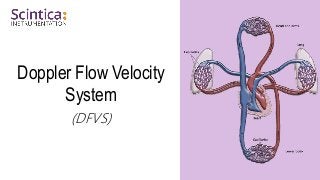
- 2. WWW.SCINTICA.COM Doppler Flow Velocity System • Noninvasively evaluate cardiovascular physiology and function. • The system consists of four components: • Pulsed Doppler Transceiver, • Doppler Signal Digitizer, • Doppler Workstation, • and Handheld Probe. • Surgical warming and real-time integration of ECG signals to the DFVS software from the Rodent Surgical Monitor+. • 2mm diameter handheld probe achieves ~15° angles. Doppler Flow Velocity System 2
- 3. WWW.SCINTICA.COM Pulsed Doppler Ultrasound • Achieving consistency and accuracy with proper measurement angle… • V = (c Δf) / (2fo cos θ) • θ = angle between velocity vector & beam vector Doppler Flow Velocity System 3 Optimal Angle = ~15°
- 4. WWW.SCINTICA.COM Using Pulsed Doppler Ultrasound 4 Advantages Noninvasive - longitudinal studies Short signal acquisition times Can be measured at various locations Signals from 2 sites can be combined Possible to achieve small angles Know-How Knowledge of anatomy Shapes and timing of waveforms Doppler Flow Velocity System
- 5. WWW.SCINTICA.COM Main Applications Systolic & Diastolic Function Confirmation and Stratification of TAC Coronary Flow Reserve Aortic/Arterial Stiffness Peripheral Flow Perfusion Doppler Flow Velocity System 5 Mouse Heart Mouse Aorta
- 6. Globally linking scientists with precision tools for research through expertise in science, engineering and support
Editor's Notes
- (Theta) is the angle between the line of blood flow and the doppler sound beam. This angle is important as anything under 15deg you wont see much difference in error, but above 15deg you’ll see exponential error increase and thus need angle correction, which also inherently has error. SV is sample volume and represents