
Recommended
More Related Content
Similar to retina and role in vision
Similar to retina and role in vision (20)
Recently uploaded
Recently uploaded (20)
retina and role in vision
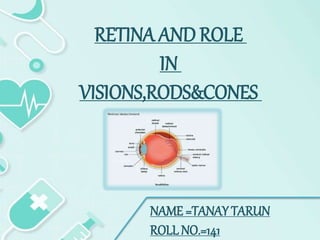
- 1. NAME =TANAY TARUN ROLL NO.=141 RETINA AND ROLE IN VISIONS,RODS&CONES
- 2. RETINA The retina is the innermost layer of the eye. It consists of photoreceptor cells that convert light energy into nerve impulses. These electrical signals are passed via the optic nerve to the visual cortex allowing us to visualise our surroundings.
- 3. RETINA LAYERS The retina consists of layers, which can be subcategorized into retinal pigmented epithelium (RPE) and neural retina. The RPE is a single layer of cuboidal epithelial cells and located in the outermost layer of the retina. It is responsible for the nourishment and support of the neural retina. The tight junctions between the RPE cells form part of the blood-retinal barrier, which helps to prevent molecules passing from the choroid into the retina. The RPE is also involved in a visual cycle as it regenerates photosensitive pigments.
- 4. LAYERS OF NEURAL RETINA The neural retina consists of multiple layers. The three main cells in the neural retina are (from the outermost to innermost): Photoreceptor cell Bipolar cell Retinal ganglion cell
- 5. The photoreceptors are involved in phototransduction, a process of converting light photons to an electrical impulse. The impulse is then relayed by the bipolar cell to the ganglion cell. The axons of the ganglion cells then form the nerve fibre layer of the retina, which exits the eye as an optic nerve. PHOTORECEPTOR CELL Types of Photoreceptor There are three main types of photoreceptors in the human eyes called Rods cones intrinsically photosensitive retinal ganglion cells
- 6. RODES Rods are much more sensitive to light than cones. They can signal the absorption of a single photon! Hence, they are mainly responsible for scotopic vision (in low-light levels). However, as the light levels increase their phototransduction cascades become saturated and are unable to reflect changes in light intensity. In terms of their distribution, rods are found on the outside of the fovea and contribute to peripheral vision. Thus, patients with degenerative changes of rod cells, such as retinitis pigmentosa, may present with a symptom of night-blindness known as nyctalopia and peripheral vision loss.
- 7. CONES In contrast, cones are concentrated in the fovea – the central part of this contains no rods. This is also the part of the retina with the highest acuity of vision. In contrast to rods, cones are much less sensitive to light. Hence, they are solely responsible for vision in the daylight. The other main function of cones is colour vision. It is mediated by three different types of cones, which are sensitive to different ranges of light wavelengths.
- 8. Intrinsically photosensitive retinal ganglion cells:- Intrinsically photosensitive retinal ganglion cells (ipRGCs), also called photosensitive retinal ganglion cells (pRGC), or melanopsin- containing retinal ganglion cells (mRGCs), are a type of neuron in the retina of the mammalian eye.
- 9. THANK YOU...