Recent advances on colo rectal carcinoma1
•
23 likes•2,058 views

On recent advances of colorectal carcinoma, basically focusing on molecular pathogenesis

Recommended
Recommended
More Related Content
What's hot
What's hot (20)
Viewers also liked
Viewers also liked (20)
Similar to Recent advances on colo rectal carcinoma1
Similar to Recent advances on colo rectal carcinoma1 (20)
Recently uploaded
Recently uploaded (20)
Recent advances on colo rectal carcinoma1
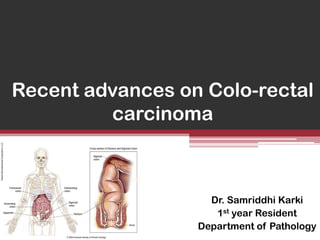
- 1. Recent advances on Colo-rectal carcinoma Dr. Samriddhi Karki 1st year Resident Department of Pathology
- 2. • • • • • • • • • • • Introduction Epidemiology Etiology MOLECULAR PATHOGENESIS Clinical features Morphology Diagnostic modalities Staging and grading Spread and Metastasis Treatment Prognosis
- 3. INTRODUCTION • Third most common type of cancer and second most frequent cause of cancer-related death. • Most curable form of carcinoma of the gastrointestinal tract. • Usually begins as a noncancerous polyp that can, over time, become a cancerous tumor. • Males and females are equally affected. • Mean age : 62 year
- 4. EPIDEMIOLOGY • Worldwide distribution • Highest incidence rates in ▫ ▫ ▫ ▫ ▫ ▫ ▫ United States Canada Australia New Zeaand Denmark Sweden, and Other developed countries
- 5. • Colorectal Carcinoma (CRC) among Nepalese young adults accounts for a high incidence (28%) of all CRC cases. • Although right sided colonic cancer has been increasing, rectum is the commonest site. Asian Pacific Journal of Cancer Prevention, Vol 13, 2012
- 7. 5. Polyps 6. Pelvic Irradiation 7. Genetic factors ▫ Hereditary non-polyposis colorectal cancer (HNPCC) Lynch syndrome ▫ Familial adenomatous polyposis (FAP)
- 9. Hereditary Non-polyposis colorectal cancer ( HNPCC) • Autosomal dominant disorder. • Cancers at several sites ▫ ▫ ▫ ▫ ▫ ▫ ▫ ▫ ▫ Colorectum Endometrium Stomach Ovary Uterus Brain Small bowel Hepatobiliary Skin
- 10. Features of CRC in HNPCC • • • • • • Young age Right –sided location Mucinous features Poor differentiation Lymphocytic infiltration Lack of necrosis
- 13. Familial adenomatous polyposis (FAP) • Autosomal dominant disorder. • Numerous ( 100- 1000) colorectal adenomas. • Mutation of the adenomatous polyposis coli (APC) gene. • In left untreated colorectal carcinoma ( 100%) , before age 30.
- 14. MOLECULAR PATHOGENESIS • TWO distinct genetic pathways . 1. APC / β- catenin pathway ▫ Associated with WNT signaling pathway and the chromosomal instability pathway. 2. Microsatellite instability pathway ▫ Associated with defects in DNA mismatch repair
- 15. APC / β- catenin pathway • APC tumor suppressor gene (5q21) • Downregulate growth promoting signals (β-catenin) • Component of WNT signaling pathway. • Catenins proteins found in complexes with cadherin cell adhesion molecules. • β-catenin participates in the WNT signaling pathway as a growth promoting signals.
- 16. WNT signaling pathway • Network of proteins that passes signals from cell surface receptors to the nucleus through cytoplasm leading to expression of target genes (transcription regulator genes- c MYC) • Major role in controlling cell fate, adhesion, and cell polarity during embryonic development. • WNT signaling is also required for self –renewal of the hematopoetic stem cells.
- 18. Chromosomal instability pathway (CIN) • Increased rate of chromosome missegregation in mitosis . • Due to – Gain / loss of chromosome ( aneuploidy) – Gross chromosomal rearrangements (GSM)
- 19. • Earliest event involved in CIN is APC gene mutation ( 80%) ▫ K-RAS mutation ▫ P53 gene mutation • Late event : DCC (deleted in colonic carcinoma) gene mutation • Advanced event : DPC4/ SMAD4 mutation (18q21)
- 20. CIN forms the basis of Adenoma -Carcinoma Sequence
- 22. MICROSATELLITE INSTABILITY pathway (MSI ) 1. 2. 3. 4. Satellite DNA? Microsatellite DNA? Why is it more liable to be unstable ? How this instability leads to colorectal carcinoma ?
- 23. Satellite DNA • Satellite DNA is composed of tandemly repeating DNA ( non - coding regions) • Tandem repeats occur in DNA when a pattern of two or more nucleotides is repeated. A-T-T-C-G-A-T-T-C-G-A-T-T-C-G
- 24. Type of DNA repeat 1. Satellite No. of nucleotide repeat 5-200 2. Minisatellite a. Hypervariable b. Telomeric 3. Microsatellite a. Monomorpic b. Polymorphic 10-60 6 1-4
- 25. • Microsatellite DNA : If the number of nucleotide repeat is 1-4 ▫ Dinucleotide repeat: When exactly two nucleotides are repeated. Eg: ACACACAC Such regions in DNA are commonly affected in HNPCC.
- 26. • Microsatellites are more prone to get unstable compared to other neutral regions of DNA • This instability is due to any errors , most likely error is ▫ Slippage during DNA replication . • Such error is normally repaired by Mismatch repair enzymes which are encoded by MisMatch repair genes.
- 27. Defect in MMR gene Reduced capacity of cells to repair specific types of DNA damage Increased rate of mutation accumulation in microsatellite DNA
- 28. Mismatch repair genes • MSH2 (2p21) , MSH3, MSH4, MSH5, MSH6 • MLH1(3p21.3), MLH2, MLH3 • PMS1, PMS2
- 29. • As majority of microsatellites are located in the non-coding region, these mutations are generally silent. • Some microsatellites which are present in the coding region of the gene are involved in the regulation of cell growth , like those encoding ▫ Type II TGF-ß receptor ▫ Proapoptotic protein BAX
- 30. Defect in MMR genes • Mutation HNPCC • CpG island Hypermethylation in MMR genes Sporadic CCR
- 31. CpG Island Hypermethylation • • • • • What is CpG? Terms like : CpG site and CpG Island? What is methylation? What is hypermethylation ? How hypermethylation leads to carcinoma?
- 32. • CpG sites: ▫ Regions of DNA where a cytosine occurs next to a guanine. ▫ DNA methylation occurs at these sites by an enzyme called DNA methyltransferases. • In humans, 80 to 90% of all CpGs are methylated. • This methylation results in the conversion of the cytosine to 5-methylcytosine.
- 33. • The remaining 10% nonmethylated CpGs are grouped in a cluster forming CpG island , and is usually located in the promoter regions towards 5’ end. • The unique property of CpG island is that it is unmethylated in the germ line. • Methylation of CpG island within the promoters of genes silencing of tumor suppressor genes Cancer
- 37. MSI testing • MSI can be detected by PCR amplification of microsatellite loci in DNA extracted from CRC specimens . • Newer tests: Nucleic acid flourescence labelling, laser scanning , flourescence PCR amplification . • To identify the risk for hereditary cancer and predict the outcome of CRC. • To detect MLH1 and MSH2 germline mutations .
- 38. The Bethesda Guidelines MSI testing is recommended in people with any of the following features : 1. Cancer in families that meet Amsterdam criteria. 2. Two HNPCC- related cancers 3. A first degree relative with CRC and/or HNPCC- related extracolonic cancer and/or colorectal adenoma diagnosed under 40yr. 4. Right-sided CRC with an undifferentiated pattern on HPE. 5. Signet-ring cell type CRC diagnosed under 45yr. 6. Adenomas diagnosed under 40 yr.
- 39. CLINICAL FEATURES • Asymptomatic for years. • Left sided colonic carcinomas ▫ occult bleeding ▫ changes in bowel habit ▫ crampy left lower quadrant discomfort • Right sided colonic carcinomas ▫ fatigue ▫ weakness ▫ iron deficiency anemia (Anemia in females may arise from gynecologic causes, but it is a clinical maxim that iron deficiency anemia in an older man means gastrointestinal cancer until proved otherwise)
- 40. MORPHOLOGY • 50% rectosigmoid area (involvement of the proximal colon is increasing) • Right-sided tumors more common in the ▫ elderly ▫ blacks ▫ patients with diverticular disease
- 41. GROSS A. PROXIMAL COLON • Polypoid: ▫ Bulky mass, well-defined/ rolled margins and a sharp dividing line with the normal bowel. • Ulcerative: ▫ Less elevated surface and is centrally ulcerated • These tumors rarely cause obstruction
- 43. B. DISTAL COLON • Annular lesions producing “napkin – ring” constrictions and luminal narrowing . • These tumors can cause obstruction.
- 45. MICROSCOPIC Tall columnar carcinomas looks similar. • •All colorectalcells resembling dysplastic epithelium as in adenomas. • Almost all – ADENOCARCINOMAS •secreting variable amounts of mucin. Inflammatory infiltrations (lymphocytes, plasma cells, eosinophils, histiocytes ) are prominent at the edge of the tumor. • Ranges from well-differentiated to undifferentiated, frankly anaplastic •masses. Poorly differentiated tumors might form few GLANDS. • Rarely, the tumor stroma may exhibit metaplastic bone formation
- 46. Well - differentiated Poorly differentiated
- 48. Mucinous adenocarcinoma • 15 % of all CRCs • Common in rectum. • Microscopically : more than 50% extracellular mucin • High association with MSI • Worse prognosis.
- 49. Signet ring adenocarcinoma • Rare • Microscopically : more than 50% intracellular mucin • About one third cases are associated with MSI • Worst prognosis.
- 50. Medullary carcinoma Rare. Common in proximal colon . Occurs in elderly. Microscopic : Sheets of malignant cells with vesicular nuclei , prominent nucleoli, abundant pink cytoplasm. • Invariably associated with MSI . • Favourable prognosis . • • • •
- 51. Serrated adenocarcinoma • 7.5% of all CRCs. • Common in proximal colon. • Derived from serrated adenoma. • Microscopic: ▫ serrated, mucinous or trabecular pattern of growth ▫ abundant eosinophilic cytoplasm ▫ chromatin condensation ▫ preserved polarity, and ▫ no necrosis.
- 52. Squamous differentiation • Common in proximal colon. • Usually associated with glandular elements (adenosquamous carcinoma) • Occasionally , seen in a pure form (squamous cell carcinoma). • Evidence for human papilloma virus 16 involvement in the pathogenesis of some rectal cases .
- 53. Trophoblastic differentiation • Can occur focally in CRCs. • hCG can be demonstrated immunohistochemically in such tumor cells. • Occasionally the entire tumor has the appearance of a choriocarcinoma. • This phenomenon should be distinguished from conventional adenocarcinomas( where hCG positivity is more common )
- 54. Immunohistochemical features • Conventional adenocarcinoma of large bowel express are MUC1 and MUC3. • Mucinous carcinoma express MUC2. • CRCs invariably positive for cytokeratin (CK) positivity for CK20 and negativity for CK7 • Positive for CEA. • Positive for CDX2, in majority of CRCs. • Tumor-associated glycoprotein (TAG-72) is present in 100 % of invasive colorectal carcinoma. • CRCs , especially poorly differentiated show loss of blood group isoantigens and of HLA A, B, and C expression.
- 55. Other markers • • • • • • • • • • Villin Cathepsin B Neurolipin-1 SRCA2 Cadherin-17 Calretinin Human chorionic gonadotropin (hCG) Placental alkaline phosphatase (PLAP) ~10% Estrogen and progesterone receptors Racemase
- 58. Biopsy • There is a need of POSITIVE BIOPSY before radical surgery for CRC. • In large lesions, several biopsies should be taken form diverse areas. • Biopsy from center only granulation tissue • Biopsy from the very periphery only hyperplastic colonic epithelium
- 59. Cytology • Its an accurate way of diagnosing CRC. • Little practical value. • Low-lying rectal lesions can be easily sampled. • Brush cytology can also be performed via the fiberoptic scope. • It is a sensitive technique, perhaps even more so than endoscopic biopsy, but it has not yet found widespread acceptance.
- 60. Various screening modalities • Colonoscopy • Virtual colonography • Sigmoidoscopy • Fecal occult blood test • Double contrast barium enema • Digital rectal examination
- 61. Staging and Grading • In 1937, Dukes proposed staging for rectal carcinoma . • In 1954, Astler and Coller proposed different staging system. • American Joint committee on Cancer(AJCC) • The Union Internationale Countre Le Cancer (UJCC)
- 62. Dukes’ Stage A • The tumor involve the wall of the bowel only. • Treatment is surgery to remove the tumor and some surrounding lymph nodes
- 63. Dukes’ Stage B • The cancer extend through the wall has not spread to the lymph nodes. • Colon cancer is treated with surgery and, in some cases, chemotherapy after surgery. • Rectal cancer is treated with surgery, radiation therapy, and chemotherapy
- 64. Dukes’ Stage C • The cancer has spread to the regional lymph nodes (lymph nodes near the colon and rectum) ▫ C1: regional L.N ▫ C2: mesenteric B.V. ligature • Colon cancersurgery and chemotherapy • Rectalcancersurgery, radiation therapy, and chemotherapy
- 65. Dukes’ Stage D • Spread outside of the colon or rectum to other areas of the body • Treatment : chemotherapy. • Surgery to remove the colon or rectal tumor may or may not be done • Additional surgery to remove metastases may also be done in carefully selected patients
- 66. Astler and Coller Staging System • Stage A ▫ Limited to mucosa • Stage B1 ▫ Involving the muscularis externa but not penetrating it • Stage B2 ▫ Penetrating through the muscularis externa • Stage C1 ▫ Confined to the bowel wall but with nodal metastasis • Stage C2 ▫ Penetrating through the wall and with nodal metastsis
- 67. • TNM Staging of Colon Cancer • • • • • • • Tumor (T) T0 = none evident Tis = in situ (limited to mucosa) T1 = invasion of lamina propria or submucosa T2 = invasion of muscularis propria T3 = invasion through muscularis propria into subserosa or nonperitonealized perimuscular tissue T4 = invasion of other organs or structures • • • • • Lymph Nodes (N) 0 = none evident 1 = 1 to 3 positive pericolic nodes 2 = 4 or more positive pericolic nodes 3 = any positive node along a named blood vessel • • • Distant Metastases (M) 0 = none evident 1 = any distant metastasis • • • • • • • • • 5-Year Survival Rates T1 = 97% T2 = 90% T3 = 78% T4 = 63% Any T; N1; M0 = 66% Any T; N2; M0 = 37% Any T; N3; M0 = data not available Any M1 = 4%
- 68. Microscopically, colorectal carcinoma can be graded into ▫ I – well differentiated ▫ II- moderately differentiated ▫ III- poorly differentiated
- 69. Spread and Metastasis •Common sites ▫Regional lymph nodes ▫Liver
- 70. Lymph node metastasis • More common in the tumors showing ▫ poorly differentiated areas ▫ highly infiltrative pattern of growth. • Minimum number of nodes recovered from a surgical specimen of colorectal carcinoma should be 14 or 15.
- 71. Liver metastases • More common in the tumors showing evidence of blood vessel invasion.
- 72. • Other relatively common metastatic sites include ▫ peritoneum ▫ lung ▫ ovaries.
- 74. PROGNOSIS • The 5-year survival rate after curative resection for CRC ranges between 40% and 60% . • Local recurrence and/or regional lymph node metastases occur in over 90% of the failure cases. • Over two-thirds of the recurrences are evident within the first 2 years and 91% by 5 years. • The prognosis of colorectal carcinoma is related to a number of clinical and pathologic parameters.
- 75. • Category I ▫ Well supported by the literature, generally used in patient management and of sufficient importance to modify TNM stage groups. • Category IIA ▫ Extensively studied biologically and/or clinically. Prognostic value for therapy, sufficient to be noted in pathology report • Category IIB ▫ Well studied but not sufficiently established for Category I or IIA • Category III ▫ Not yet established to meet criteria for Category I or II • Category IV ▫ Studied and shows no consistent prognostic significance
- 76. Age P Perforation P Sex M-P Category I CEA seum levels P Obstruction P Prognosis Tumor edge NP-P Category III Tumor location +/- Tumor size +/Local extent P Tumor multiplicity (S)
- 77. III Presence of neuroendocrine cells +/- Tumor margins/ inflm rxn G Category IIA Tumor budding P Acinar morphology P Vascular invasion P PROGNOSIS Microscopic tumor type (Mu/S/A -P, Me -G) Category IIB Tumor thickness +/- Pericolonic tumor deposits P Surgical Margins (R - P) Perineurial invasion P Category IIA
- 80. THANK YOU
Editor's Notes
- Colorectal cancer is the third most common cancer in both men and women. Colorectal cancer incidence rates have been decreasing for most of the past two decades, which has largely been attributed to increases in the use of colorectal cancer screening tests that allow the detection and removal of colorectal polyps before they progress to cancer. From 2004 to 2008, annual declines in white men were much larger than those in African American men, 2.9% versus 0.8%, respectively; whereas, among women, declines among whites (2.2% per year) and African Americans (1.7% per year) were similar. In contrast to the overall declines, colorectal cancer incidence rates have been increasing by 1.7% per year since 1992 among adults younger than 50 years of age, for whom screening is not recommended for those at average risk.
- Diet: the exact mechanism is not well established . However, it has been theorized that reduced fiber content decreased stool bulk and altered composition of the intestinal microbiota increase in the synthesis of toxic oxidative metabolites by bacterial metabolism ( which will remain in contact with colonic mucosa for a longer time as a result of reduced stool bulk )High fat intake increase hepatic synthesis of cholesterol and bile acids , which can be converted into carcinogens by intestinal bacteria
- The most imp neoplastic polyps that are precursore to the colonic adenoca are the colonic adenomas. These can be small , pedunculated to large ,sessile. 50% of the adults living in the western world develop colonic adenomas by age 50.Size of the adenomas is the most imp factor that corresponds to the risk of malignancy.Polyps (precancerous growth associated with aging)Irradiationusually for carcinoma of cervix
- Rare forms of CRC syndromes:Torre Muir syndrome : Multiple CRC + multiple sebaceous tumor + keratoacanthomas
- Involves a series of molecular alterations.Both pathways involve stepwise accumulation of multiple mutations, but the genes involved and the mechanisms by which the mutations accumulate differs.
- The Wnt signaling pathway is a network of proteins that passes signals from receptors on the surface of the cell through the cytoplasm and ultimately to the cell's nucleus where the signaling cascade leads to the expression of target genes. It controls cell-cell communication in the embryo and adult Through these signaling pathways, Wnt proteins play a variety of important roles in embryonic development, cell differentiation, and cell polarity generation.
- K-RAS mutation , usually in larger polyps
- Clear cell ca Accumulation of glycogen results in a clear appearance of the cytoplasm.Micropapillary 20% ;Greater frequency of lymphovascular invasion and lymph node metastases;Poor prognosisBasaloid Similar to its counterpart in the anal canal.Rare in colorectum . Oncocytic Occurs after preoperative chemoradiation or developing from a villous adenoma with oncocytic changes.Glassy cell carcinomasimilar to its counterpart in the uterine cervixAnaplastic Similar to that of its counterpart in many other organs and its behavior is very aggressiveHepatoid Similar to that of its gastric counterpart.
- CDX2 is a caudal-type homeobox gene which encodes a transcription factor that plays an important role in the proliferation and differentiation of intestinal epithelial cells. It is found by immunohistochemistry in the overwhelming majority of colorectal carcinomas. it can also be expressed in primary mucin-producing carcinomas of ovary, bladder, and lung, as well as pancreaticobiliary adenocarcinomas.
- Villina cytoskeletal protein associated with the axial microfilament bundles of brush border microvilliCathepsinBalysosomal cysteine proteinaseNeuropilin a molecule normally present in the developing nervous systemSRCA 2an ATPase crucial to many cell functionsCadherinalso known as liver-intestine cadherinCalretinincan be expressed by a minority of colorectal adenocarcinomas (especially the undifferentiated oneshuman chorionic gonadotropin (hCG) common in mucinous and poorly differentiated tumors ( high reactivity) Placental alkaline phosphatase (PLAP) ~ 10% of all colorectal carcinomas Estrogen and progesterone receptors are usually absent or are present in a small minority of tumor cells Racemase, a marker for prostatic adenocarcinoma, is expressed in over half of large bowel adenocarcinomas, a potential source of misdiagnosis
- EM: Presence of prominent collections of microfilaments running perpendicular to the cell membrane and entering the brush border.This feature, although helpful, is not diagnostic. It can also be found in intestinal-type carcinomas of the stomach, small bowel, gallbladder, and pancreas
- the technique employed to obtain the specimen – which involves extensive cleansing of the colon followed by a diagnostic enema with manipulation of the patient – has led to an unenthusiastic response from clinicians.