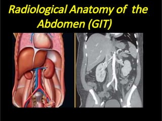
4. Imaging Modali es for the Abdomen and Pelvis.
•
•
•
•
•
•
•
•
•
•
Commonly u lized:
Ultrasound
CT (computed tomography)
Radiography
Abdominal plain film
Fluoroscopy
– Hysterosalpingography
Other modali es:
MRI
– Magne c resonance imaging
Nuclear medicine
– Gallium scan
Positron Emission Tomography (PET).
5. X - RAY --- FOUR BASIC DENSITIES
Air.
So ssue.
Fat.
Bone.
6. Fluoroscopy
•
–
–
–
–
–
•
•
•
Dynamic radiography
Permits real- me evalua on
of the gastrointes nal tract
Barium Swallow (esophagus)
Upper GI Series (stomach)
Small Bowel Follow-through
Barium Enema (colon)
Barium (& air) is introduced
by enema or swallowing
Barium appears white on the
images (high density
a enuates the x-ray beam)
Can assess both intrinsic
(mucosal) and some extrinsic
(mass-effect) abnormali es.
7. Nuclear Medicine - GI Bleeding Scan
•
•
•
•
Evaluates bleeding, par cularly from the lower GI tract.
Radiopharmaceu cal = Tc99m in vitro labelled RBCs.
Sequen al 5 minute images acquired over an hour.
Looking for progressive accumula on of tracer.
Bleeding on the cecum.
8. Reading the Abdominal Plain Film.
•Also known as the
•
–
–
–
•
•
“KUB” (kidney, ureter, &
bladder).
Use a systema c approach
to Interpreta on.
Lung bases & diaphragms.
Bones.
So ssues.
Abnormal calcifica ons.
Organs.
Stomach
11. What’s Up on an Abdominal Film?
•
•
Always check the lung bases for an infiltrate.
Look for free air on the upright film: commonly beneath
the right hemidiaphragm.
Free air under right hemidiaphragm
due to perforated duodenal ulcer
Diaphragm
Liver edge