
Recommended
Recommended
More Related Content
Similar to Bacteriophages come in different sizes and shapes but most of them.docx
Similar to Bacteriophages come in different sizes and shapes but most of them.docx (20)
More from rock73
More from rock73 (20)
Recently uploaded
Recently uploaded (20)
Bacteriophages come in different sizes and shapes but most of them.docx
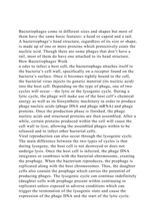
- 1. Bacteriophages come in different sizes and shapes but most of them have the same basic features: a head or capsid and a tail. A bacteriophage’s head structure, regardless of its size or shape, is made up of one or more proteins which protectively coats the nucleic acid. Though there are some phages that don’t have a tail, most of them do have one attached to its head structure. How Bacteriophages Work n oder to infect a host cell, the bacteriophage attaches itself to the bacteria’s cell wall, specifically on a receptor found on the bacteria’s surface. Once it becomes tightly bound to the cell, the bacterial virus injects its genetic material (its nucleic acid) into the host cell. Depending on the type of phage, one of two cycles will occur – the lytic or the lysogenic cycle. During a lytic cycle, the phage will make use of the host cell’s chemical energy as well as its biosynthetic machinery in order to produce phage nucleic acids (phage DNA and phage mRNA) and phage proteins. Once the production phase is finished, the phage nucleic acids and structural proteins are then assembled. After a while, certain proteins produced within the cell will cause the cell wall to lyse, allowing the assembled phages within to be released and to infect other bacterial cells. Viral reproduction can also occur through the lysogenic cycle. The main difference between the two types of cycles is that during lysogeny, the host cell is not destroyed or does not undergo lysis. Once the host cell is infected, the phage DNA integrates or combines with the bacterial chromosome, creating the prophage. When the bacterium reproduces, the prophage is replicated along with the host chromosomes. Thus, the daughter cells also contain the prophage which carries the potential of producing phages. The lysogenic cycle can continue indefinitely (daughter cells with prophage present within continuing to replicate) unless exposed to adverse conditions which can trigger the termination of the lysogenic state and cause the expression of the phage DNA and the start of the lytic cycle.
- 2. These adverse conditions include exposure to UV or mutagenic chemicals and desiccation. http://phages.org/bacteriophage/ Patients in hospitals, especially those on breathing machines, those with devices such as catheters, and patients with wounds from surgery or from burns are potentially at risk for serious, life-threatening infections. n hospitals, where the most serious infections occur, Pseudomonas can be spread on the hands of healthcare workers or by equipment that gets contaminated and is not properly cleaned. https://www.cdc.gov/hai/organisms/pseudomonas.html P. aeruginosa can develop resistance to antibacterials either through the acquisition of resistance genes on mobile genetic elements (i.e., plasmids) or through mutational processes that alter the expression and/or function of chromosomally encoded mechanisms. Both strategies for developing drug resistance can severely limit the therapeutic options for treatment of serious infections. https://www.ncbi.nlm.nih.gov/pmc/articles/PMC2772362/ Multidrug-resistant Pseudomonas can be deadly for patients in critical care. An estimated 51,000 healthcare-associated P. aeruginosa infections occur in the United States each year. More than 6,000 (13%) of these are multidrug-resistant, with roughly 400 deaths per year attributed to these infections. Multidrug-resistant Pseudomonas was given a threat level of serious threat in the CDC AR Threat report. https://www.cdc.gov/hai/organisms/pseudomonas.html Antibacterial Resistance Trends Presented in Table Table11 are rates of P. aeruginosa resistance
- 3. to several antipseudomonal drugs (54, 95, 99, 100, 178, 211, 212). This summary is not meant to be inclusive of all of the published literature, but rather highlights data reported for isolates from several U.S. surveillance studies since January 2000. If multiple years were included in a study, the resistance rates for the most recent year are presented in Table Table11. Rates of antibacterial resistance among P. aeruginosa isolates from hospitals and ICUs https://www.ncbi.nlm.nih.gov/pmc/articles/PMC2772362/ Each set must be modified twice a year to retain its ability to lyse a large proportion of the target species [5], [11]. P. aeruginosa bacteriophages are numerous, and current knowledge of their diversity shows that they are distributed in at least 7 genera of purely lytic phages (T7-like, ΦKMV-like, LUZ24- like, N4-like, PB1-like, ΦKZ-like, JG004-like) in addition to a similar number of temperate genera [12], [13]. Within each genus, phages with a variety of different host spectra are observed, in part at least reflecting differences in their tail- associated adhesins http://journals.plos.org/plosone/article?id=10.1371/journal.pone .0060575 In- vitro and In- vivo Phage Trials Running head: BACTERIOPHAGE THERAPY 1 BACTERIOPHAGE THERAPY 2
- 4. BACTERIOPHAGE THERAPY ON Pseudomonas aeruginosa NAME: Roaya Alhawsawi LIU 11/13/2016 BACTERIOPHAGE THERAPY ON Pseudomonas aeruginosa Introduction Bacteriophages are viruses that infect bacteria and replicate within the bacterial cell wall. Most have double-stranded DNA genomes found in heads with icosahedral symmetry, and their tails vary in length. All bacteriophages are classified in the order Caudovirales and belong to the families Myoviridae (long, contractile tail), Siphoviridae (long, non-contractile tail)and Podoviridae (short, non-contractile tail) (Harper & Enright, 2011). Bacteriophages were first discovered by Fredrick Twort and Felix D’Herelle in 1915. Since then, bacteriophages started being widely used for treating bacterial infections. This, however, did not last long as chemical antibiotics were discovered and preferred because bacteriophages were not well understood and their efficacy was controversial. Things have now changed due to the development of antibiotic-resistant bacterial strains and bacteriophages have started being used
- 5. again. Comment by Daniel Ginsburg: You need more background on phage. Life cycle. Lytic vs. lysogenic, etc. Pseudomonas aeruginosa is a multidrug-resistant Gram-negative bacteria that causes infections in the lungs of cystic fibrosis patients and is a regular cause of hospital-acquired bacterial pneumonia and ventilator-associated pneumonia. Since bacteriophages are capable of reaching bacteria protected within biofilms (such as those found in the lungs of patients with cystic fibrosis), they are being considered as an alternative treatment to antibiotics. Comment by Daniel Ginsburg: You need more about P. aeruginosa. How many people get it every year? It is lethal? Who is infected? You should get some statistics from the CDC or WHO. I would also start with this and then talk about phage. You should also talk about the developent of drug resistance. What are they resistant to? Types of P.aeruginosa bacteriophages Many P.aeruginosa bacteriophages have been discovered, and they are classified into at least seven genera of lytic phages. These include T7-like, ɸKMV-like, LUZ24- like, N4- like, PB1-like, ɸKZ-like and JG004-like including a similar number of temperate genera.(Essoh et al., 2013). Therapeutic bacteriophage cocktails such as “pyophage” have also been formulated. “Pyophage” contains many different phages that target streptococcus, staphylococcus, Escherichia coli, proteus and P. aeruginosa. Comment by Daniel Ginsburg: How are these different from each other? In- vitro and In- vivo Phage Trials Many in-vitro and in-vivo phage trials have been conducted on animal models and human patients. In in-vitro trials, the potential of phages against P. aeruginosa strains in planktonic cultures or biofilms isolates has been evaluated. Some of the trials that have been done include: (1.) Fu et al. study of the effect of lytic phages in the prevention of P. aeruginosa biofilm formation in hydrogel-coated catheters, (2.) Pires et al. study
- 6. of biofilm control using a broad- host- range phage for P. aeruginosa, (3.) Torres-Barceló et al. report on treatment using a combination of Podoviridae phage LUZ7 and streptomycin against P. aeruginosa PAO1 (Pires, Vilas Boas, Sillankorva, & Azeredo, 2015). Comment by Daniel Ginsburg: Don’t just list these. You need to talk a little bit about each of them. What phage was used? What kind of system was it done in? How were the phage delivered? How well did they work? Some of the in- vivo trials that have been conducted on human patients include: (1.) Wright et al. study of the the efficacy and safety of a therapeutic phage preparation (Biophage-PA), (2.) Sivera Marza et al. report on the successful topical use of phage to treat a burn patient who had been colonized by P. aeruginosa months after skin grafts had been applied and (3.) Merabishvili et al. description of a quality-controlled small-scale production of a phage cocktail (BFC-1) for use in human clinical practice(Pires et al., 2015). Discussion Many bacteriophages have been isolated and their effects against P. aeruginosa documented. A cocktail of ɸMR299-2 and ɸNH-4 was effective in eliminating P. aeruginosa NH57388A (mucoid) and P. aeruginosa MR299 (non- mucoid) strains when growing as a biofilm on a cystic fibrosis bronchial epithelial CFBE41o- cell line (Alemayehu et al., 2012). PAK-P or P3- CHA reduced mortality and lung damage in mice with lethal pneumonia caused by MDR P. aeruginosa (Rolain, Hraiech, & Bregeon, 2015). Cocktail from “pyophage” showed lytic activity against 70% of P. aeruginosa strains cultured in growth medium, while PAK-P1 reduced mortality and lung inflation in mice with lethal pneumonia caused by PAK bioluminescent P. aeruginosa strain. Phage LUZ7 used with streptomycin inhibited growth of the P. aeruginosa PAOI strain, while Engineered T7 phage with aiiA gene inhibited biofilm formation in the P. aeruginosa PAOI strain. Phage PB-1 and tobramycin reduced resistance to tobramycin in the P. aeruginosa PAOI strain (Rolain et al., 2015). Phage P2-10Ab01 isolated from sewage
- 7. water in Abidjan could lyse two pyophage-resistant strains, C7- 6 and C9-5, and PAP3 was capable of lysogenization (Essoh et al., 2013). Comment by Daniel Ginsburg: How effective? Comment by Daniel Ginsburg: How were the phage delivered? Comment by Daniel Ginsburg: What is this? Comment by Daniel Ginsburg: You need a little more detail about each of these. How did they work? How was the experiment done? Quorum Sensing Inhibition Since hydrolyzing acyl homoserine lactonases can decrease in vivo virulence of P. aeruginosa and in vitro biofilm production, bacteriophages can be modified genetically to produce lactonase, which would facilitate inhibition of P.aeruginosa biofilm production (Rolain et al., 2015). … Comment by Daniel Ginsburg: How do these work? I don’t understand what this has to do with quoroum sensing. Mechanism of Bacteriophage Resistance Even though bacteriophage therapy has made headway in the treatment of bacterial infections, the issue of bacteria that are resistant to phages has come up. The mechanisms of bacterial resistance to phages drive the evolution of both bacteria and bacteriophages, and ongoing isolation of new bacteriophages targeting various hosts and host receptors is, therefore, necessary. Bacterial resistance to phages may involve three mechanisms: inhibition of the adsorption of phages on the bacteria and injection of DNA, use of restriction enzymes to degrade phage DNA and the CRISPR- Cas system that gives bacteria immunity against the phages (Essoh et al., 2013). The CRISPR-Cas mediates interference against certain types of temperate phages. However, some phages found in the Mu- like genus have been found to carry genes that can inactivate the system. Challenges Facing Use of Bactereophages The potential disadvantages of bacteriophages can be categorized into four: phage selection, phage-host- range
- 8. limitation uniqueness of phages as pharmaceuticals and unfamiliarity with phages (Loc-Carrillo & Abedon, 2011). Not all phages are good for therapeutics since some of them may cause the development of immunogenic reactions due to large uncontrolled amounts of phages in circulation (Paul et al., 2011). However, use of bacteriophages devoid of endotoxins should not induce a strong stimulation of the pro-inflammatory markers (Morello et al., 2011). Conclusion In this therapy, isolation of bacteriophage using the strain from the patient is preferred over using readymade phages (Henry, Lavigne, & Debarbieux, 2013). Moreover, bioluminescent bacteria can be used to compare several bacteriophages, so as to establish candidates for therapeutics based on their real in vivo efficacy instead of they are in vitro performance (Debarbieux et al., 2010). With a combination of proper selection of phages, proper formulation and improved clinical understanding of how phages work, bacteriophage could easily become the most effective way for treating bacterial infections. Comment by Daniel Ginsburg: As I see it, everything up to this point is Body. This would be your Discussion and it needs to be expanded. Can you talk more about isolating phage from patients, using bioluminescent bacteria, etc.? References Alemayehu, D., Casey, P. G., McAuliffe, O., Guinane, C. M., Martin, J. G., Shanahan, F., … Hill, C. (2012). Bacteriophages φMR299-2 and φNH-4 can eliminate Pseudomonas aeruginosa in the murine lung and on cystic fibrosis lung airway cells. mBio, 3(2), e00029-12. http://doi.org/10.1128/mBio.00029-12 Debarbieux, L., Leduc, D., Maura, D., Morello, E., Criscuolo, A., Grossi, O., … Touqui, L. (2010). Bacteriophages can treat and prevent Pseudomonas aeruginosa lung infections. The Journal of Infectious Diseases, 201(7), 1096–104. http://doi.org/10.1086/651135 Essoh, C., Blouin, Y., Loukou, G., Cablanmian, A., Lathro, S., Kutter, E., … Qimron, U. (2013). The Susceptibility of
- 9. Pseudomonas aeruginosa Strains from Cystic Fibrosis Patients to Bacteriophages. PLoS ONE, 8(4), e60575. http://doi.org/10.1371/journal.pone.0060575 Harper, D. R., & Enright, M. C. (2011). Bacteriophages for the treatment of Pseudomonas aeruginosa infections. Journal of Applied Microbiology, 111(1), 1–7. http://doi.org/10.1111/j.1365-2672.2011.05003.x Henry, M., Lavigne, R., & Debarbieux, L. (2013). Predicting in vivo efficacy of therapeutic bacteriophages used to treat pulmonary infections. Antimicrobial Agents and Chemotherapy, 57(12), 5961–5968. http://doi.org/10.1128/AAC.01596-13 Loc-Carrillo, C., & Abedon, S. T. (2011). Pros and cons of phage therapy. Bacteriophage, 1(2), 111–114. http://doi.org/10.4161/bact.1.2.14590 Morello, E., Saussereau, E., Maura, D., Huerre, M., Touqui, L., & Debarbieux, L. (2011). Pulmonary bacteriophage therapy on Pseudomonas aeruginosa cystic fibrosis strains: first steps towards treatment and prevention. PloS One, 6(2), e16963. http://doi.org/10.1371/journal.pone.0016963 Paul, V., Sundarrajan, S., Rajagopalan, S., Hariharan, S., Kempashanaiah, N., Padmanabhan, S., … Watson, J. (2011). Lysis-deficient phages as novel therapeutic agents for controlling bacterial infection. BMC Microbiology, 11(1), 195. http://doi.org/10.1186/1471-2180-11-195 Pires, D. P., Vilas Boas, D., Sillankorva, S., & Azeredo, J. (2015). Phage Therapy: a Step Forward in the Treatment of Pseudomonas aeruginosa Infections. Journal of Virology, 89(15), 7449–56. http://doi.org/10.1128/JVI.00385-15 Rolain, J.-M., Hraiech, S., & Bregeon, F. (2015). Bacteriophage-based therapy in cystic fibrosis-associated Pseudomonas aeruginosa infections: rationale and current status. Drug Design, Development and Therapy, Volume 9, 3653. http://doi.org/10.2147/DDDT.S53123