
Recommended
More Related Content
What's hot
What's hot (20)
Similar to SURGICAL ANATOMY OF THE INGUINAL CANAL
Similar to SURGICAL ANATOMY OF THE INGUINAL CANAL (20)
More from rks sivasankar
Recently uploaded
Recently uploaded (20)
SURGICAL ANATOMY OF THE INGUINAL CANAL
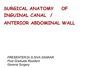
- 1. SURGICAL ANATOMY OF INGUINAL CANAL / ANTERIOR ABDOMINAL WALL PRESENTER:Dr.S.SIVA SANKAR Post Graduate Resident General Surgery
- 2. Anterior Abdominal wall ● extends from the thoracic cage to the pelvis bounded superiorly by the cartilages of the 7th to 10th ribs and the xiphoid process of sternum (level of T9) ● inferiorly by the inguinal ligament and the superior margins of the anterolateral aspects of the pelvic girdle (iliac crests ,pubic crests,and pubic symphsis )
- 3. L5 L1
- 4. Transpyloric plane passes horizontally midway between the xiphoid process and the umblicus at the level of L1 Transtubercular plane passes horizontally through the tubercle of the iliac crest ,about 5cm behind the asis ,at the level of L5 Two vertical midclavicular line passes through the midinguinal point.
- 5. Layers of Abdominal wall ● skin ● superficial fascia ● External oblique muscle ● Internal oblique muscle ● Transversus abdominis muscle ● Transversalis fascia ● preperitoneal adipose tissue ● peritoneum
- 6. Superficial fascia ● In two layers ● fascia Camper - superficial fatty layer. ● fascia Scarpa - deep membranous layer, superficial fascia
- 8. Superficial fatty layer (fascia camper) It is continuous with the superficial fat over the rest of the body.
- 9. Deep membranous layer(fascia scarpa) ● deeper ,dense layer of fibrous connective tissuue fuses with the fascia lata of the thigh,medial part of inguinal ligament,and pubic tubercle in the fold of each groin ● fascia contains cutaneous nerves ,vessels and superficial lymphatics
- 11. Muscles of Anterolateral Abdominal wall ● external oblique ● internal oblique ● transverse abdominis ● rectus abdominis ● pyramidalis
- 13. External oblique • Origin: external surfaces and inferior borders of 5-12 ribs • Insertion: iliac crest, linea alba, f • Direction: inferiomedial direction • Nerve supply: T7 – L1
- 14. External oblique mucle ● originate from the lower 7 ribs and course in inferomedial direction ● at the mid clavicular line give rise to flat ,strong aponeurosis that passes anteriorly to the rectus sheath to insert medially into the linea alba. ● the most posterior of the fibres run vertically dounward to insert into the anterioor half of the iliac crest ● the lower portion of the aponeurosis is rolled posteriorly and superiorly to form a groove extends from the anterior superior iliac spine to pubic tubercle and is termed the Inguinal ligament. ● The shelving edge of the inguinal ligament is used in various repairs of inguinal hernia.
- 16. Internal oblique • Origin: thoracolumbar fascia, iliac crest, lat 2/3 of inguinal ligament • Insertion: pectan pubis (conjoint tendon), linea alba, 10-12 ribs • Direction: superomedial • Nerve supply: T7 – L1
- 17. Internal oblique muscle ● originates from the iliopsoas fascia ,from the anterior 2/3rd of the iliac crest and lumbo dorsal fascia ● course in a direction opposite of the external oblique muscle-superomedial ● uppermost fibres insert into the lower five ribs and their cartilage ● central fibres form an aponeurosis runs medially which envelops the rectus abdominis muscle as part of rectus sheath ● some of the lower muscle fascicles accompany the spermatic cord into the scrotum as the cremasteric muscle
- 19. tranverse abdominis muscle ● arises from the lower 6 costal cartilage ,spines of the lumbar vertebra,iliac crest and ilopsoas fascia beneath the lateral ⅓ rd of the inuinal ligament ● course transversely to give rise to flat aponeurotic sheet that passes posterior to the rectus abdominis above the semicircular line (of douglas) and anterior to the muscle below it. ● inferior most fibres pass infero medially along with the lower fibres of the internal oblique and form aponeurotic arch of the transverse abdominis muscle.
- 21. transversalis fascia ● covers the deep surface of the transversus abdominis muscle and ,with its various extensions , forms a complete fascial envelope across the abdominal cavity ● binds together the muscle and aponeurotic fascicles into a continuoous layer and reinforces weak areas. ● responsible for the structural integrity of the abdominal wall ● By definition ,a hernia results from a defect in the transversalis fascia.
- 22. Rectus abdominis • Origin: costal cartilage 5-7, xiphoid process • Insertion:Pubic crest ,pubic symphysis • Direction: vertical • Nerve supply: T7- T11 • Tendinous intersections
- 23. Rectus sheath
- 25. linea alba ● the rectus abdominis muscles are held closely in apposition near the anterior midline by the linea alba . ● consists of a band of dense,crisscrossedfibres of the aponeurosis of the abdominal muscles extends from the xiphoid to the pubic symphysis. ● wider above the umblicus than below thus facilitating the placement of surgical incisions in the midline without entering the right or left rectus sheath
- 26. Pyramidalis • Inconsistent muscle, within rectus sheath • Origin: pubic symphysis and pubic crest • Insertion: linea alba • Nerve supply: T12
- 27. PREPERITONEAL ADIPOSE TISSUE lies between the fascia transversalis and the parietal peritoneum and contain adipose and areolar tissue. PARIETAL PERITONEUM ● innermost layer of the abdominal wall.. ● consists of a thin layer of dense ,irregular connective tissue covered on its inner surfae by a single layer of squamous mesothelium.
- 29. BLOOD SUPPLY superior and the inferior epigastric arteries. The skin near the midline is supplied by branches of the The skin of the flanks is supplied by branches of the ● Deep circumflex iliac arteries ● intercostal arteries ● lumbar arteries
- 30. ● The superior epigastric artery ,one of the terminal branches of the internal thoracic artery enters the upper part of the rectus sheath supplies the upper central part of the anterior abdominal wall and anastomoses with the inferior epigastric artery. ● The inferior epigastric artery ,branch of the external iliac artery just above the inguinal ligament supppling the lower central part of the anterior abdominal wall and anastomoses with the superior epigastric artery
- 31. Below the umblicus the skin and superficial fascia are supplied by three small branches from femoral artery • Superficial epigastric artery • Superficial external pudendal artery • Superficial circumflex iliac artery
- 32. VENOUS DRAINAGE SUPERFICIAL VEINS The superficial veins form a network that radiates out from the umbilicus. Above the umblicus , drain into the axillary vein via the lateral thoracic vein. Below the umblicus ,drain into the femoral vein via the superficial epigastric and great saphenous veins.
- 33. DEEP VEINS The deep veins of the abdominal wall, the superior epigastric, inferior epigastric, and deep circumflex iliac veins, follow the arteries of the same name and drain into the internal thoracic and external iliac veins.
- 34. lymphatics above the umblicus, drain into the axillary lymph nodes. below the umblicus, drain into the superficial inguinal nodes The deep lymphatics follow the arteries and drain into the internal thoracic ,external iliac ,posterior mediastinal and para-aortic nodes superficial lymphatics
- 36. • Nerve supply • Cutaneous nerves • Lower 5 intercostal nerves • subcostal nerve • 1st lumbar nerve • 1st lumbar nerve is represented by Iliohypogastric and Ilioinguinal nerves. • They supply the skin of the anterior abdominal wall,the muscles and the parietal peritoneum • Lower six thoracic nerves pierce the posterior Wall of the rectus sheath and supply the rectus muscle and pyramidalis • Dermatome • Xyphoid process T7 • Pubic symphysis L1
- 37. Nerve supply
- 38. skin Incisions
- 39. INGUINAL CANAL ● it is an intermuscular passage of about 4cm in length in lower part of abdominal wall located parallel with and immediately superior to the medial half of the inguinal ligament. ● extends between the deep inguinal ring and superficial inguinal ring.
- 41. superficial inguinal ring ● it is an ovoid opening of the external oblique aponeurosis that is positioned superiorly and slightly lateral to the pubic tubercle. ● bounded by superomedial and inferolateral crus joined by the crisscross intercrural fibres
- 42. Deep inguinal ring ● it is an U shaped condensation of the transversalis fascia. ● lies 1.25 cm above the inguinal ligament ,midway between the symphysis pubis and the anterior superior iliac spine ● the inferior epiastric vessels lie posteriorly and medial to the deep inguinal ring
- 43. Inguinal ligament ● Inguinal ligament is the inferior edge of the ext. oblique aponeurosis extends from the anterior superior iliac spine to the pubic tubercle and turning posteriorly to form the shelving edge thus forming the inferior wall of the inguinal canal
- 45. Boundaries of Inguinal canal ● anteriorly external oblique aponeurosis. ● superiorly arched fibres of the internal oblique and transverse abdominis musculoaponeurosis. ● inferiorly grooved upper surface of the inguinal ligament and medial most by lacunar ligament
- 46. ● posteriorly transversalis fascia and conjoined tendon medially lower parallel fibres of the aponeurosis of transverse abdominis and internal oblique fuse to form the conjoint tendon and gets attached to the pubic crest and pecten pubis thus forms strong posterior wall for the medial part of the canal .
- 49. structures passing through the inguinal canal ● The spermatic cord in males and the round ligament of the uterus in females ,enters the inguinal canal through the deep inguinal ring and passes out through the superficial inguinal ring. ● The ilioinguinal nerve enters the canal through the interval between the external and internal oblique muscles runs anterior to the spermatic cord in the inguinal canal and branches at the superficial inguinal ring.
- 50. coverings of spermatic cord ● internal spermatic fascia derived from the transversalis fascia when it enters the deep inguinal ring cremasteric muscle and fascia arise from the lowermost fibres of the internal oblique muscle and encompasses the spermatic cord and attach to the tunica vaginalis of the testis . ● external spermatic fascia while passing out from the superficial inguinal ring spermatic cord receives its outer coverin from external oblique aponeursis.
- 52. contents of spermatic cord ● vas deferens ● artery to vas deferens , ● testicular artery ● pampiniform plexus of veins ● cremasteric vessels ● genital branch of genitofemoral nerve ● remains f processus vaginalis ● lymphatics ● sympathetic plexus around the artery to vas deferens
- 54. hernia ● an abnormal protrusion of an organ or tissue through a defect in its surrounding wall. • • Indirect inguinal hernia • Direct inguinal hernia
- 55. ● it is the site for direct hernias. The triangle has the following borders: 1) Medial border of rectus abdominus(medially) 2) Inguinal ligament (inferiorly) 3) Inferior epigastric vessels(laterally) HESSELBACH’S TRIANGLE