SlideShare a Scribd company logo
1 of 10
Download to read offline
MRI of LEFT KNEE
Protocol:
Multiplanar MRI of the left knee joint performed in the sagittal, coronal and
transverse planes using T1 weighted spin echo, T2 and proton-density weighted fast
spin echo, fatsaturated & T2* weighted gradient echo sequences.
Observations:
There is complete tear of the patellar tendon from its attachment at the inferior pole
of patella with a large defect between the torn ends and resultant high riding patella
i.e. superior dislocation of patella.
There is a fluid collection seen anterior to and inferior to the superiorly displaced
patella.
The visualised part of Quadriceps tendon appears normal.
Patella and its cartilage are normal.
There is thinning of the articular cartilage in the medial compartment of the
femorotibial joint space.
Few small patchy subarticular PD fatsat hyperintense areas are seen in the
subarticular marrow of the medial femoral and tibial condyle.
Rest of the articular surfaces and subarticular marrow are normal.
There is mild extrusion of the body and anterior horn of the medial meniscus with
mild deformation of their shape and reduction of size.
Grade III Hyperintense signal (oblique tear) reaching inferior articular surface is seen
in the posterior horn of medial meniscus.
Grade II hyperintense signal (horizontal tear) is seen in the body of medial meniscus.
Grade I hyperintense signal is seen through entire substance of lateral meniscus.
Hyperintensity is seen on PDFS sequence along the Medial collateral ligament,
however no disruption of its fibres.
The cruciate and lateral collateral ligaments are uniform in the outline and do not
display any focal signal abnormality.
Mild knee joint effusion.
Conclusion:
 Complete tear of the patellar tendon from its attachment at the
inferior pole of patella with a large defect between the torn ends
and resultant superior dislocation of patella (patella alta).
 Fluid collection inferior to and anterior to the superiorly displaced
patella.
 Changes of osteoarthritis involving the medial compartment of
the knee (femorotibial) joint.
 Mild extrusion of the body and anterior horn of the medial
meniscus.
 Oblique tear reaching inferior articular surface in the posterior
horn of the medial meniscus.
 Horizontal tear in body of medial meniscus.
 Grade I hyperintense signal in the entire substance of lateral
meniscus.
 Grade I sprain of the MCL.
Dr. Rahul Hegde
MBBS, MD, FRCR, USMLE
Feb 21,2015 12:47
MRI OF LUMBAR SPINE
Clinical Profile: Low back pain
Prior: None
Technique: Multiplanar multiecho MRI of the lumbosacral spine was performed.
Findings:
Loss of normal lumbar lordosis is seen with mild degenerative changes.
The lumbar vertebral bodies demonstrate normal height, alignment and marrow
signal. Posterior elements are intact with normal ligamentum flavum and facets.
L1/2 disc - The disc reveals no significant bulge or herniation. Neural foramina
and exiting nerve roots appear normal.
L2/3 disc - The disc reveals no significant bulge or herniation. Neural foramina
and exiting nerve roots appear normal.
L3/4 disc – The disc reveals no significant bulge or herniation. Neural foramina
and exiting nerve roots appear normal.
L4/5 disc – The disc reveals no significant bulge or herniation. Neural foramina
and exiting nerve roots appear normal.
L5/S1 disc – There is mild loss of disc height and hydration. Mild diffuse
intervertebral disc bulge is seen with mild mass effect over the thecal sac.
Neural foraminae are mildly compromised.
The lower thoracic cord and conus appear normal.
The pre and paravertebral soft tissue appear normal.
Impression:
1. Mild degenerative changes at lumbar spine.
2. Disc bulge at L5/S1 level causing mild mass effect over the thecal sac.
Dr. Kedar Athawale
Feb 06,2015 08:51
DMRD, DNB
Consultant Radiologist
MRI of PELVIS
Technique: Multiplanar, multiecho MRI of pelvis was performed.
Findings:
Marrow signal abnormality is seen in both femoral heads with serpiginous areas of low
signal on T1 and heterogeneously hyperintense signal on T2/STIR. No collapse or
flattening of the femoral heads is seen. Minimal bilateral hip effusion is seen.
The other visualized bones demonstrate normal marrow signal intensity. The iliac crests
appear normal.
The muscles, tendons and ligaments around the hip joint demonstrate normal signal
intensity. No evidence of muscle tear, strain or contusion.
The visualized pelvic viscera are unremarkable.
Impression:
Findings are suggestive of avascular necrosis of bilateral femoral heads (stage 2) as
described.
No other significant abnormality detected.
Please correlate with patient’s clinical findings and laboratory studies.
Dr. Preshit Javadekar
Feb 12,2015 00:45
MBBS, DMRE(Bom), DNB
Consultant Radiologist
MRI and MRA OF BRAIN
Technique: Multiplanar and multiecho MRI of brain was performed.
Findings:
Multiple small hyperintensities are noted involving periventricular white matter in T2W &
Flair images. These do not show diffusion restriction.
Hyperintensity is noted involving central pons on T2W & Flair images. It does not show
diffusion restriction.
Rest of both cerebral parenchyma show normal gray-white matter differentiation with normal
sulci, gyri and basal cisterns.
Centrally located gray matter nuclei appear normal in size, shape and intensity.
Diffusion imaging does not reveal evidence of acute infarct.
Ventricular system appears normal in size & shape. No evidence of periventricular oozing.
Midline structures like interhemispheric fissure, 3rd ventricle, pineal region and rest of brain
stem appear normal.
Posterior fossa structures appear normal.
Mild tortuosity of left optic nerve is noted. Right optic nerve appears normal in the given
images.
MR Brain Angiography:
The internal carotid arteries show normal course and caliber and are symmetrically disposed.
Mild irregularity is noted involving intracranial part of left ICA. No significant luminal
narrowing is noted.
Both middle cerebral arteries arise normally from the internal carotid on either side and forms
normal insular loops. Mild irregularity is noted involving M1 part of left MCA. No
significant luminal narrowing is noted.
The anterior cerebral artery shows no signs of narrowing or displacement. The basilar artery
shows a normal course and caliber and divides into normal size posterior cerebral arteries.
The anterior and posterior communicating arteries appear unremarkable.
IMPRESSION:
• Chronic white matter ischemia.
• Hyperintensity involving central pons on T2W & Flair images. It does not show diffusion
restriction.
• Mild tortuosity of left optic nerve.
• Mild irregularity involving intracranial part of left ICA and M1 part of left MCA. No
significant luminal narrowing.
Dr. Arti Iyer. Patil
Dec 29,2014 11:33
MBBS, MD
MRI OF HIP JOINTS (PLAIN AND CONTRAST)
Clinical information: Known case of Sickle cell anemia. Previously AVN of Right
Hip. Presently, pain in Left Hip.
Protocol: Multiplanar MRI of the both hip joints was performed using T1weighted
spin echo, T2 weighted fast spin, echo, T1 and PD Fatsaturated sequences. Post
contrast multiplanar T1 weighted sequence obtained after injection of gadolinium.
Observations:
LEFT HIP:
The left femoral head is spherical in shape with no articular collapse.
The left femoral head, neck and posterior part of the left acetabulum show diffuse
hyperintense signal on T2 and T2 fatsaturated sequences which is isointense on T1
sequence. On post contrast sequence, a large part of the femoral head shows no
enhancement with thick peripheral enhancement around the non-enhancing area.
There is thick synovial enhancement with minimal joint effusion.
There is periarticular hyperintense soft tissue on T2 and T2 fatsaturated sequences
seen around the left hip joint involving the left obturator externus, pectinues muscles
and the origin of the left adductor muscles which shows post contrast enhancement
with few non-enhancing irregular necrotic areas within.
Multifocal areas of marrow hyperintensity on T2 and T2 fatsaturated sequences is
seen in both upper femoral shafts, intertrochanteric regions, right iliac blade, right
ischial tuberosity which show heterogeneous post contrast enhancement with
peripheral contrast enhancement in upper femoral shafts.
RIGHT HIP:
The right femoral head is small in size with collapse of articular surface on the
superolateral aspect. Small subchondral defects seen in femoral head and the in the
acetabulum which show no post contrast enhancement.
No synovial thickening or effusion seen.
No periarticular abnormal soft tissue seen around the right hip joint.
Conclusion:
 Diffuse signal abnormality in the left femoral head and neck as described
above with synovial thickening and enhancement, minimal left hip joint
effusion and abnormal periarticular inflammatory soft tissue involving
the left obturator externus, pectinues muscles and the origin of the left
adductor muscles. These findings are most likely suggestive of left hip
septic arthritis with periarticular infective/ inflammatory soft tissue. Less
likely possibility of AVN with superadded infection.
 Multifocal areas of signal abnormality in both upper femoral shafts,
intertrochanteric regions, right iliac blade, right ischial tuberosity as
described above is most likely suggestive of multifocal osteomyelitis.
 Findings in Right hip joint suggestive of Stage IV AVN.
Correlate with clinical findings, lab investigations and other imaging investigations.
Dr. Rahul Hegde
MBBS, MD, FRCR, USMLE
Feb 21,2015 12:47
CONTRAST ENHANCED MRI OF THE ABDOMEN
Clinical information: Known case of prostatic carcinoma. APR and Colostomy
done. Evidence of sarcoma on biopsy of the colostomy site.
Prior Imaging: None
Protocol: Multiplanar imaging of the abdomen was performed using T1 weighted
spin echo, T2 weighted fast spin echo, gradient echo, fatsaturated sequences. Post
contrast T1 weighted fat saturated sequences obtained after injection of gadolinium.
Observations:
The sigmoid colostomy is seen extending though the inferior part of the left rectus
muscle in the left lower qudrant of the abdomen.
The stoma opening is edematous and shows moderate enhancing thickening.
Rest of the sigmoid colon and visualised bowel loops are unremarkable.
Mild T2 hyperintensity is seen at in the subcutaenous fat and in the adbominal wall at
the stoma site which show mild post contrast enhancement.
Few small bowel loops beneath the stoma site also show mild wall enhancement
which is most likely related to post op inflammation.
No significant lymphadenopathy.
No free fluid in the abdomen.
Liver is normal in size, signal intensity, shows normal contrast enhancement with no
focal lesions.
Gallbladder is distended with normal wall thickness. CBD is normal.
Pancreas is normal in size, intensity and enhancement.
Spleen is normal in size, intensity and shows normal contrast enhancement.
Both adrenals are normal.
Both kidneys are normal in size, intensity and show normal density nephrogram.
A small T2 hyperintense and T1 hypointense simple cortical cyst seen in the upper
part of the left kidney.
A small 1.2 cm sized T1 hypointense, T2 hyperintense lesion is seen in posterior part
of right iliac bone adjacent to the right SI joint.
Rest of the visualized bones show normal marrow signal intensity.
Conclusion:
 Enhancing thickening at the stoma opening of the sigmoidostomy, however
no obvious mass noted in the abdominal wall or intra-abdominally. Correlate
with Histopathology reports.
 Mild edema and granulation tissue in the subcutaneous fat and abdominal
wall around the stoma.
 A small lesion in posterior part of right iliac bone could be suggestive of bone
metastasis in this known case of prostatic carcinoma. This needs correlation
with radiographs/ bone scan.
Dr. Rahul Hegde
MBBS, MD, FRCR, USMLE
Feb 21,2015 12:47

More Related Content

What's hot

Presentation1.pptx radiological imaging of mediastinal masses .
Presentation1.pptx radiological imaging of mediastinal masses .Presentation1.pptx radiological imaging of mediastinal masses .
Presentation1.pptx radiological imaging of mediastinal masses .Abdellah Nazeer
 
Presentation1.pptx. ultrasound examination of the ankle joint.
Presentation1.pptx. ultrasound examination of the ankle joint.Presentation1.pptx. ultrasound examination of the ankle joint.
Presentation1.pptx. ultrasound examination of the ankle joint.Abdellah Nazeer
 
Liver segments on ultrasound
Liver segments on ultrasoundLiver segments on ultrasound
Liver segments on ultrasoundDurre Sabih
 
Radiology Spotters mixed collection ppt
Radiology Spotters mixed collection pptRadiology Spotters mixed collection ppt
Radiology Spotters mixed collection pptDr pradeep Kumar
 
Imaging neurology spotters
Imaging   neurology spottersImaging   neurology spotters
Imaging neurology spottersNeurologyKota
 
Radiological Anatomy of pharynx and esophagus abdul final
Radiological Anatomy of pharynx and esophagus abdul finalRadiological Anatomy of pharynx and esophagus abdul final
Radiological Anatomy of pharynx and esophagus abdul finalabduljelil nejmu
 
Ascites and Pleural Effusion
 Ascites and Pleural Effusion Ascites and Pleural Effusion
Ascites and Pleural EffusionMedia Genie
 
Venous Doppler upper limb
Venous Doppler upper limb Venous Doppler upper limb
Venous Doppler upper limb Milan Silwal
 
Presentation1.pptx, radiological imaging of lower limb ischemia.
Presentation1.pptx, radiological imaging of lower limb ischemia.Presentation1.pptx, radiological imaging of lower limb ischemia.
Presentation1.pptx, radiological imaging of lower limb ischemia.Abdellah Nazeer
 
Normal Anatomy of Buccal mucosa and cancer
 Normal Anatomy of  Buccal mucosa and cancer Normal Anatomy of  Buccal mucosa and cancer
Normal Anatomy of Buccal mucosa and cancerKanhu Charan
 
Doppler ultrasound of carotid arteries
Doppler ultrasound of carotid arteriesDoppler ultrasound of carotid arteries
Doppler ultrasound of carotid arteriesSamir Haffar
 
Dr ahmed esawy imaging of adeniod enlargment
Dr ahmed esawy imaging of adeniod enlargmentDr ahmed esawy imaging of adeniod enlargment
Dr ahmed esawy imaging of adeniod enlargmentAHMED ESAWY
 
Presentation1.pptx, ultrasound examination of the knee joint.
Presentation1.pptx, ultrasound examination of the knee joint.Presentation1.pptx, ultrasound examination of the knee joint.
Presentation1.pptx, ultrasound examination of the knee joint.Abdellah Nazeer
 
Doppler ultrasound of the kidneys
Doppler ultrasound of the kidneysDoppler ultrasound of the kidneys
Doppler ultrasound of the kidneysSamir Haffar
 
Presentation1.pptx, radiological anatomy of the knee joint.
Presentation1.pptx, radiological anatomy of the knee joint.Presentation1.pptx, radiological anatomy of the knee joint.
Presentation1.pptx, radiological anatomy of the knee joint.Abdellah Nazeer
 
Presentation1.pptx, ct normal anatomy of the abdomen and pelvis.
Presentation1.pptx, ct normal anatomy of the abdomen and pelvis.Presentation1.pptx, ct normal anatomy of the abdomen and pelvis.
Presentation1.pptx, ct normal anatomy of the abdomen and pelvis.Abdellah Nazeer
 

What's hot (20)

Presentation1.pptx radiological imaging of mediastinal masses .
Presentation1.pptx radiological imaging of mediastinal masses .Presentation1.pptx radiological imaging of mediastinal masses .
Presentation1.pptx radiological imaging of mediastinal masses .
 
Shoulder ultrasound
Shoulder ultrasoundShoulder ultrasound
Shoulder ultrasound
 
MRI of Shoulder anatomy
MRI of Shoulder anatomyMRI of Shoulder anatomy
MRI of Shoulder anatomy
 
Presentation1.pptx. ultrasound examination of the ankle joint.
Presentation1.pptx. ultrasound examination of the ankle joint.Presentation1.pptx. ultrasound examination of the ankle joint.
Presentation1.pptx. ultrasound examination of the ankle joint.
 
Radiology Spotters
Radiology Spotters Radiology Spotters
Radiology Spotters
 
Liver segments on ultrasound
Liver segments on ultrasoundLiver segments on ultrasound
Liver segments on ultrasound
 
Radiology Spotters mixed collection ppt
Radiology Spotters mixed collection pptRadiology Spotters mixed collection ppt
Radiology Spotters mixed collection ppt
 
Imaging neurology spotters
Imaging   neurology spottersImaging   neurology spotters
Imaging neurology spotters
 
Radiological Anatomy of pharynx and esophagus abdul final
Radiological Anatomy of pharynx and esophagus abdul finalRadiological Anatomy of pharynx and esophagus abdul final
Radiological Anatomy of pharynx and esophagus abdul final
 
Ascites and Pleural Effusion
 Ascites and Pleural Effusion Ascites and Pleural Effusion
Ascites and Pleural Effusion
 
Venous Doppler upper limb
Venous Doppler upper limb Venous Doppler upper limb
Venous Doppler upper limb
 
Presentation1.pptx, radiological imaging of lower limb ischemia.
Presentation1.pptx, radiological imaging of lower limb ischemia.Presentation1.pptx, radiological imaging of lower limb ischemia.
Presentation1.pptx, radiological imaging of lower limb ischemia.
 
Normal Anatomy of Buccal mucosa and cancer
 Normal Anatomy of  Buccal mucosa and cancer Normal Anatomy of  Buccal mucosa and cancer
Normal Anatomy of Buccal mucosa and cancer
 
Renal doppler
Renal dopplerRenal doppler
Renal doppler
 
Doppler ultrasound of carotid arteries
Doppler ultrasound of carotid arteriesDoppler ultrasound of carotid arteries
Doppler ultrasound of carotid arteries
 
Dr ahmed esawy imaging of adeniod enlargment
Dr ahmed esawy imaging of adeniod enlargmentDr ahmed esawy imaging of adeniod enlargment
Dr ahmed esawy imaging of adeniod enlargment
 
Presentation1.pptx, ultrasound examination of the knee joint.
Presentation1.pptx, ultrasound examination of the knee joint.Presentation1.pptx, ultrasound examination of the knee joint.
Presentation1.pptx, ultrasound examination of the knee joint.
 
Doppler ultrasound of the kidneys
Doppler ultrasound of the kidneysDoppler ultrasound of the kidneys
Doppler ultrasound of the kidneys
 
Presentation1.pptx, radiological anatomy of the knee joint.
Presentation1.pptx, radiological anatomy of the knee joint.Presentation1.pptx, radiological anatomy of the knee joint.
Presentation1.pptx, radiological anatomy of the knee joint.
 
Presentation1.pptx, ct normal anatomy of the abdomen and pelvis.
Presentation1.pptx, ct normal anatomy of the abdomen and pelvis.Presentation1.pptx, ct normal anatomy of the abdomen and pelvis.
Presentation1.pptx, ct normal anatomy of the abdomen and pelvis.
 

Similar to Statim healthcare MRI Sample Reports

What are the CPT and ICD10 codes Exam CT scan of lumba.pdf
What are the CPT and ICD10 codes     Exam CT scan of lumba.pdfWhat are the CPT and ICD10 codes     Exam CT scan of lumba.pdf
What are the CPT and ICD10 codes Exam CT scan of lumba.pdfzubermsm1
 
Lubar Arthritis : Lumbar Stenosis by Pablo Pazmino, MD
Lubar Arthritis : Lumbar Stenosis by Pablo Pazmino, MDLubar Arthritis : Lumbar Stenosis by Pablo Pazmino, MD
Lubar Arthritis : Lumbar Stenosis by Pablo Pazmino, MDPablo Pazmino
 
Spinal tuberculosis
Spinal tuberculosisSpinal tuberculosis
Spinal tuberculosisAlwan Hussen
 
Presentation1.pptx, ultrasound of the hand and fingers.
Presentation1.pptx, ultrasound of the hand and fingers.Presentation1.pptx, ultrasound of the hand and fingers.
Presentation1.pptx, ultrasound of the hand and fingers.Abdellah Nazeer
 
What to do if the spine x-ray shows a ---? – Part 2
What to do if the spine x-ray shows a ---? – Part 2What to do if the spine x-ray shows a ---? – Part 2
What to do if the spine x-ray shows a ---? – Part 2SpinePlus
 
Presentation1, radiological film reading of the hip joint.
Presentation1, radiological film reading of the hip joint.Presentation1, radiological film reading of the hip joint.
Presentation1, radiological film reading of the hip joint.Abdellah Nazeer
 
Presentation1.pptx, ultrasound examination of the elbow joint.
Presentation1.pptx, ultrasound examination of the elbow joint.Presentation1.pptx, ultrasound examination of the elbow joint.
Presentation1.pptx, ultrasound examination of the elbow joint.Abdellah Nazeer
 
Presentation1.pptx mri of elbow joint
Presentation1.pptx mri of elbow jointPresentation1.pptx mri of elbow joint
Presentation1.pptx mri of elbow jointAbdellah Nazeer
 
Syringomyelia
SyringomyeliaSyringomyelia
SyringomyeliaSubbu Raj
 
MR Imaging of shoulder and knee joints
MR Imaging of shoulder and knee jointsMR Imaging of shoulder and knee joints
MR Imaging of shoulder and knee jointsSahil Chaudhry
 
Spinal dysraphism
Spinal dysraphismSpinal dysraphism
Spinal dysraphismairwave12
 
Presentation1, MD MCQ Cases..pptx
Presentation1, MD MCQ Cases..pptxPresentation1, MD MCQ Cases..pptx
Presentation1, MD MCQ Cases..pptxAbdellah Nazeer
 
Imaging of neurocutaneous syndrome overview
Imaging of neurocutaneous syndrome overviewImaging of neurocutaneous syndrome overview
Imaging of neurocutaneous syndrome overviewcharusmita chaudhary
 
Presentation1, mri quiz cases.
Presentation1, mri quiz cases.Presentation1, mri quiz cases.
Presentation1, mri quiz cases.Abdellah Nazeer
 
Presentation1 Short cases MD..pptx
Presentation1 Short cases MD..pptxPresentation1 Short cases MD..pptx
Presentation1 Short cases MD..pptxAbdellah Nazeer
 

Similar to Statim healthcare MRI Sample Reports (20)

What are the CPT and ICD10 codes Exam CT scan of lumba.pdf
What are the CPT and ICD10 codes     Exam CT scan of lumba.pdfWhat are the CPT and ICD10 codes     Exam CT scan of lumba.pdf
What are the CPT and ICD10 codes Exam CT scan of lumba.pdf
 
Lubar Arthritis : Lumbar Stenosis by Pablo Pazmino, MD
Lubar Arthritis : Lumbar Stenosis by Pablo Pazmino, MDLubar Arthritis : Lumbar Stenosis by Pablo Pazmino, MD
Lubar Arthritis : Lumbar Stenosis by Pablo Pazmino, MD
 
Spinal tuberculosis
Spinal tuberculosisSpinal tuberculosis
Spinal tuberculosis
 
Presentation1.pptx, ultrasound of the hand and fingers.
Presentation1.pptx, ultrasound of the hand and fingers.Presentation1.pptx, ultrasound of the hand and fingers.
Presentation1.pptx, ultrasound of the hand and fingers.
 
Spots with keys
Spots with keysSpots with keys
Spots with keys
 
What to do if the spine x-ray shows a ---? – Part 2
What to do if the spine x-ray shows a ---? – Part 2What to do if the spine x-ray shows a ---? – Part 2
What to do if the spine x-ray shows a ---? – Part 2
 
Presentation1, radiological film reading of the hip joint.
Presentation1, radiological film reading of the hip joint.Presentation1, radiological film reading of the hip joint.
Presentation1, radiological film reading of the hip joint.
 
Presentation1.pptx, ultrasound examination of the elbow joint.
Presentation1.pptx, ultrasound examination of the elbow joint.Presentation1.pptx, ultrasound examination of the elbow joint.
Presentation1.pptx, ultrasound examination of the elbow joint.
 
Presentation1.pptx mri of elbow joint
Presentation1.pptx mri of elbow jointPresentation1.pptx mri of elbow joint
Presentation1.pptx mri of elbow joint
 
Syringomyelia
SyringomyeliaSyringomyelia
Syringomyelia
 
MR Imaging of shoulder and knee joints
MR Imaging of shoulder and knee jointsMR Imaging of shoulder and knee joints
MR Imaging of shoulder and knee joints
 
Mri of knee
Mri of kneeMri of knee
Mri of knee
 
LUMBER CANAL STENOSIS ppt (5)
LUMBER CANAL STENOSIS ppt (5)LUMBER CANAL STENOSIS ppt (5)
LUMBER CANAL STENOSIS ppt (5)
 
Spinal dysraphism
Spinal dysraphismSpinal dysraphism
Spinal dysraphism
 
Presentation1, MD MCQ Cases..pptx
Presentation1, MD MCQ Cases..pptxPresentation1, MD MCQ Cases..pptx
Presentation1, MD MCQ Cases..pptx
 
Presentation1, MD MCQ Cases..pptx
Presentation1, MD MCQ Cases..pptxPresentation1, MD MCQ Cases..pptx
Presentation1, MD MCQ Cases..pptx
 
Osteochondroma
OsteochondromaOsteochondroma
Osteochondroma
 
Imaging of neurocutaneous syndrome overview
Imaging of neurocutaneous syndrome overviewImaging of neurocutaneous syndrome overview
Imaging of neurocutaneous syndrome overview
 
Presentation1, mri quiz cases.
Presentation1, mri quiz cases.Presentation1, mri quiz cases.
Presentation1, mri quiz cases.
 
Presentation1 Short cases MD..pptx
Presentation1 Short cases MD..pptxPresentation1 Short cases MD..pptx
Presentation1 Short cases MD..pptx
 

Recently uploaded

Hot Call Girl In Ludhiana 👅🥵 9053'900678 Call Girls Service In Ludhiana
Hot  Call Girl In Ludhiana 👅🥵 9053'900678 Call Girls Service In LudhianaHot  Call Girl In Ludhiana 👅🥵 9053'900678 Call Girls Service In Ludhiana
Hot Call Girl In Ludhiana 👅🥵 9053'900678 Call Girls Service In LudhianaRussian Call Girls in Ludhiana
 
VIP Call Girls Sector 67 Gurgaon Just Call Me 9711199012
VIP Call Girls Sector 67 Gurgaon Just Call Me 9711199012VIP Call Girls Sector 67 Gurgaon Just Call Me 9711199012
VIP Call Girls Sector 67 Gurgaon Just Call Me 9711199012Call Girls Service Gurgaon
 
💚😋Kolkata Escort Service Call Girls, ₹5000 To 25K With AC💚😋
💚😋Kolkata Escort Service Call Girls, ₹5000 To 25K With AC💚😋💚😋Kolkata Escort Service Call Girls, ₹5000 To 25K With AC💚😋
💚😋Kolkata Escort Service Call Girls, ₹5000 To 25K With AC💚😋Sheetaleventcompany
 
No Advance 9053900678 Chandigarh Call Girls , Indian Call Girls For Full Ni...
No Advance 9053900678 Chandigarh  Call Girls , Indian Call Girls  For Full Ni...No Advance 9053900678 Chandigarh  Call Girls , Indian Call Girls  For Full Ni...
No Advance 9053900678 Chandigarh Call Girls , Indian Call Girls For Full Ni...Vip call girls In Chandigarh
 
indian Call Girl Panchkula ❤️🍑 9907093804 Low Rate Call Girls Ludhiana Tulsi
indian Call Girl Panchkula ❤️🍑 9907093804 Low Rate Call Girls Ludhiana Tulsiindian Call Girl Panchkula ❤️🍑 9907093804 Low Rate Call Girls Ludhiana Tulsi
indian Call Girl Panchkula ❤️🍑 9907093804 Low Rate Call Girls Ludhiana TulsiHigh Profile Call Girls Chandigarh Aarushi
 
Bangalore call girl 👯‍♀️@ Simran Independent Call Girls in Bangalore GIUXUZ...
Bangalore call girl  👯‍♀️@ Simran Independent Call Girls in Bangalore  GIUXUZ...Bangalore call girl  👯‍♀️@ Simran Independent Call Girls in Bangalore  GIUXUZ...
Bangalore call girl 👯‍♀️@ Simran Independent Call Girls in Bangalore GIUXUZ...Gfnyt
 
VIP Call Girl Sector 88 Gurgaon Delhi Just Call Me 9899900591
VIP Call Girl Sector 88 Gurgaon Delhi Just Call Me 9899900591VIP Call Girl Sector 88 Gurgaon Delhi Just Call Me 9899900591
VIP Call Girl Sector 88 Gurgaon Delhi Just Call Me 9899900591adityaroy0215
 
Dehradun Call Girls Service ❤️🍑 8854095900 👄🫦Independent Escort Service Dehradun
Dehradun Call Girls Service ❤️🍑 8854095900 👄🫦Independent Escort Service DehradunDehradun Call Girls Service ❤️🍑 8854095900 👄🫦Independent Escort Service Dehradun
Dehradun Call Girls Service ❤️🍑 8854095900 👄🫦Independent Escort Service DehradunNiamh verma
 
💚😋Chandigarh Escort Service Call Girls, ₹5000 To 25K With AC💚😋
💚😋Chandigarh Escort Service Call Girls, ₹5000 To 25K With AC💚😋💚😋Chandigarh Escort Service Call Girls, ₹5000 To 25K With AC💚😋
💚😋Chandigarh Escort Service Call Girls, ₹5000 To 25K With AC💚😋Sheetaleventcompany
 
Call Girls Service Chandigarh Gori WhatsApp ❤7710465962 VIP Call Girls Chandi...
Call Girls Service Chandigarh Gori WhatsApp ❤7710465962 VIP Call Girls Chandi...Call Girls Service Chandigarh Gori WhatsApp ❤7710465962 VIP Call Girls Chandi...
Call Girls Service Chandigarh Gori WhatsApp ❤7710465962 VIP Call Girls Chandi...Niamh verma
 
Call Girls Chandigarh 👙 7001035870 👙 Genuine WhatsApp Number for Real Meet
Call Girls Chandigarh 👙 7001035870 👙 Genuine WhatsApp Number for Real MeetCall Girls Chandigarh 👙 7001035870 👙 Genuine WhatsApp Number for Real Meet
Call Girls Chandigarh 👙 7001035870 👙 Genuine WhatsApp Number for Real Meetpriyashah722354
 
VIP Kolkata Call Girl New Town 👉 8250192130 Available With Room
VIP Kolkata Call Girl New Town 👉 8250192130  Available With RoomVIP Kolkata Call Girl New Town 👉 8250192130  Available With Room
VIP Kolkata Call Girl New Town 👉 8250192130 Available With Roomdivyansh0kumar0
 
Chandigarh Call Girls 👙 7001035870 👙 Genuine WhatsApp Number for Real Meet
Chandigarh Call Girls 👙 7001035870 👙 Genuine WhatsApp Number for Real MeetChandigarh Call Girls 👙 7001035870 👙 Genuine WhatsApp Number for Real Meet
Chandigarh Call Girls 👙 7001035870 👙 Genuine WhatsApp Number for Real Meetpriyashah722354
 
Call Girl Price Amritsar ❤️🍑 9053900678 Call Girls in Amritsar Suman
Call Girl Price Amritsar ❤️🍑 9053900678 Call Girls in Amritsar SumanCall Girl Price Amritsar ❤️🍑 9053900678 Call Girls in Amritsar Suman
Call Girl Price Amritsar ❤️🍑 9053900678 Call Girls in Amritsar SumanCall Girls Service Chandigarh Ayushi
 
Russian Escorts Aishbagh Road * 9548273370 Naughty Call Girls Service in Lucknow
Russian Escorts Aishbagh Road * 9548273370 Naughty Call Girls Service in LucknowRussian Escorts Aishbagh Road * 9548273370 Naughty Call Girls Service in Lucknow
Russian Escorts Aishbagh Road * 9548273370 Naughty Call Girls Service in Lucknowgragteena
 
Russian Call Girls Kota * 8250192130 Service starts from just ₹9999 ✅
Russian Call Girls Kota * 8250192130 Service starts from just ₹9999 ✅Russian Call Girls Kota * 8250192130 Service starts from just ₹9999 ✅
Russian Call Girls Kota * 8250192130 Service starts from just ₹9999 ✅gragmanisha42
 

Recently uploaded (20)

Hot Call Girl In Ludhiana 👅🥵 9053'900678 Call Girls Service In Ludhiana
Hot  Call Girl In Ludhiana 👅🥵 9053'900678 Call Girls Service In LudhianaHot  Call Girl In Ludhiana 👅🥵 9053'900678 Call Girls Service In Ludhiana
Hot Call Girl In Ludhiana 👅🥵 9053'900678 Call Girls Service In Ludhiana
 
VIP Call Girls Sector 67 Gurgaon Just Call Me 9711199012
VIP Call Girls Sector 67 Gurgaon Just Call Me 9711199012VIP Call Girls Sector 67 Gurgaon Just Call Me 9711199012
VIP Call Girls Sector 67 Gurgaon Just Call Me 9711199012
 
💚😋Kolkata Escort Service Call Girls, ₹5000 To 25K With AC💚😋
💚😋Kolkata Escort Service Call Girls, ₹5000 To 25K With AC💚😋💚😋Kolkata Escort Service Call Girls, ₹5000 To 25K With AC💚😋
💚😋Kolkata Escort Service Call Girls, ₹5000 To 25K With AC💚😋
 
No Advance 9053900678 Chandigarh Call Girls , Indian Call Girls For Full Ni...
No Advance 9053900678 Chandigarh  Call Girls , Indian Call Girls  For Full Ni...No Advance 9053900678 Chandigarh  Call Girls , Indian Call Girls  For Full Ni...
No Advance 9053900678 Chandigarh Call Girls , Indian Call Girls For Full Ni...
 
indian Call Girl Panchkula ❤️🍑 9907093804 Low Rate Call Girls Ludhiana Tulsi
indian Call Girl Panchkula ❤️🍑 9907093804 Low Rate Call Girls Ludhiana Tulsiindian Call Girl Panchkula ❤️🍑 9907093804 Low Rate Call Girls Ludhiana Tulsi
indian Call Girl Panchkula ❤️🍑 9907093804 Low Rate Call Girls Ludhiana Tulsi
 
Bangalore call girl 👯‍♀️@ Simran Independent Call Girls in Bangalore GIUXUZ...
Bangalore call girl  👯‍♀️@ Simran Independent Call Girls in Bangalore  GIUXUZ...Bangalore call girl  👯‍♀️@ Simran Independent Call Girls in Bangalore  GIUXUZ...
Bangalore call girl 👯‍♀️@ Simran Independent Call Girls in Bangalore GIUXUZ...
 
Call Girl Guwahati Aashi 👉 7001305949 👈 🔝 Independent Escort Service Guwahati
Call Girl Guwahati Aashi 👉 7001305949 👈 🔝 Independent Escort Service GuwahatiCall Girl Guwahati Aashi 👉 7001305949 👈 🔝 Independent Escort Service Guwahati
Call Girl Guwahati Aashi 👉 7001305949 👈 🔝 Independent Escort Service Guwahati
 
#9711199012# African Student Escorts in Delhi 😘 Call Girls Delhi
#9711199012# African Student Escorts in Delhi 😘 Call Girls Delhi#9711199012# African Student Escorts in Delhi 😘 Call Girls Delhi
#9711199012# African Student Escorts in Delhi 😘 Call Girls Delhi
 
Russian Call Girls in Dehradun Komal 🔝 7001305949 🔝 📍 Independent Escort Serv...
Russian Call Girls in Dehradun Komal 🔝 7001305949 🔝 📍 Independent Escort Serv...Russian Call Girls in Dehradun Komal 🔝 7001305949 🔝 📍 Independent Escort Serv...
Russian Call Girls in Dehradun Komal 🔝 7001305949 🔝 📍 Independent Escort Serv...
 
VIP Call Girl Sector 88 Gurgaon Delhi Just Call Me 9899900591
VIP Call Girl Sector 88 Gurgaon Delhi Just Call Me 9899900591VIP Call Girl Sector 88 Gurgaon Delhi Just Call Me 9899900591
VIP Call Girl Sector 88 Gurgaon Delhi Just Call Me 9899900591
 
College Call Girls Dehradun Kavya 🔝 7001305949 🔝 📍 Independent Escort Service...
College Call Girls Dehradun Kavya 🔝 7001305949 🔝 📍 Independent Escort Service...College Call Girls Dehradun Kavya 🔝 7001305949 🔝 📍 Independent Escort Service...
College Call Girls Dehradun Kavya 🔝 7001305949 🔝 📍 Independent Escort Service...
 
Dehradun Call Girls Service ❤️🍑 8854095900 👄🫦Independent Escort Service Dehradun
Dehradun Call Girls Service ❤️🍑 8854095900 👄🫦Independent Escort Service DehradunDehradun Call Girls Service ❤️🍑 8854095900 👄🫦Independent Escort Service Dehradun
Dehradun Call Girls Service ❤️🍑 8854095900 👄🫦Independent Escort Service Dehradun
 
💚😋Chandigarh Escort Service Call Girls, ₹5000 To 25K With AC💚😋
💚😋Chandigarh Escort Service Call Girls, ₹5000 To 25K With AC💚😋💚😋Chandigarh Escort Service Call Girls, ₹5000 To 25K With AC💚😋
💚😋Chandigarh Escort Service Call Girls, ₹5000 To 25K With AC💚😋
 
Call Girls Service Chandigarh Gori WhatsApp ❤7710465962 VIP Call Girls Chandi...
Call Girls Service Chandigarh Gori WhatsApp ❤7710465962 VIP Call Girls Chandi...Call Girls Service Chandigarh Gori WhatsApp ❤7710465962 VIP Call Girls Chandi...
Call Girls Service Chandigarh Gori WhatsApp ❤7710465962 VIP Call Girls Chandi...
 
Call Girls Chandigarh 👙 7001035870 👙 Genuine WhatsApp Number for Real Meet
Call Girls Chandigarh 👙 7001035870 👙 Genuine WhatsApp Number for Real MeetCall Girls Chandigarh 👙 7001035870 👙 Genuine WhatsApp Number for Real Meet
Call Girls Chandigarh 👙 7001035870 👙 Genuine WhatsApp Number for Real Meet
 
VIP Kolkata Call Girl New Town 👉 8250192130 Available With Room
VIP Kolkata Call Girl New Town 👉 8250192130  Available With RoomVIP Kolkata Call Girl New Town 👉 8250192130  Available With Room
VIP Kolkata Call Girl New Town 👉 8250192130 Available With Room
 
Chandigarh Call Girls 👙 7001035870 👙 Genuine WhatsApp Number for Real Meet
Chandigarh Call Girls 👙 7001035870 👙 Genuine WhatsApp Number for Real MeetChandigarh Call Girls 👙 7001035870 👙 Genuine WhatsApp Number for Real Meet
Chandigarh Call Girls 👙 7001035870 👙 Genuine WhatsApp Number for Real Meet
 
Call Girl Price Amritsar ❤️🍑 9053900678 Call Girls in Amritsar Suman
Call Girl Price Amritsar ❤️🍑 9053900678 Call Girls in Amritsar SumanCall Girl Price Amritsar ❤️🍑 9053900678 Call Girls in Amritsar Suman
Call Girl Price Amritsar ❤️🍑 9053900678 Call Girls in Amritsar Suman
 
Russian Escorts Aishbagh Road * 9548273370 Naughty Call Girls Service in Lucknow
Russian Escorts Aishbagh Road * 9548273370 Naughty Call Girls Service in LucknowRussian Escorts Aishbagh Road * 9548273370 Naughty Call Girls Service in Lucknow
Russian Escorts Aishbagh Road * 9548273370 Naughty Call Girls Service in Lucknow
 
Russian Call Girls Kota * 8250192130 Service starts from just ₹9999 ✅
Russian Call Girls Kota * 8250192130 Service starts from just ₹9999 ✅Russian Call Girls Kota * 8250192130 Service starts from just ₹9999 ✅
Russian Call Girls Kota * 8250192130 Service starts from just ₹9999 ✅
 

Statim healthcare MRI Sample Reports

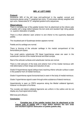
  • 1. MRI of LEFT KNEE Protocol: Multiplanar MRI of the left knee joint performed in the sagittal, coronal and transverse planes using T1 weighted spin echo, T2 and proton-density weighted fast spin echo, fatsaturated & T2* weighted gradient echo sequences. Observations: There is complete tear of the patellar tendon from its attachment at the inferior pole of patella with a large defect between the torn ends and resultant high riding patella i.e. superior dislocation of patella. There is a fluid collection seen anterior to and inferior to the superiorly displaced patella. The visualised part of Quadriceps tendon appears normal. Patella and its cartilage are normal. There is thinning of the articular cartilage in the medial compartment of the femorotibial joint space. Few small patchy subarticular PD fatsat hyperintense areas are seen in the subarticular marrow of the medial femoral and tibial condyle. Rest of the articular surfaces and subarticular marrow are normal. There is mild extrusion of the body and anterior horn of the medial meniscus with mild deformation of their shape and reduction of size. Grade III Hyperintense signal (oblique tear) reaching inferior articular surface is seen in the posterior horn of medial meniscus. Grade II hyperintense signal (horizontal tear) is seen in the body of medial meniscus. Grade I hyperintense signal is seen through entire substance of lateral meniscus. Hyperintensity is seen on PDFS sequence along the Medial collateral ligament, however no disruption of its fibres. The cruciate and lateral collateral ligaments are uniform in the outline and do not display any focal signal abnormality. Mild knee joint effusion. Conclusion:  Complete tear of the patellar tendon from its attachment at the inferior pole of patella with a large defect between the torn ends and resultant superior dislocation of patella (patella alta).
  • 2.  Fluid collection inferior to and anterior to the superiorly displaced patella.  Changes of osteoarthritis involving the medial compartment of the knee (femorotibial) joint.  Mild extrusion of the body and anterior horn of the medial meniscus.  Oblique tear reaching inferior articular surface in the posterior horn of the medial meniscus.  Horizontal tear in body of medial meniscus.  Grade I hyperintense signal in the entire substance of lateral meniscus.  Grade I sprain of the MCL. Dr. Rahul Hegde MBBS, MD, FRCR, USMLE Feb 21,2015 12:47
  • 3. MRI OF LUMBAR SPINE Clinical Profile: Low back pain Prior: None Technique: Multiplanar multiecho MRI of the lumbosacral spine was performed. Findings: Loss of normal lumbar lordosis is seen with mild degenerative changes. The lumbar vertebral bodies demonstrate normal height, alignment and marrow signal. Posterior elements are intact with normal ligamentum flavum and facets. L1/2 disc - The disc reveals no significant bulge or herniation. Neural foramina and exiting nerve roots appear normal. L2/3 disc - The disc reveals no significant bulge or herniation. Neural foramina and exiting nerve roots appear normal. L3/4 disc – The disc reveals no significant bulge or herniation. Neural foramina and exiting nerve roots appear normal. L4/5 disc – The disc reveals no significant bulge or herniation. Neural foramina and exiting nerve roots appear normal. L5/S1 disc – There is mild loss of disc height and hydration. Mild diffuse intervertebral disc bulge is seen with mild mass effect over the thecal sac. Neural foraminae are mildly compromised. The lower thoracic cord and conus appear normal. The pre and paravertebral soft tissue appear normal. Impression: 1. Mild degenerative changes at lumbar spine. 2. Disc bulge at L5/S1 level causing mild mass effect over the thecal sac. Dr. Kedar Athawale Feb 06,2015 08:51 DMRD, DNB Consultant Radiologist
  • 4. MRI of PELVIS Technique: Multiplanar, multiecho MRI of pelvis was performed. Findings: Marrow signal abnormality is seen in both femoral heads with serpiginous areas of low signal on T1 and heterogeneously hyperintense signal on T2/STIR. No collapse or flattening of the femoral heads is seen. Minimal bilateral hip effusion is seen. The other visualized bones demonstrate normal marrow signal intensity. The iliac crests appear normal. The muscles, tendons and ligaments around the hip joint demonstrate normal signal intensity. No evidence of muscle tear, strain or contusion. The visualized pelvic viscera are unremarkable. Impression: Findings are suggestive of avascular necrosis of bilateral femoral heads (stage 2) as described. No other significant abnormality detected. Please correlate with patient’s clinical findings and laboratory studies. Dr. Preshit Javadekar Feb 12,2015 00:45 MBBS, DMRE(Bom), DNB Consultant Radiologist
  • 5. MRI and MRA OF BRAIN Technique: Multiplanar and multiecho MRI of brain was performed. Findings: Multiple small hyperintensities are noted involving periventricular white matter in T2W & Flair images. These do not show diffusion restriction. Hyperintensity is noted involving central pons on T2W & Flair images. It does not show diffusion restriction. Rest of both cerebral parenchyma show normal gray-white matter differentiation with normal sulci, gyri and basal cisterns. Centrally located gray matter nuclei appear normal in size, shape and intensity. Diffusion imaging does not reveal evidence of acute infarct. Ventricular system appears normal in size & shape. No evidence of periventricular oozing. Midline structures like interhemispheric fissure, 3rd ventricle, pineal region and rest of brain stem appear normal. Posterior fossa structures appear normal. Mild tortuosity of left optic nerve is noted. Right optic nerve appears normal in the given images. MR Brain Angiography: The internal carotid arteries show normal course and caliber and are symmetrically disposed. Mild irregularity is noted involving intracranial part of left ICA. No significant luminal narrowing is noted. Both middle cerebral arteries arise normally from the internal carotid on either side and forms normal insular loops. Mild irregularity is noted involving M1 part of left MCA. No significant luminal narrowing is noted. The anterior cerebral artery shows no signs of narrowing or displacement. The basilar artery shows a normal course and caliber and divides into normal size posterior cerebral arteries. The anterior and posterior communicating arteries appear unremarkable. IMPRESSION: • Chronic white matter ischemia.
  • 6. • Hyperintensity involving central pons on T2W & Flair images. It does not show diffusion restriction. • Mild tortuosity of left optic nerve. • Mild irregularity involving intracranial part of left ICA and M1 part of left MCA. No significant luminal narrowing. Dr. Arti Iyer. Patil Dec 29,2014 11:33 MBBS, MD
  • 7. MRI OF HIP JOINTS (PLAIN AND CONTRAST) Clinical information: Known case of Sickle cell anemia. Previously AVN of Right Hip. Presently, pain in Left Hip. Protocol: Multiplanar MRI of the both hip joints was performed using T1weighted spin echo, T2 weighted fast spin, echo, T1 and PD Fatsaturated sequences. Post contrast multiplanar T1 weighted sequence obtained after injection of gadolinium. Observations: LEFT HIP: The left femoral head is spherical in shape with no articular collapse. The left femoral head, neck and posterior part of the left acetabulum show diffuse hyperintense signal on T2 and T2 fatsaturated sequences which is isointense on T1 sequence. On post contrast sequence, a large part of the femoral head shows no enhancement with thick peripheral enhancement around the non-enhancing area. There is thick synovial enhancement with minimal joint effusion. There is periarticular hyperintense soft tissue on T2 and T2 fatsaturated sequences seen around the left hip joint involving the left obturator externus, pectinues muscles and the origin of the left adductor muscles which shows post contrast enhancement with few non-enhancing irregular necrotic areas within. Multifocal areas of marrow hyperintensity on T2 and T2 fatsaturated sequences is seen in both upper femoral shafts, intertrochanteric regions, right iliac blade, right ischial tuberosity which show heterogeneous post contrast enhancement with peripheral contrast enhancement in upper femoral shafts. RIGHT HIP: The right femoral head is small in size with collapse of articular surface on the superolateral aspect. Small subchondral defects seen in femoral head and the in the acetabulum which show no post contrast enhancement. No synovial thickening or effusion seen. No periarticular abnormal soft tissue seen around the right hip joint. Conclusion:  Diffuse signal abnormality in the left femoral head and neck as described above with synovial thickening and enhancement, minimal left hip joint effusion and abnormal periarticular inflammatory soft tissue involving the left obturator externus, pectinues muscles and the origin of the left
  • 8. adductor muscles. These findings are most likely suggestive of left hip septic arthritis with periarticular infective/ inflammatory soft tissue. Less likely possibility of AVN with superadded infection.  Multifocal areas of signal abnormality in both upper femoral shafts, intertrochanteric regions, right iliac blade, right ischial tuberosity as described above is most likely suggestive of multifocal osteomyelitis.  Findings in Right hip joint suggestive of Stage IV AVN. Correlate with clinical findings, lab investigations and other imaging investigations. Dr. Rahul Hegde MBBS, MD, FRCR, USMLE Feb 21,2015 12:47
  • 9. CONTRAST ENHANCED MRI OF THE ABDOMEN Clinical information: Known case of prostatic carcinoma. APR and Colostomy done. Evidence of sarcoma on biopsy of the colostomy site. Prior Imaging: None Protocol: Multiplanar imaging of the abdomen was performed using T1 weighted spin echo, T2 weighted fast spin echo, gradient echo, fatsaturated sequences. Post contrast T1 weighted fat saturated sequences obtained after injection of gadolinium. Observations: The sigmoid colostomy is seen extending though the inferior part of the left rectus muscle in the left lower qudrant of the abdomen. The stoma opening is edematous and shows moderate enhancing thickening. Rest of the sigmoid colon and visualised bowel loops are unremarkable. Mild T2 hyperintensity is seen at in the subcutaenous fat and in the adbominal wall at the stoma site which show mild post contrast enhancement. Few small bowel loops beneath the stoma site also show mild wall enhancement which is most likely related to post op inflammation. No significant lymphadenopathy. No free fluid in the abdomen. Liver is normal in size, signal intensity, shows normal contrast enhancement with no focal lesions. Gallbladder is distended with normal wall thickness. CBD is normal. Pancreas is normal in size, intensity and enhancement. Spleen is normal in size, intensity and shows normal contrast enhancement. Both adrenals are normal. Both kidneys are normal in size, intensity and show normal density nephrogram. A small T2 hyperintense and T1 hypointense simple cortical cyst seen in the upper part of the left kidney.
  • 10. A small 1.2 cm sized T1 hypointense, T2 hyperintense lesion is seen in posterior part of right iliac bone adjacent to the right SI joint. Rest of the visualized bones show normal marrow signal intensity. Conclusion:  Enhancing thickening at the stoma opening of the sigmoidostomy, however no obvious mass noted in the abdominal wall or intra-abdominally. Correlate with Histopathology reports.  Mild edema and granulation tissue in the subcutaneous fat and abdominal wall around the stoma.  A small lesion in posterior part of right iliac bone could be suggestive of bone metastasis in this known case of prostatic carcinoma. This needs correlation with radiographs/ bone scan. Dr. Rahul Hegde MBBS, MD, FRCR, USMLE Feb 21,2015 12:47