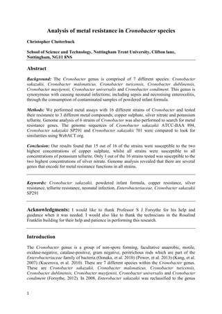
Analysis of metal resistance in Cronobacter species
1. 1
Analysis of metal resistance in Cronobacter species
Christopher Clutterbuck
School of Science and Technology, Nottingham Trent University, Clifton lane,
Nottingham, NG11 8NS
Abstract
Background: The Cronobacter genus is comprised of 7 different species: Cronobacter
sakazakii, Cronobacter malonaticus, Cronobacter turicensis, Cronobacter dublinensis,
Cronobacter muytjensii, Cronobacter universalis and Cronobacter condiment. This genus is
synonymous with causing neonatal infections; including sepsis and necrotising enterocolitis,
through the consumption of contaminated samples of powdered infant formula.
Methods: We performed metal assays with 16 different strains of Cronobacter and tested
their resistance to 3 different metal compounds; copper sulphate, silver nitrate and potassium
tellurite. Genome analysis of 6 strains of Cronobacter was also performed to search for metal
resistance genes. The genome sequences of Cronobacter sakazakii ATCC-BAA 894,
Cronobacter sakazakii SP291 and Cronobacter sakazakii 701 were compared to look for
similarities using WebACT.org.
Conclusion: Our results found that 15 out of 16 of the strains were susceptible to the two
highest concentrations of copper sulphate, whilst all strains were susceptible to all
concentrations of potassium tellurite. Only 1 out of the 16 strains tested was susceptible to the
two highest concentrations of silver nitrate. Genome analysis revealed that there are several
genes that encode for metal resistance functions in all strains.
Keywords: Cronobacter sakazakii, powdered infant formula, copper resistance, silver
resistance, tellurite resistance, neonatal infection, Enterobacteriaceae, Cronobacter sakazakii
SP291
Acknowledgments: I would like to thank Professor S J Forsythe for his help and
guidance when it was needed. I would also like to thank the technicians in the Rosalind
Franklin building for their help and patience in performing this research.
Introduction
The Cronobacter genus is a group of non-spore forming, facultative anaerobic, motile,
oxidase-negative, catalase-positive, gram negative, peritrichous rods which are part of the
Enterobacteriaceae family of bacteria (Oonaka, et al. 2010) (Power, et al. 2013) (Kang, et al.
2007) (Kucerova, et al. 2010). There are 7 different species within the Cronobacter genus.
These are Cronobacter sakazakii, Cronobacter malonaticus, Cronobacter turicensis,
Cronobacter dublinensis, Cronobacter muytjensii, Cronobacter universalis and Cronobacter
condiment (Forsythe, 2012). In 2008, Enterobacter sakazakii was reclassified to the genus
2. 2
Cronobacter spp. which originally contained 4 species; Cronobacter sakazakii, Cronobacter
malonaticus, Cronobacter turicensis and, Cronobacter dublinensis (Strydom, et al. 2012).
Cronobacter spp. have been linked to causing infections in both adults and neonatal babies
(Wang, et al. 2009). Although adults are more often infected by these bacterial species,
babies are at the most risk of infection and death from these species of bacteria and the
diseases they cause. Neonatal babies that have a compromised immune system or are of a low
birth weight are at the most risk of infection (Forsythe, 2012) (Strydom, et al. 2012).
Cronobacter spp. are an opportunistic species, but out of the whole genus, only C. sakazakii,
C. malonaticus and C. turicensis have been isolated from cases of neonatal meningitis, which
is an acute inflammation of the membranes surrounding the brain and the spinal cord
(Strydom, et al. 2012). These species have also been found to cause septicaemia, necrotising
enterocolitis; which causes an infection of the intestines, bloody diarrhoea and brain
abscesses. These diseases occur within a few days of infection. They are very server and have
a mortality rate between 10 and 80% (Caubilla-Barron, et al. 2007) (Strydom, et al. 2012).
The main source of infection has been linked to powdered infant formula (PIF). Other
sources of contamination have also been found, including; soil, rats, flies, beer, mugs, milk
powder factories, a chocolate factory, and even houses (Forsythe 2005). Neonates are most at
risk of infection due to their sterile gastrointestinal tract; which is quickly colonised though
oral ingestion, and their immature immune system (Strydom, et al. 2012) (Forsythe, 2005).
These factors leave them vulnerable to infections and diseases.
Possible routes of contamination for the powdered infant formula could be in the
development process. It is developed to mimic human breast milk rather than cow’s milk. To
do this, the cow’s milk is modified in various ways. The levels of protein, minerals and fat
are reduced. The levels of whey protein, carbohydrates and the calcium to phosphate ration
are increased, and extra vitamins are added to it as well.
All of the ingredients are added to the milk in 3 different methods; a wet, dry or combined
method. The wet method combines all the ingredients in a liquid phase. This liquid is then
heat treated and spray dried to get the final powdered formula. The dry method involves
preparing the ingredients separately and heat treating them prior to being combined in a dry
form. The dry method has a higher chance of contamination than the wet method, which is
why some manufacturers use a combination method instead. This involves the combination
of all the soluble ingredients during a liquid phase, which is then heat treated. The less
soluble ingredients are then added after to the spray dried powder (Strydom, et al. 2012).
Cronobacter spp. are extremely durable. They are generally agreed to be thermo-tolerant and
can grow at temperatures from 6°c to 47°c, with their optimum temperature being 39°c. They
are also acid resistant, being able to endure pHs between 3.5 and 5, with survival at pH 3
being transitory. They have also been shown at grow at a ph of 7 as well. Some members of
the genus have been shown to have even greater osmotic and desiccation tolerance than
Escherichia coli. Cronobacter spp. has been shown to do better in dry condition at lower
temperatures (4°c) when compared to higher temperatures (21-31°c). Some are even able to
survive in desiccation for 2 years and then multiply rapidly again once the bacteria is
rehydrated (Strydom, et al. 2012). Once this bacterium is rehydrated, it’s doubling time
increases. At 10°c, it is every 14 hours, but at room temperature, it is only 45 minutes. The
risk of infection also increases with the temperature. If the bacterium is left at 25°c for 6
3. 3
hours, the risk of infection 30 times greater than at 0 hours. If the bacterium is left at 25° for
10 hours, the risk of infection is 30,000 times greater than at 0 hours (Forsythe, 2005).
Cronobacter spp. are also able to form biofilms on multiple surfaces, including glass,
stainless steel, latex and polycarbonate. They are able to form biofilms quicker on a
hydrophobic surface compared to a hydrophilic one. Biofilms help to increase the bacteria
resistance to environmental stress, detergents and antibiotics. The growth of the biofilm is
enhanced by the presence of a novel heteropolysaccharide (Strydom, et al. 2012). This is
comprised of glucuronic acid (29-32%), D-glucose (23-30%), D-galactose (19-24%), L-
fucose (13-22%) and D-mannose (0-8%) (Harris and Oriel, 1989).
According to work by Hurrel et al, 2009; biofilms can also build up on neonatal feeding
tubes. Their work shows that C. sakazakii was isolated from biofilms inside of neonatal
feeding tubes that were from babies that were being fed breast milk and ready to feed
formula; but less frequently isolated compared to other Enterobacteriaceae.
Biofilms in neonatal feeding tubes pose a problem for neonates that have to be fed through
them. This could lead to a possible source of infection for the neonate. This is where our
experiment comes in. There is research into the use of metals as possible antimicrobials.
These include copper, silver and tellurite. We will look at multiple Cronobacter spp. and
determine with the use of metal assays and bioinformatics research whether these metals
could be used as possible antimicrobials.
Analysis of the genome of C. sakzakii ATCC-BAA 894 revealed genes that are associated
with the invasion of brain microvascular endothelial cells. These were cusCFBA and cusR.
These genes have been identified in E. coli as encoding an RND type copper efflux system
(Elguindi, et al. 2012). E. coli uses 3 systems for copper resistance. There is the P-type
ATPase CopA; which pumps excess copper out of the cytoplasm, a multicopper oxidase
CueO, and Cus determinants that confers copper and silver resistance (Franke, et al. 2003).
Cus has 2 operons; cusRS and cusCFBA. CusCFBA encodes the proteins for an efflux
system for copper and its transcription is dependent on the copper or silver concentration.
Knock out models revealed that in E. coli, deletion of cusA or cusCFBA lead to silver
sensitivity; but only lead to copper sensitivity under anaerobic conditions. To achieve copper
sensitivity under aerobic conditions, deletion of cueO (multicopper oxidase) was also needed.
All of the cus genes are needed for complete copper resistance. Even a single nucleotide
deletion in all 4 cus structural genes leads to a decrease in copper resistance.
Tellurite resistance genes are widely found in pathogenic organisms; and analysis of the C.
sakazakii ATCC-BAA 894 genome by Joseph et al, 2012; revealed tellurite resistance genes
(terACDYZ). Homologies of these genes have also been found and analysed in E. coli. In E.
coli, the terBCDEF gene is essential for tellurite resistance, but its mechanism for providing
the resistance is still unknown. However, it is known that the tellurite metal is reduced and
deposited inside the resistant bacterial cell. It has been suggested that tellurite resistance helps
the cell with resistance to strong oxidative agents; with tellurite salts being strong oxidative
agents themselves. This could lead to the explanation of how cells survive in macrophages
and in mammalian hosts (Vavrova, et al. 2006).
4. 4
Methods
Inoculating bacterial strains:
Strains of C. sakazakii 20, 658, 669, 695, 700, 701, 702, 703, 715, 730, 767, 1218, C.
turicensis 507 and 564, C. dublinensis 582 and C. malonaticus 681 were streaked onto
Tryptone Soya Agar (TSA) plates and incubated at 37°c for 24 hours to get single colonies.
Single colonies from these plates were then used to inoculate TSA slopes, which were then
left again to incubate for 24 hours at 37°c. The slopes were used to store the bacteria at 5°c
whilst it wasn’t in use.
The agar slopes where used to inoculate bottles containing 10ml of Tryptone Soya Broth
(TSB). These were left to incubate at 37°c for 24 hours. These inoculated bottles were used to
aseptically inoculate TSA plates with 0.1ml (100μl) of broth. This was evenly spread over the
plate with a sterile glass spreader and then left for the bacterial broth to soak into the agar.
Metal solutions:
1 molar copper sulphate (pentahydrate) and potassium tellurite and 2 molar, silver nitrate
were created by dissolving the required mass in 1 ml of sterile distilled water. To create the
lower concentrations of metal solutions; serial dilution was used. The concentrations used
were 1 molar, 0.1 molar, 0.01 molar and 0.001 molar for copper sulphate and potassium
tellurite. Silver nitrate was used at concentrations of 2 molar, 1 molar, 0.1 molar and 0.01
molar.
Metal assays:
Once the bacterial broth was dried into
the agar plates, 13mm filer paper disks
were added to each plate (4 per plate),
one for each metal concentration. 7μl
of each metal solution was then added
to each disk on the same day the agar
plates were inoculated with bacterial
broth (image 1). These were then left
to incubate for 24 hours at 37°c. The
zone of inhibition was then measured
and recorded.
Bioinformatics:
Only 4 of the strains tested with the
metal assays had gene sequences
available for analysis online. These
where C. sakazakii 701 (accession
number: CALE01000001-
CALE01000768), C. malonaticus 681
(accession number: CALC01000001-
CALC01000171), C. turicensis 564
(accession number: CALB01000001-
CALB01000114) and C. dublinensis
582 (accession number:
CALA01000001-CALA01000427).
Two other species of Cronobacter were used for comparison. These were C. sakazakii
Image 1: Diagram showing the set up of the metal assays and how the
concentrations were distributed. Blue writing = Silver nitrate
concentrations. Red writing = Copper sulphate and Potassium nitrate
concentrations
2 molar
1 molar
1 molar
0.1 molar
0.1 molar
0.01 molar
0.01 molar
0.001 molar
Inoculated
Agar plate
Filter paper
disks
5. 5
ATCC-BAA 894 (accession number: NC_009778.1) and C. sakazakii SP291 (accession
number: NC_020260.1). C. sakazakii ATCC-BAA 894 was used as the main bacteria to
locate the metal resistance genes and compare their sequence to. Genes pertaining to metal
resistances in these strains were found using NCBI protein searches and through the analysis
of the genome of C. sakazakii ATCC-BAA 894 using BioCyc.org. Other genes that pertain to
metal resistance, but were not found to be a part of the genome of C. sakazakii ATCC-BAA
894; such as TerC, were compared using the sequence found in C. turicensis 564.
Once the genes were selected, they were compared against the other strains using BLASTP.
Then name of the protein most related to metal resistance; and its identities score, was noted
down. The genomes of C. sakazakii ATCC-BAA 894, C. sakazakii SP291 and C. sakazakii
701 were also compared against each other using WebACT.org.
Results
Metal Assay:
The results in table 1 show that all the strains tested were resistant to concentrations of copper
sulphate below 0.01 molar. 3 strains were resistant to 0.1 molar copper sulphate (C. sakazakii
702, C. sakazakii 730 and C. sakazakii 767); whilst C. sakazakii 695 was completely resistant
to all copper sulphate concentrations.
As shown in table 2, only 4 of the strains were resistant to 0.001 molar potassium tellurite (C.
sakazakii 658, C. sakazakii 695, C. sakazakii 730, and C. sakazakii 1218). All of the strains
showed no resistance to potassium tellurite above a concentration of 0.01 molar.
Comparatively, all of the strains; except for C. sakazakii 730, were resistant to silver nitrate.
C. sakazakii 730 was also resistant to concentrations below 0.01 molar; as seen in table 3.
Bioinformatics:
The genomes of the different bacterial strains have varying lengths and plasmid numbers. C.
sakzakii ATCC-BAA 894 has a genome that is 4.53 Mb in size (GC% of 56.8%) and 2
plasmids; pESA2 (31,208 bps) and pESA3 (131,196 bps). C. sakazakii SP291 has a genome
that is 4.52 Mb in size (GC% of 56.8%) and 3 plasmids; pSP291-1 (118,136 bps), pSP291-2
(52,134 bps) and pSP291-3 (4,422 bps). C. sakazakii 701 has a genome that is 4.85 Mb in
size (GC% of 55.8%) and no known plasmids. C. turicensis 564 has a genome that is 4.57
Mb in size (GC% of 57.2%) and no known plasmids. C. dublinensis 582 has a genome that is
4.76 Mb in size (GC% of 57.3%) and no known plasmids. C. malonaticus 681 has a genome
that is 4.55 Mb in size (GC% of 56.5%) and no known plasmids. The genome sequences used
for C. sakazakii 701, C. turicensis 564, C. dublinensis 582 and C. malonaticus 681 are as of
this publication, incomplete.
Analysis of the genomes of C. sakazakii 701, C. malonaticus 681, C. turicensis 564, C.
dublinensis 582, C. sakazakii ATCC-BAA 894 and C. sakazakii SP291 revealed the presence
or absence of metal resistance genes. All of the genes were found to be on the main
chromosomes of the bacteria and not encoded in the plasmids.
Copper and silver resistance genes:
Table 4 shows the results for copper and silver, metal resistance genes amongst the strains
analysed. C. sakazakii SP291 was found to contain 4 out of the 5 genes related to copper and
silver resistance. All four of the related gene also had a high identities percentage; CusR
(99%), cusC (100%), cusB (97%) and periplasmic copper-binding protein (97%).
6. 6
It had a different gene in place of cusS; copper resistant sensor kinase PcoS. PcoS was the
only gene found to have an identities percentage of 36%, which is significantly lower when
compared to the rest of its compared genes.
C. sakazakii 701 again had 3 out of the 5 same genes for copper and silver resistance
compared to C. sakazakii ATCC-BAA 894. The gene found to be different in C. sakazakii
701 was the CzcB (cobalt/zinc/cadmium efflux RND transporter, membrane fusion protein)
gene. This did have an identities percentage of 97% when its amino acid sequence was
compared to that of cusB of C. sakazakii ATCC-BAA 894.
CusF (cation efflux system protein cusF precursor) was found to have an identities score of
97% when compared to periplasmic copper-binding protein of C. sakazakii ATCC-BAA 894.
The three other genes that were compared all had high identities; cusS (99%), cusR (100%),
and cusC (98%). C. malonaticus 681 was found to contain the same gene results as C.
sakazakii 701, but with slightly different identity scores.
C. dublinensis 582 and C. turicensis 564, was found to contain none of the genes that were
searched for. Some related genes were found however. CusS comparison resulted in the gene
CpxA (copper sensory histidine kinase CpxA) for both species. CusR comparison resulted in
the gene CpxR (copper-sensing two-component system response regulator CpxR) for both
species.
For cusC, C. dublinensis 582 had hypothetical protein BN133_2129 as a result and C.
turicensis 564 had FIG00554734: hypothetical protein as a result. For cusB, C. dublinensis
582 had a result for membrane fusion component of tripartite multidrug resistance system and
C. turicensis 564 had probable RND efflux membrane fusion protein as a result. For
periplasmic copper-binding protein, C. dublinensis 582 did not have any viable resistance
genes available, whilst C. turicensis 564 had FIG00554092: hypothetical protein as a result.
All of the copper and silver resistance genes found in C. dublinensis 582 and C. turicensis
564 had low levels of identities, with a maximum score of only 35% when compared to the
sequences of C. sakazakii ATCC-BAA 894.
CopA in both C. sakazakii ATCC-BAA 894 and C. sakazakii SP291 encodes a copper
exporting ATPase. In the other 4 strains tested, it encoded for a lead, cadmium, zinc and
mercury transporting ATPase; copper-translocating P-type ATPase with all species having a
high identities score. However, only a small portion of the sequence was found to match with
C. dublinensis 582 (57/60 amino acids).
NCBI protein searches for CueO with C. sakazakii ATCC-BAA 894 gave the result of a
hypothetical protein ESA_03209. When this was put into BLASTP against the other bacterial
strains, it results in a high identities match for C. sakazakii SP291 of a multicopper oxidase.
For the other 4 strains, high identity matches were found for blue copper oxidase CueO
precursor.
Tellurite resistance genes:
Table 5 shows the genes that were found that pertain to tellurite resistance. For TehB, C.
sakazakii ATCC-BAA-894 was used for the reference sample, whilst C. turicensis 564 was
used as the reference sample for genes TehA and TerC. TehB was found in all the samples
tested with high percentages of identities.
7. 7
Average diameter of the zones of inhibition for bacterial strains per copper sulphate concentration (cm)
C. sak 20 C. sak 658 C. sak 669 C. sak 695 C. sak 700 C. sak 701 C. sak 702 C. sak 703 C. sak 715 C. sak 730 C. sak 767 C. sak 1218 C. tur 507 C. tur 564 C. mal 681 C. dub 582
1 molar 2.20 2.07 2.37 0.00 2.07 2.13 2.10 1.93 2.00 1.37 1.50 2.13 1.83 2.03 2.17 2.13
0.1 molar 1.50 1.37 1.40 0.00 1.33 1.37 0.00 1.40 1.30 0.00 0.00 1.40 0.43 0.93 1.37 1.40
0.01 molar 0.00 0.00 0.00 0.00 0.00 0.00 0.00 0.00 0.00 0.00 0.00 0.00 0.00 0.00 0.00 0.00
0.001 molar 0.00 0.00 0.00 0.00 0.00 0.00 0.00 0.00 0.00 0.00 0.00 0.00 0.00 0.00 0.00 0.00
Table 1: Size of the zone of inhibition using the metal copper sulphate
Average diameter of the zones of inhibition for bacterial strains per potassium tellurite concentration (cm)
C. sak 20 C. sak 658 C. sak 669 C. sak 695 C. sak 700 C. sak 701 C. sak 702 C. sak 703 C. sak 715 C. sak 730 C. sak 767 C. sak 1218 C. tur 507 C. tur 564 C. mal 681 C. dub 582
1 molar 4.13 3.97 4.83 4.50 3.90 4.57 4.10 3.60 3.83 2.40 3.90 4.20 3.47 4.93 4.67 3.80
0.1 molar 3.53 3.13 3.93 3.30 3.47 3.77 3.67 3.07 3.37 0.97 3.35 3.33 3.17 3.67 3.77 2.97
0.01 molar 2.60 1.97 3.00 2.47 2.60 2.87 2.77 2.23 2.47 0.47 2.50 2.13 2.47 2.60 2.90 2.10
0.001 molar 2.00 0.00 1.63 0.00 1.63 1.70 1.73 1.63 1.47 0.00 1.50 0.00 1.43 1.87 1.83 1.63
Table 2: Size of the zone of inhibition using the metal potassium tellurite
Average diameter of the zones of inhibition for bacterial strains per silver nitrate concentration (cm)
C. sak 730
2 molar 2.40
1 molar 0.97
0.1 molar 0.47
0.01 molar 0.00
Table 3: Size of the zone of inhibition using the metal silver nitrate
8. 8
BLAST search results for copper and silver resistance genes in Cronobacter species
Gene name: Species that the gene
came from:
C. sakazakii SP291 C. sakazakii 701 C. malonaticus 681 C. dublinensis 582 C. turicensis 564
cusS
(Accession number:
AGE88743),
491 A.A’s long
C. sakazakii ATCC-
BAA 894
Copper resistant sensor
kinase PcoS (Identities
= 110/308 [36%])
Copper sensory histidine
kinase cusS
(Identities = 487/490
[99%])
Copper sensory histidane
kinase cusS (Identities =
488/490 [99%])
Copper sensory
histidine kinase CpxA
(Identities = 29/82
[35%])
Copper sensory histidine
kinase CpxA (Identities =
78/271 [29%])
cusR
(Accession number:
YP_001440254.1),
226 A.A’s long
C. sakazakii ATCC-
BAA 894
DNA-binding
transcriptional activator
CusR (Identities =
225/226 [99%])
Copper-sensing two-
component system
response regulator CusR
(Identies = 226/226
[100%])
Copper-sensing two-
component system
response regulator CusR
(Identities = 226/226
[100%])
Copper-sensing two-
component system
response regulator
CpxR (Identities =
34/121 [28%])
Copper-sensing two-
component system response
regulator CpxR (Identities =
80/228 [35%])
cusC
(Accession number:
YP_007442924),
461 A.A’s long
C. sakazakii ATCC-
BAA 894
Copper/silver efflux
system outer membrane
protein CusC (Identities
= 461/461 [100%])
Cation efflux system
protein CusC precursor
(Identities = 452/461
[98%])
Cation efflux system
protein CusC precursor
(Identities = 450/461
[98%])
Hypothetical protein
BN133_2129 (Identities
= 56/175 [32%])
FIG00554734: hypothetical
protein (Identities = 90/294
[31%])
cusB
(Accession number:
YP_001440257),
430 A.A’s long
C. sakazakii ATCC-
BAA 894
Copper/silver efflux
system membrane
fusion protein CusB
(Identities = 366/376
[97%])
Cobalt/zinc/cadmium
efflux RND transporter,
membrane fusion protein,
CzcB family (Identities =
421/430 [98%])
Cobalt/zinc/cadmium
efflux RND transporter,
membrane fusion protein,
CzcB family (Identities =
419/430 [97%])
Membrane fusion
component of tripartite
multidrug resistance
system (Identities =
19/73 [26%])
Probable RND efflux
membrane fusion protein
(identities = 47/187 [25%])
Periplasmic copper-
binding protein
(Accession number:
ABU79420)
117 A.A’s long
C. sakazakii ATCC-
BAA 894
Periplasmic copper-
binding protein
(Identities = 113/117
[97%])
Cation efflux system
protein CusF precursor
(Identities = 113/117
[97%])
Cation efflux system
protein CusF precursor
(Identities = 115/117
[98%])
No related genes found FIG00554092: hypothetical
protein (Identities = 20/84
[24%])
CopA
(Accession number:
YP_001438847),
835 A.A’s long
C. sakazakii ATCC-
BAA 894
Copper exporting
ATPase (Identities =
826/835 [99%])
Lead, cadmium, zinc and
mercury transporting
ATPase; Copper-
translocating P-type
ATPase (Identities =
238/246 [97%])
Lead, cadmium, zinc and
mercury transporting
ATPase; Copper-
translocating P-type
ATPase (Identities =
225/228 [99%])
Lead, cadmium, zinc
and mercury
transporting ATPase;
Copper-translocating P-
type ATPase (Identities
= 57/60 [95%])
Lead, cadmium, zinc and
mercury transporting
ATPase; Copper-
translocating P-type ATPase
(identities = 480/490
[98%])
hypothetical protein
ESA_03209
(Accession number:
ABU78431),
529 A.A’s long
C. sakazakii ATCC-
BAA 894
Multcopper oxidase
(Identities = 513/520
[99%])
Blue copper oxidase
CueO precursor
(Identities = 522/529
[99%])
Blue copper oxidase
CueO precursor
(Identities = 517/529
[98%])
Blue copper oxidase
CueO precursor
(Identities = 495/529
[94%])
Blue copper oxidase CueO
precursor (Identities =
496/520 [95%])
Table 4: List of genes found that are related to copper and silver metal resistance. Identities show the number of amino acids that the sequences have in common. Genes in red in indicate the closest related
gene that matched the searched sequence
9. 9
BLAST search results for tellurite resistance genes in Cronobacter species
Gene name: Species that the
gene came from:
C. sakazakii ATCC-
BAA 894
C. sakazakii SP291 C. sakazakii 701 C. malonaticus 681 C. dublinensis 582 C. turicensis 564
TehB
(Accession number:
YP_001437811),
197 A.A’s long
C. sakazakii ATCC-
BAA 894
Tellurite resistance
protein TehB
(Identities = 191/197
[97%])
Tellurite resistance
protein TehB
(Identities = 115/121
[95%])
Tellurite resistance
protein TehB (Identities
= 62/62 [100%])
Tellurite resistance
protein TehB (Identities
= 73/78 [94%])
Tellurite resistance
protein TehB
(Identities = 111/119
[93%])
TehA
(Accession number:
ZP_19165219),
335 A.A’s long
C. turicensis 564 Hypothetical protein:
ESA_03549
(Identities = 15/39
[38%])
Hypothetical protein:
CSSP291_09175
(Identities = 19/44
[43%])
FIG00553343:
hypothetical protein
(Identities = 19/44
[43%])
FIG00553343:
hypothetical protein
(Identities = 19/44
[43%])
Tellurite resistance
protein TehA (Identities
= 94/105 [90%])
TerC
(Accession number:
ZP_19165014),
335 A.A’s long
C. turicensis 564 Hypothetical protein
ESA_03499
(Identities = 312/322
[97%])
Inner membrane
protein Alx (identities
= 314/322 [98%])
Integral membrane
protein TerC
(Identities = 36/36
[100%])
Integral membrane
protein TerC (Identities
= 287/303 [95%])
Integral membrane
protein TerC (Identities
= 270/281 [96%])
Table 5: List of genes found that are related to copper and silver metal resistance. Identities show the number of amino acids that the sequences have in common. Genes in red in indicate the closest related
gene that matched the searched sequence. Black squares indicate that that strain was used as the sequence sample for the BLAST search.
10. 10
TehA was found with a high percentage of identities in only C. dublinensis 582. For C.
sakazakii 701and C. malonaticus 681, FIG00553343: hypothetical protein was found, with an
identities percentage of 43% for both species. In C. sakazakii SP291, hypothetical protein
CSSP291_09175 was found with an identities percentage of 43%. For C. sakazakii ATCC-
BAA894, hypothetical protein: ESA_03549 was found with an identities percentage of 38%.
TerC was found with high identity percentages in C. sakazakii 701, C. malonaticus 681 and
C. dublinensis 582. For C. sakazakii SP291, inner membrane protein Alx was found with an
identities percentage of 98%. For C. sakazakii ATCC-BAA894, hypothetical protein:
ESA_03499 was found with an identities percentage of 97%.
Discussion
The metal assays show tellurite is a more efficient antimicrobial than copper against
Cronobacter spp. All strains tested were resistant to 0.01 molar and 0.001 molar copper
sulphate; with C. sakazakii 695 being completely resistant to 0.1 and 1 molar copper sulphate
as well (figure S1). Comparatively, only 4 strains tested were resistant to 0.001 molar
potassium tellurite. All the other strains showed some inhibition of growth at this
concentration (figure S2).
Silver nitrate only had 1 strain of bacteria that was not resistant to it; C. sakazakii 730. This
could have been due to the use of too low a concentration for the silver to have any
antimicrobial effect. Silver has been shown to be very effective as an antimicrobial agent
(Zheng, et al. 2012) (Długosz, et al. 2012). It could be that silver isn’t as good an
antimicrobial when it is part of a compound compared to when it is purer. This area would
require further investigation and analysis.
Comparing the metal assay results of the 4 strains that had their genomes analysed shows
some interesting results. For potassium tellurite, C. dublinensis 582 has the smallest zones of
inhibition across all concentrations, indicating the strongest resistance (figure S4). This graph
also shows that C. turicensis 564 had the second strongest resistance to tellurite
concentrations of 0.1 molar and 0.01 molar. This correlates with the information found about
the tellurite resistance genes. Only C. dublinensis 581 and C. turicensis 564 had all 3 of the
tellurite resistance genes searched for.
The results for the metal assay and the gene information found do not correlate for copper
resistance. In the metal assay, C. turicensis 564 was found to have the strongest resistance by
having the smallest zones of inhibition for 1 molar and 0.1 molar concentrations of copper
sulphate (figure S3). However, the copper resistance gene analysis showed that C. turicensis
564 only contained 4 out of the 7 copper resistance genes searched for. This result could be
caused by C. turicensis 564 having more efficient CueO and CopA genes, in order to
compensate for its lack of copper efflux genes; cusF, cusC and cusB. This area of research
involving Cronobacter spp. and their metal resistance mechanisms, and genes that encode
metal resistance requires further investigation.
Even though these Cronobacter spp. show metal resistance genes, they are still susceptible to
being killed by high enough concentrations of the metals used. Copper sulphate and
potassium tellurite were both good at preventing growth of Cronobacter spp., with potassium
tellurite being more efficient that copper sulphate. Work by Elguindi et al, 2012; showed that
the survival times of C. sakazakii on 88.6% and 99% copper alloys rapidly killed the bacteria
11. 11
on contact under all experimental conditions tested. Research into combining copper sulphate
into powdered infant formula has also shown some promising results. The addition of
50μg/ml of copper (II) suphate with 0.2% lactic acid for 6 hours at 21°c resulted in complete
elimination of Cronobacter spp. (Strydom, et al. 2012). The addition of 50-100μg/ml of
copper (II) suphate showed a 1-2 decrease in C. sakazakii survival when added to powdered
infant formula. The addition of 100μg/ml of copper (II) suphate with 0.2% lactic acid for 2
hours completely eliminated C. sakazakii (Elguindi, et al. 2012).
Tellurite (TeO3
2-
) has also been shown to be an effective antimicrobial. Tellurite has been
shown to be much more toxic when it is in the form of a simple salt; such as sodium tellurite
(Na2TeO3) than ordinary tellurite (TeO4
2-
). Tellurite is highly toxic to bacteria, with it being
toxic at concentrations as low as 1μg/ml. Bacteria that are resistant to this chemical reduce
the toxic TeO3
2-
to the less toxic Te0
. This leads to black deposits inside of the cell (Chasteen,
et al. 2009). Evidence of this can be seen in image S1. The image shows black deposits inside
and at the edges of the zones of inhibition, indicating where potassium tellurite was reduced
inside the bacterial cell, but then released when the cell was destroyed.
There is already research in progress for the use of copper, silver and tellurite as
antimicrobials. Copper is being tested as an antimicrobial surface. This involved either
creating the surface with copper in it from the beginning; or using a cheaper alternative, such
as spraying the surface with copper using one of the following techniques, plasma spray, arc
spray or cold spray. These processes would be useful in hospitals where microbes could build
up on surfaces that haven’t been cleaned recently. If the product had an antimicrobial surface
to it, it would help to reduce infections and bacteria spreading in hospitals (Champagne and
Helfritch, 2013). This technique of copper surface coating could also be applied to neonatal
feeding tubes to help reduce the build up of biofilms between feedings.
Silver has been shown to have excellent antimicrobial properties when it is in a nanoparticle
form, which is then incorporated into titanium or calcium carbonate microparticles (Zheng, et
al. 2012) (Długosz, et al. 2012). Elguindi et al, 2012; performed experiments with flexiline
impregnated with silver, but the silver showed no sign of decreasing the biofilm formation.
As mentioned previously, this could be due to the silver either not being at a high enough
concentration to have an antimicrobial effect. The combination of these two technologies,
flexiline impregnated with titanium or calcium carbonate microparticles containing silver
nanoparticles could help to decrease biofilms by increasing the antimicrobial effects of silver.
Tellurite has been tested as a possible addition to antibiotics for bacterial infections. Bacteria
are becoming more resistant to modern antibiotics and over the last 40 years, only 2 new
antibiotics have been produced, oxazolidinone and daptomycin. Tellurite has been chosen to
be used in conjunction with antibiotics to help increase their effectiveness due to its
numerous cell targets and its low effect on eukaryotic cells. Eukaryotic cells have been tested
with concentrations of up to 50μM tellurite (TeO3
2-
) and have shown no sign of being
affected. Death of eukaryotic cells by tellurite has been recorded at 160-1,600μM
concentrations. The amount that is needed to kill an E. coli cell is 4μM (40 times smaller than
the minimal concentration needed to kill a eukaryotic cell (Molina-Quiroz, et al. 2012).
Copper has also been used to fight and destroy cancer cells. TiO2 nanoparticles had their
surface functionalised with GABA, phosphate groups, amine and sulphate so that they would
be able to attach to the cell surface of eukaryotic cells. The nanoparticles contained copper
acetate or copper acetylacetonate. These copper compounds were then released into the cell
12. 12
and passed through the cell membrane, through the intracellular spaces. The copper
complexes then interacted with the cell in an unknown way to eventually kill it (Lopez, et al.
2013). If this technology could be adapted so that the TiO2 would attach to bacterial cells
rather than eukaryotic cells, it could greatly improve the effectiveness of antibiotics.
There is clear evidence to show that C. sakazakii sequence type 4 (ST4) is associated with
causing neonatal meningitis (Cruz-Córdova, et al. 2012) (Hariri, et al. 2013). C. sakazakii
SP291; whose genome was recently sequenced, is also an ST4 bacteria. Comparison of its
genome to C. sakazakii 701 (ST4) and C. sakazakii ATCC-BAA 894 (ST1) will help to
provide information that will help us to understand this new ST4 strain better. Image S2 is the
WebACT comparison of the genomes between these 3 species.
The genome comparison shows that C. sakazakii SP291 has a lot of genes in common with
both C. sakazakii ATCC-BAA 894 and C. sakazakii 701. When compared to C. sakazakii
701, the majority of the sequences that it shares are reverse sequences, even though it is the
same sequence type as C. sakazakii 701. When it is compared to C. sakazakii ATCC-BAA
894, it shares a lot of near identical sequence, without them being reversed; indicating that
it’s sequence is more closely related to C. sakazakii ATCC-BAA 894 than to C. sakazakii
701. The relationship between C. sakazakii ATCC-BAA 894 and C. sakazakii 701 looks
almost identical to the relationship between C. sakazakii 701 and C. sakazakii SP291. This
also indicates that the sequence C. sakazakii SP291 is more closely related to C. sakazakii
ATCC-BAA 894 than to C. sakazakii 701
This also correlates with the results of the metal resistance genes search. For copper and
silver resistance, C. sakazakii SP291 shared 6 of the 7 genes searched for with C. sakazakii
ATCC-BAA 894 and none with C. sakazakii 701. For tellurite resistance, C. sakazakii SP291
shared 1 out 3 of the genes searched for with both C. sakazakii ATCC-BAA 894 and C.
sakazakii 701.
Conclusion
Our results show that copper and tellurite would be effective antimicrobial metals. They also
show that silver would not be a good antimicrobial if used in the same form as in our
experiments. Other research shows that silver in an effective antimicrobial metal when used
in a pure, nanoparticle form. Analysis of the metal resistance genes have revealed that all of
the strains that had genomes we could test, had some of the metal resistance genes to copper,
silver and tellurite. The genome comparison of C. sakazakii SP291 with C. sakazakii ATCC-
BAA 894 and C. sakazakii 701 revealed that C. sakazakii SP291’s genome has more in
common with C. sakazakii ATCC-BAA 894, even though C. sakazakii ATCC-BAA 894 is
ST1; whilst C. sakazakii SP291 is ST4. Further research and analysis of the bacterial
genomes of the other strains that were used as part of the metal assay will be required to fully
understand the full capabilities and mechanisms of their metal resistance.
References
CAUBILLA-BARRON, J., HURRELL, E., TOWNSEND, S., CHEETHAM, P., LOC-
CARRILLO, C., FAYET, O., PRÈRE, M.F. and FORSYTHE, S.J., 2007. Genotypic and
Phenotypic Analysis of Enterobacter sakazakii Strains from and Outbreak Resulting in
Fatalities in a Neonatal Intensive Care Unit in France. Journal of Clinical Microbiology, 45
(12), Pg: 3979-3985.
13. 13
CHAMPAGNE, V.K. and HELFRITCH, D.J., 2013. A demonstration of the antimicrobial
effectiveness of various copper surfaces. Journal of Biological Engineering, 7 (1), Pg: 1-6.
CHASTEEN, T.G., FUENTES, D.E., TANTALEÁN, J.C. and VÁSQUEZ, C.C., 2009.
Tellurite: history, oxidative stress, and molecular mechanisms of resistance. FEMS
Microbiology Reviews, 33 (4), Pg: 820-832.
CRUZ-CÓRDOVA, A., ROCHA-RAMÍREZ, L.M., OCHOA, S.A., GÓNZALEZ-
PEDRAJO, B., ESPINOSA, N., ESLAVA, C., HERNÁNDEZ-CHIÑAS, U., MENDOZA-
HERNÁNDEZ, G., RODRÍGUEZ-LEVIZ, A., VALENCIA-MAYORAL, P.,
SADOWINSKI-PINE, S., HERNÁNDEZ-CASTRO, R., ESTRADA-GARCÍA, I., MUÑOZ-
HERNÁNDEZ, O., ROSAS, I. and XICOHTENCATL-CORTES, J., 2012. Flagella from
Five Cronobacter Species Induce Pro-Inflammatory Cytokines in Macrophage Derivatives
from Human Monocytes. PloS One, 7 (12), Pg: 1-13.
DŁUGOSZ, M., BULWAN, M., KANIA, G., NOWAKOWSKA, M. and ZAPOTOCZNY,
S., 2012. Hybrid calcium carbonate/polymer microparticles containing silver nanoparticles as
antibacterial agents. Journal of Nanoparticle Research: An Interdisciplinary Forum of
Nanoscale Science and Technology, 14 (12), Pg: 1-8.
ELGUINDI, J., ALWATHNANI, H.A. and RENSING, C., 2012. Rapid inactivation
of Cronobacter sakazakii on copper alloys following periods of desiccation stress. World
Journal of Microbiology and Biotechnology, 28 (4), Pg: 1837-1841.
FORSYTHE, S.J., 2012. Myths and Legends of Cronobacter - A new bacterial pathogen of
babies? Microbiology Today, Pg: 30-33.
FORSYTHE, S.J., 2005. Enterobacter sakazakii and other bacteria in powdered infant milk
formula. Maternal and Child Nutrition, 1 (1), Pg: 44-50.
FRANKE, S., GRASS, G., RENSING, C. and NIES, D.H., 2003. Molecular Analysis of the
Copper-transporting Efflux System CusCFBA of Escherichia coli. Journal of Bacteriology,
185 (13), Pg: 3804-3812.
HARIRI, S., JOSEPH, S. and FORSYTHE, S.J., 2013. Cronobacter sakazakii ST4 Strains
and Neonatal Meningitis, United States. Emerging Infectious Diseases, 19 (1), Pg: 175-177.
HARRIS, L.S. and ORIEL, P.J., 1989. Heteropolysaccharide produced by Enterobacter
sakazakii. United States 806636. Patent Application 4. Feb 21 1989.
HURREL, E., KUCEROVA, E., LOUGHLIN, M., CAUBILLA-BARRON, J., HILTON, A.,
ARMSTRONG, R., SMITH, C., GRANT, J., SHOO, S. and FORSYTHE, S.J., 2009.
Neonatal enteral feeding tubes as loci for colonisation by members of the
Enterobacteriaceae. BMC Infectious Diseases, 9 (146), Pg: 1-9.
JOSEPH, S., DESAI, P., JI, Y., CUMMINGS, C.A., SHIH, R., DEGORICIJA, L., RICO, A.,
BRZOSKA, P., HAMBY, S.E., MASOOD, N., HARIRI, S., SONBOL, H.,
CHUZHANOVA, N., MCCLELLAND, M., FURTADO, M.R. and FORSYTHE, S.J., 2012.
Comparative Analysis of Genome Sequences Covering the Seven Cronobacter species. PloS
One, 7 (11), Pg: 1-13.
14. 14
KANG, S.E., NAM, Y.S. and HONG, K.W., 2007. Rapid Detection of Enterobacter
sakazakii Using TaqMan Real-Time PCR Assay. Journal of Microbiology and
Biotechnology, 17 (3), Pg: 516-519.
KUCEROVA, E., CLIFTON, S.W., XIA, X.Q., LONG, F., PORWOLLIK, S., FULTON, L.,
FRONICK, C., MINX, P., KYUNG, K., WARREN, W., FULTON, R., FENG, D.,
WOLLAM, A., SHAH, N., BHONAGIRI, V., NASH, W.E., HALLSWORTH-PEPIN, K.,
WILSON, R.K., MCCLELLAND, M. and FORSYTHE, S.J., 2010. Genome Sequence
of Cronobacter sakazakii BAA-894 and Comparative Genomic Hybridization Analysis with
Other Cronobacter Species. PloS One, 5 (3), Pg: 1-10.
LOPEZ, T., ORTIZ-ISLAS, E., GUEVARA, P. and GÓMEZ, E., 2013. Catalytic
nanomedicine technology: copper complexes loaded on titania nanomaterials as cytotoxic
agents of cancer cell. International Journal of Nanomedicine, 8, Pg: 581-592.
MOLINA-QUIROZ, R.C., MUÑOZ-VILLAGRÁN, C.M., DE LA TORRE, E.,
TANTALEÁN, J.C., VÁSQUEZ, C.C. and PÉREZ-DONOSO, J.M., 2012. Enhancing the
Antibiotic Antibacterial Effect by Sub Lethal Tellurite Concentrations: Tellurite and
Cefotaxime Act Synergistically in Escherichia coli. PloS One, 7 (4), Pg: 1-6.
OONAKA, K., FURUHATA, K., HARA, M. and FUKUYAMA, M., 2010. Powder Infant
Formula Milk Contaminated with Enterobacter sakazakii. Japanese Journal of Infectious
Diseases, 63 (2), Pg: 103-107.
POWER, K.A., YAN, Q., FOW, E.M., COONEY, S. and FANNING, S., 2013. Genome
sequence of Cronobacter sakazakii SP291, a Persistent Thermotolerant Isolate Derived from
a Factory Producing Powdered Infant Formula. Genome Announcements, 1 (2), Pg: 1.
STRYDOM, A., CAWTHORN, D., CAMERON, M. and WITTHUHN, R.C., 2012. Species
of Cronobacter - A review of recent advances in the genus and their significance in infant
formula milk. International Dairy Journal, 27 (1-2), Pg: 3-12.
VAVROVA, S., VALKOVA, D., DRAHOVSKA, H., KOKAVEC, J., MRAVEC, J. and
TURNA, J., 2006. Analysis of the tellurite resistance determinant on the pNT3B derivative of
the pTE53 plasmid for uropathogenic Escherichia coli. Biometals: An International Journal
on the Role of Metal Ions in Biology, Biochemistry and Medicine, 19 (5), Pg: 453-460.
WANG, M., CAO, B., GAO, Q., SUN, Y., LIU, P., FENG, L. and WANG, L., 2009.
Detection of Enterobacter sakazakii and Other Pathogens Associated with Infant Formula
Powder by Use of a DNA Microarray. Journal of Clinical Microbiology, 47 (10), Pg: 3178-
3184.
ZHENG, Y., LI, J., LIU, X. and SUN, J., 2012. Antimicrobial and osteogenic effect of Ag-
implanted titanium with a nanostructured surface. International Journal of Nanomedicine, 7,
Pg: 875-884.
15. 15
Appendix:
0.00
0.50
1.00
1.50
2.00
2.50
1 molar 0.1 molar 0.01 molar 0.001 molar
Zoneofinhibition(cm)
Copper sulphate concentrations
C. sak 20
C. sak 658
C. sak 669
C. sak 695
C. sak 700
C. sak 701
C. sak 702
C. sak 703
C. sak 715
C. sak 730
C. sak 767
C. sak 1218
C. tur 507
C. tur 564
Figure S1: Graph comparing the size of the zone of inhibition at different concentrations of copper sulphate
0.00
1.00
2.00
3.00
4.00
5.00
6.00
1 molar 0.1 molar 0.01 molar 0.001 molar
Zoneofinhibition(cm)
Potassium tellurite concentrations
C. sak 20
C. sak 658
C. sak 669
C. sak 695
C. sak 700
C. sak 701
C. sak 702
C. sak 703
C. sak 715
C. sak 730
C. sak 767
C. sak 1218
C. tur 507
C. tur 564
C. dub 582
Figure S2: Graph comparing the size of the zone of inhibition at different concentrations of potassium tellurite
16. 16
Figure S4: Graph comparing the size of the zone of inhibition at different concentrations of potassium tellurite for strains
which had their genomes analysed
0.00
0.50
1.00
1.50
2.00
2.50
1 molar 0.1 molar 0.01 molar 0.001 molar
Zoneofinhibition(cm)
Copper sulphate concentrations
C. sak 701
C. tur 564
C. mal 681
C. dub 582
Figure S3: Graph comparing the size of the zone of inhibition at different concentrations of copper sulphate for strains that
had their genomes analysed
0.00
1.00
2.00
3.00
4.00
5.00
6.00
1 molar 0.1 molar 0.01 molar 0.001 molar
Zoneofinhibition(cm)
Potassium tellurite concentrations
C. sak 701
C. tur 564
C. mal 681
C. dub 582
17. 17
Image S1: Metal assay of C. sakazakii 700 against potassium tellurite
18. 18
D
A
C
B
Image S2: Genome comparison using WebACT.org. Genomes A and D are C. sakazakii SP291, genome B is C.
sakazakii ATCC-BAA 894 and genome C is C. sakazakii 701.