
Recommended
More Related Content
What's hot
What's hot (20)
Similar to Dog Tapeworm Cyst
Similar to Dog Tapeworm Cyst (20)
Recently uploaded
Recently uploaded (20)
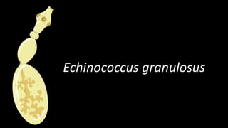
Dog Tapeworm Cyst
- 2. Common name The dog tapeworm The hydatid worm
- 3. Habitat • Adult worm resides in the small intestine of dog and other canine animals (wolf, fox and jackal). • Larval form is seen in man and other intermediate hosts (sheep, goat, cattle, pig and horse). • The dog and sheep are optimum definitive and intermediate hosts respectively and the cycle of transmission is maintained between them
- 4. Morphology • Adult worm: It is a small tapeworm measuring 36 mm in length. It consists of a scolex, neck and strobila • Scolex: It is pyriform in shape and measures about 300 um in diameter. It possesses four suckers and a protrusible rostellum with two Circular rows of hooklets. • Neck: It is short and thick. • Strobila: It consists of three segments (occasionally four). The first segment is immature, the second is mature and the third (and the fourth when present) is gravid. • Eggs: These are indistinguishable from those of other Taenia species. these are spherical, brown in color, and measure 31-43 um in diameter, and contain hexacanth embryos With three pairs of hooklets. • Larval form: This is found within the hydatid cyst which develops in the intermediate host
- 5. Morphology
- 7. Life Cycle • E. granulosus passes its life cycle in two hosts. • The adult worm lives attached to the mucosa of small intestine of dog and other canine animals. • The eggs are discharged in the feces. These are swallowed by the intermediate hosts while grazing in the fields. • Man acquires infection by a direct contact with infected dog or by allowing the dog to feed from the same dish or by ingesting water or food contaminated with dog’s feces containing eggs of E. granulosus. • In the duodenum the hexacanth embryos called oncospheres hatch out. These penetrate the intestinal wall and enter into the radicles of portal vein and are carried to the liver. • The liver acts as the first filter where 60-70% of human infections are located. Some embryos may pass through the hepatic capillaries and enter the pulmonary circulation. • Lungs act as the second filter. A few of these embryos may pass pulmonary circulation too and enter general circulation and may lodge in various organs like brain, heart, spleen, kidneys, genital organs, muscles, bones, etc. • Wherever the embryos settle, an active cellular reaction consisting of monocytes, giant cells and eosinophils takes place around the parasite.
- 8. • A large number of the parasites may thus be destroyed by host defense mechanism. Some of the embryos, however, escape destruction and develop into hydatid cysts. The cellular reaction in these cases gradually disappears, followed by the appearance of fibroblasts and the formation of new blood vessels • Fibroblasts lay fibrous tissue, which envelops the growing embryo. This is known as peri-cyst. • This merges With surrounding normal tissue. The parasite derives its nutrition through this layer. In old cysts the peri-cyst may become sclerosed or calcified and parasite within it may die. • Inside the peri-cyst, the embryo develops into a fluid- filled bladder known as hydatid cyst. From inner side of the cyst, brood capsules with a number of scolices are developed.
- 11. • When animals that serve as intermediate hosts are slaughtered, the viscera may not be disposed off properly and may be consumed by animals that serve as definitive hosts. The adult worms then develop in the intestine of definitive host. • These lay eggs which are passed in the feces of infected animals and the cycle is repeated. Since dog has no access to the hydatid cysts developed in viscera of man, therefore, the life Cycle of the parasite comes to a dead end
- 13. Hydatid Cyst
- 14. Pathogenicity • E. granulosus causes cystic echinococcosis or hydatidosis or hydatid disease or hydatid cyst in man. • It represents larval form of the parasite. The disease is generally acquired during childhood though it does not manifest before adult life. The cyst wall secreted by the embryo consists of two layers
- 15. • Ectocyst • It is outer layer. It is tough, acellular, laminated, hyaline membrane up to 1mm in thickness. • It resembles white of a hard-boiled egg. It is elastic, therefore, when excised or ruptured, it curls on itself thus exposing the inner layer containing brood capsules, scolices and daughter cysts. • Endocyst • It is inner or germinal layer. It consists of a number of nuclei embedded in a protoplasmic mass. It measures 22-25 um in thickness. It gives rise to ectocyst on outside and brood capsules and scolices on inside. It also secretes hydatid fluid. • Hydatid fluid is clear, colourless or pale yellow. • It has specific gravity of 1.005-1.010. It is sightly acidic (pH 6.7) and contains sodium chloride, sodium sulphate, sodium phosphate and sodium and calcium salts of succinic acid. • It is antigenic, therefore, it is used for Casoni's test and when absorbed it leads to anaphylactic shock. • Centrifuged deposit of the hydatid fluid shows hydatid sand which consists of brood capsules, free scolices and hooklets.
- 16. • If the cyst ruptures, either spontaneously in the body or during surgery, the danger from death from anaphylactic shock is high. • Acephalocysts • Some cysts are sterile and never produce brood capsules; some become sterile by bacterial invasion or calcification. In other cysts the brood capsules never produce scolices; hence they are called acephalocysts. If ingested by definitive host these cysts do not lead to infection. • Endogenous daughter cyst • Sometimes a fragment of the germinal layer may detach and develop into daughter cyst inside the mother cyst. This is known as endogenous daughter cyst. It also has both ectocyst and endocyst with brood capsules and scolices. Grand daughter cysts may also be formed. • Exogenous cyst • In case of hydatid disease of bone, because of high intracystic pressure, herniation or rupture of germinal and laminated layer may occur through some weaker part of the bone resulting in formation of exogenous cyst.
- 17. Clinical disease • Hydatid disease in humans is potentially dangerous; however, cyst size and organ location will greatly influence the outcome. In untreated or in inadequately treated patients mortality is> 90% within 10-15 years of diagnosis. • Liver cysts • The majority of hydatid cysts occur in the liver, causing symptoms that may include chronic abdominal discomfort, occasionally with a palpable or visible abdominal mass. • Liver cysts tend to occur more frequently in the right lobe. If a cyst becomes infected with bacteria, it resembles an abscess. If the cyst ruptures, either spontaneously, from trauma, or during surgery, there may be serious allergic reactions including skin rash, anaphylactic shock, or death.
- 18. • Lung cysts • Cysts in the lungs are usually asymptomatic until they become large enough to cause cough, shortness of breath, or chest pain. Cyst rupture may lead to expectoration of hydatid fluid or membranes, followed by the development of infection and a lung abscess. If rupture occurs into the lung, it may cause pneumo- thorax and empyema, allergic reactions, and even anaphylactic shock. • Other sites • Other organs which may also be involved include spleen (3-5%), central nervous system and heart (1- 1.5%), kidneys, bones, muscles, female genital tract, eyes, etc. leading to visible swelling and pressure effects.
- 19. Laboratory Diagnosis It can be carried out by the following methods: 1. Casoni's test • It is an immediate hypersensitivity skin test which was introduced by Casoni in 1911. • Antigen for the Casoni’s test is sterile hydatid fluid drawn from unilocular hydatid cysts from sheep. pig, cattle or man. • The fluid is filtered, tested for sterility and stored in sealed ampoules under refrigeration. For the test, 0.2 ml of the antigen is injected intradermally in one arm. For control, an equal amount of sterile normal saline is injected intradermally on the other arm. • The control fades almost immediately, while the tested site in positive case develops a large wheal measuring 5 cm or more in diameter with multiple pseudopodia within 30 minutes. • This test has a low sensitivity (55-70) and gives false positive reactions in patients suffering from other cestode infections.
- 20. 2. Differential leucocyte count • Differential leucocyte count may reveal eosinophilia (20-259%). 3. Serological tests • Serodiagnosis of hydatid cyst may be camied out by, • Enzyme-linked immunosorbent assay (ELISA), • Radio-immunoaSsay (RIA), • Complement Fixation, • Indirect haem-agglutination (lHA), • Bentonite Flocculation • latex agglutination tests.
- 21. 4. Examination of cyst fluid • Examination of cyst fluid reveals Scolices, brood capsules and hooklets. Because leakage of fluid in the adjoining tissue may lead to anaphylactic shock, therefore, the fluid aspirated from surgically removed cyst should be examined and diagnostic puncture cyst is not recommended. 5. Histological examination • Histological examination of surgically removed cyst reveals different layers of the hydatid cyst, i.e., pericyst, ectocyst and endocyst 6. Radiodiagnosis • X-ray, ultrasound and CT scan are also helpful in the diagnosis of hydatid cyst.
- 22. Treatment • Removal of the cysts by surgery is the preferred treatment, but leakage from ruptured cysts may spread disease to other organs. • Prolonged regimens of albendazole, either alone or as an adjunct to surgery, are reportedly of some benefit. • Benzimidazole treatment should be administered in the perioperative period.
- 23. Prophylaxis • E. granulosus infection can be prevented by: • Strict personal hygiene. • Dogs should not be allowed to eat the carcasses of slaughtered animals in endemic areas. • Reduction of stray dog population