2. Geographical distribution:
- world wide in distribution.
- it occurs in person with unhygienic habits.
Habitat:
The adult worms lives in the lumen of small intestine
(jejunum ) of man.
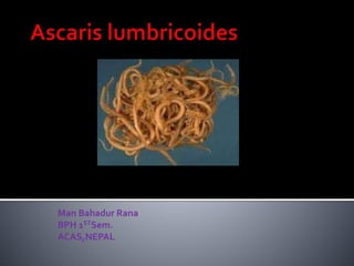
3. It is elongated and rounded in shape and
tapers at both ends.
The mouth opens at the anterior end and
possesses three finely toothed lips, one
dorsal and two ventral.
Male:
- It measures 15-25 cm in length and 3-4mm
thickness.
4. - The tail end of the male is curved ventrally in the
form of hook and contains two curved spicules for
copulatory.
Female:
- It is longer and stouter than the male and
measures 25-40cm in length and 5mm thickness.
-The posterior end is neither curved nor pointed.
-The vulva opens in the middle third of the body,
this section of worm is narrower and is called a vulvar
waist.
5.
6.
7. - The egg laying capacity of the female worm has
been estimated to be approximately 200000 eggs
daily.
Eggs:
It is of two types:
- Fertilised eggs
- Unfertilised eggs.
8. Fertilised egg:
- It is round or oval in shape measuring 60-75um in length and
40-50um in breadth.
- It is bile stained and thus brownish in colour.
- It is surrounded by a thick translucent shell with an outer
albuminous coat.
- It contains a large unsegmented ovum.
- It floats in saturated solution of common salt.
9. Unfertilised egg:
The female even if not fertilised, can produce
unfertilised eggs.
- It is narrower and longer measuring 80um in length and
55um in breadth.
- It is bile stained and brownish in colour.
- It has an thinner shell with an irregular coating of albumin.
- It contains a small atrophied ovum.
- It does not float in saturated solution of common salt
(heaviest of all helminthic egg).
10.
11. The worm passes its life cycle in one host
and no intermediate host is required.
Man is the only known definitive host of Ascaris
lumbricoides.
The various stages of life cycle are as follows.
Stage I : Eggs in faeces:
Fertilised eggs are passed
with the faeces they are not infective to man
when freshly passed.
12. Stage II : Development in soil :
The rhabditiform larvae
developed within egg shell in 10-40days time
depending on the atmospheric temperature.
Stage III : Infection by ingestion and liberation
of larvae: When ingested with food, drinks or
raw vegetables, the embryonated eggs reach to
the duodenum and digestive juices split the egg
shell, the rhabditiform larva liberated in the
upper part of small intestine.
13. Stage IV : Migration through lungs : The
larvae liberated in the small intestine burrow
their way through mucous membrane of
small intestine carried by the portal
circulation to the liver. Finally, they pass out
the liver via right heart enter the pulmonary
circulation while in the lungs they grow much
bigger and breaking the capillary wall of lungs
they reach the lungs alveoli.
14. StageV : Re entry into stomach and small
intestine: From the lung alveoli, the larvae
reach the bronchi and trachea and by the
ciliated epithelium of the respiratory tract
they are propelled to the larynx and pharynx
and are once more swallowed .The larvae
pass down the oesophagus to the stomach
and localise in the small intestine, their
normal abode.
15. StageVI : Sexual maturity and egg
liberation : The larvae on reaching their
habitat grow into adult worms and become
sexually mature.The gravid females begins to
discharge eggs in the stool with in about 2
months from the time of infection.The cycle
is again repeated.
16.
17. Infection is affected by swallowingAscaris
eggs (embryonated eggs) with raw vegetable
cultivated in a soil fertilised by infected
human excreta.Water supplies may be
contaminated and infection may occur by
drinking such water.
Infecting agent: Embryonated egg.
Portal of entry: Alimentary canal.
Migration of larvae:Through lungs
Site of location: Small intestine.
18. Infection of Ascaris lumbricoides is known as
Ascariasis.
The Symptoms of Ascaris Infection may be
divided into two groups:
i)Those produced by migrating
larvae.
ii)Those produced by adult worms.
19. i)Those produced by migrating larvae:
. Larvae in the lungs:
Causes Ascaris Pneumonia (Loefflers
Syndrome).
Symptoms of pneumonia such as fever,
cough and dyspnoea.
Sputum which is often blood-tinged may
containAscaris larvae.
Urticarial rash and eosinophilia may be seen.
20. . Larvae in general circulation:
If the Ascaris larvae reach other organs, they
may set up clinical symptoms related to those
organs. E.g brain, spinal cord, heart, kidneys.
ii)Those produced by Adult worms:
1. Spoliative action: By robbing the host of its
nutrition (protein and vitamin content of the
worm is high) and may contribute to protein
energy malnutrition.
Ascaris infection may causeVitaminA
deficiency.
21. 2.Toxic action:
The body fluid of Ascaris when
absorbed is toxic and may give rise to
typhoid like fever ; also responsible for
various allergic manifestation such as
ulticaria, oedema of the face ,
conjunctivitis and irritation of the upper
respiratory tract.
22. 3. Mechanical Effect :
i) Intussucception
ii) It may penetrate through the
ulcers of the alimentary canal.
iii) Intestinal obstruction.
23.
24.
25.
26.
27.
28. . Direct evidence:
a) Finding of the adult worms.
In the stool or vomit.
X-ray diagnosis with barium meal.
b) Findings of eggs:
In the stool: Direct microscopic examination
of a saline emulsion of the stool.
In the bile: Microscopic examination of the
bile.
29. . Indirect evidences:
c) Blood exmination - Eosinophilia
d) Derma reaction (Allergic) - ‘
Scratch test’ with powdered
Ascaris antigen.
e) Antibodies against the parasite
can be detected.