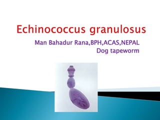
4. Adult worm:
- It is a small tapeworm, measuring 3-6mm
in length.
- It comprises of Scolex (head), neck and
Strobila consisting of 3 segments .
1st: Immature
2nd : Mature
3rd : Gravid
5. - The terminal segment is biggest, measuring
2-3 mm in length and 0.6mm width.
- The Scolex bears four suckers and a
protrusible rostellum.
Neck :
The neck is short and thick.
6.
7.
8. - It is spherical in shape and
measures 32-36umx 25-
32um.
- It contains hexacanth embryo
with 3 pairs of hooks.
- Eggs are indistinguishable
from those of other Taenia sp.
9. - This is found with in the hydatid cyst and
represent the scolex of the future adult
worm.
- It remains invaginated with in a vesicular
body.
- When it enters the definitive host, the scolex
with four suckers and hooklet, becomes
evaginated and develop into an adult worm.
10.
11.
12.
13. The worm passes its life cycle in two host.
Definitive host : Dog, wolf, fox and
jackal.
Intermediate host : sheep, goat, man.
The eggs are discharged with the faeces of
the definitive host(dogs and allied animals).
These eggs are swallowed by the
intermediate host sheep and other domestic
animals while grazing in the field and also by
man.
14. The hexacanth embryo hatched in
the duodenum.
After 8 hours after ingestion, the
embryo penetrate the gut wall and is
carried to the liver through the portal
vein.
About 70% of the onchosphere are
lodged (arrested) in the liver itself
(liver acts as a first filter).
15. Some of the embroys may pass through
hepatic capillaries, enter the pulmonary
circulation and filter out in the lungs (lungs
act as a second filter).
Few of the embroys enter the blood stream
and lodge in various organs.
Wherever the embryo (oncosphere) settles, it
forms a hydatid cyst.
16. From the inner side of the cyst, brood
capsules with a number of scolices are
developed.
Upon ingestion of carcasses of intermediate
host by the definitive host, each scolices
grows into an adult worm in about 2 months.
The cycle is repeated except with human
host.
17. As the dogs have no access to the hydatid
cyst developed in the viscera of man, the life
cycle of parasite comes to a dead end.
Life span of adult worm is 6 months and life
span of larval worm may continue to develop
for many years.
18. - The adult worms of E. granulosus in dogs do
not cause much inconvenience.
- They are found in large number (by 100 or
even 1000) in small intestine of infected dogs
where they lie embedded in the mucous
membrane and on postmortem appears as
small white specks.
- The larval worm of E. granulosus in man
causes unilocular hydatid disease
(hydatidosis).
19. - Infection is acquired by the ingestion of eggs
in the dogs faeces. This occurs in following
ways.
i) by direct contact (handling) with infected
dogs.
ii) by allowing the dog to feed from the same
dish.
iii) by taking vegetables contaminated with
infected dog faeces.
20. - Infection through contaminated water is not
common as the eggs being heavier sink to
the bottom.
Infecting agent : eggs in dog faeces.
Portal of entry : Alimentary tract.
Site of localisation : Viscera ( liver,
lungs and other organs)
21. Hydatid cyst :
The cyst wall consist of two layers:
- Outer layer (ectocyst).
- Inner or germinal layer ( endocyst).
Ectocyst:
. It is a laminated hyaline membrane,
thickness of 1mm
. To the naked eye, the ectocyst has the
appearance of the white of hard boiled eggs.
22.
23. . Elastic in nature and when incised or ruptured
curls itself back, exposing its inner surface.
Inner or germinal layer (endocyst)
:
. It is a cellular layer and it give rise to ectocyst
on outer side and brood capsule and scolices on
inner side.
. It is very thin and about 25um in thickness.
. It is a vital layer of cyst and secrete the specific
hydatid fluid.
24. It is a clear colourless fluid (may be pale
yellow in colour).
It has a low specific gravity (1.005-1.010).
It is slightly acidic (pH 6.7) in nature.
It contains sodium chloride, sodium
sulphate, sodium phosphate, and calcium
salts of succinic acids.
Antigenic – being used for immunological
test.
It is highly toxic, when absorbed give rise to
anaphylactic shock.
25. Acephalocyst: Sometimes brood capsules
are not formed and if formed are without
scolices. These types of cyst are sterile and are
called Acephalocyst. These cyst when ingested by
definitive host do not lead to infection.
Endogenous daughter cyst
formation: After a long period of growth,
daughter cyst are formed within the mother cyst
and may arise from the detached fragment of the
germinal layer. The endogenous daughter cyst
also have both ectocyst and endocyst with brood
capsule and scolices.
26. Exogenous cyst formation: In case of
hydatid disease of bone, herniation or
rupture of germinal and laminated layer may
occur through some weaker part of bone and
result in formation of exogenous cyst.
Distribution of Hydatid cyst:
Liver (75%), lungs (8%), muscle (5%), brain
(2%), kidney (2%), heart (1%), bone (1%) and
other parts of body (4%).
27.
28. Rate of growth of hydatid cyst:
- The development of hydatid cyst in man is
very slow.
- At the end of year, it is approximately 4cm in
diameter.
Clinical features :
- The chief clinical manifestation are dependent
upon local signs and if cyst is situated
superficially it may cause visible swelling.
- The disease remain symptomless for many
years and its presence is only detected at
autopsy.
29. Parasitological : Detection of scolices in cyst fluid
or centrifuged deposit (hydatid cyst) is 100%
confirmatory.
Casoni test:
- It is an immediate hypersensitivity (type-I)
skin test introduced by Casoni in 1911.
- Intradermal injection of 0.2ml of fresh sterile
hydatid fluid into the volar space of forearm.
- Rxn is seen after 30 min, a large wheal (5cm
in diameter) multiple pseudopodia is seen.
30. Detection of antibodies:
Enzyme Linked Immunosorbent Assay
(ELISA), Complement Fixation Test (CFT),
Indirect haemagglutination Assay (IHA).
Detection of Antigen :
ELISA can be used for detection of antigen
in the serum.
Blood Examination:
Eosinophilia (20-25)% may be present.
31. Radiological : This is often helpful in the
diagnosis of hydatid cyst of lungs and liver.
- USG of whole abdomen is useful in locating
the site of hydatid cyst of the abdominal
organ.
- CT-scan is more helpful.
Histological examination: Surgically,
removed cyst may be examined to reveal
different layers of the hydatid cyst i.e
ectocyst and endocyst.
32.
33. - Surgical removal of the hydatid cyst can be
performed but there may be recurrences in
2-25% cases of surgery.
- Hence postoperative chemotherapy may be
given for at least two years after surgery.
- Praziquantel and Albendazole are the
chemotherapeutic agent used for the
treatment of hydatid cyst.
34. - Personal hygiene including
avoidance of close contact with dogs.
- Control in movement of dogs.
- General health Education.
- Not allowing the dogs to eat
carcasses of slaughtered animals in
endemic areas.