tuberculosis of the abdominal
•Download as PPTX, PDF•
1 like•315 views

1. Abdominal tuberculosis is the third most common form of extrapulmonary tuberculosis, affecting the gastrointestinal tract, peritoneum, and solid organs. 2. It is usually caused by ingesting infected food or milk, or from hematogenous or contiguous spread from active pulmonary lesions. Common symptoms include abdominal pain, weight loss, and fever. 3. Diagnosis involves ascitic fluid analysis, imaging studies, biopsy, and culture of the bacteria. Treatment consists of a multi-drug antibiotic regimen over 6-9 months, with surgery sometimes needed for complications like strictures or perforations.

Recommended
More Related Content
What's hot
What's hot (19)
Similar to tuberculosis of the abdominal
Similar to tuberculosis of the abdominal (20)
Recently uploaded
Recently uploaded (20)
tuberculosis of the abdominal
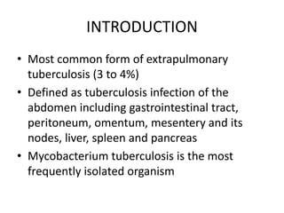
- 1. INTRODUCTION • Most common form of extrapulmonary tuberculosis (3 to 4%) • Defined as tuberculosis infection of the abdomen including gastrointestinal tract, peritoneum, omentum, mesentery and its nodes, liver, spleen and pancreas • Mycobacterium tuberculosis is the most frequently isolated organism
- 2. PATHOPYSIOLOGY • Ingestion of milk or infected food • Swallowing of sputum in active PTB • Hematogenous spread from active pulmonary lesion, miliary tuberculosis • Contiguous spread from infected foci like fallopian tubes, mesenteric lymph node • Very rarely as a consequence of peritoneal dialysis
- 3. CLASSIFICATION OF THE ABDOMINAL TUBERCULOSIS • Gastrointestinal tuberculosis Ulcerative Hypertrophic Sclerotic or fibrous Diffuse colitis Peritoneal tuberculosis Acute Chronic • 1. Ascitic form • 2. Encysted form • 3. Fibrous form
- 4. . • Tuberculosis of the mesentery and its contents • Tuberculosis of the solid viscera Liver Pancreas Spleen • Miscellaneous Retroperitoneal lymph node tuberculosis
- 5. GASTROINTESTINAL TB • Constitutes 70 to80% of abdominal tuberculosis • Any region of the gastro intestinal tract from mouth to anus can be involved • Ileoceacalarea most commonly affected • It can be of ulcerative, hypertrophic, diffuse colitis, ulcerohypertrophic, and sclerotic forms • Entero-enteric, entero-vesicaland entero- cutaneousfistula can occur • Luminal narrowing is often caused by adjacent lymphadenitis which results in traction diverticulaformation, narrowing and sinus tract formation
- 7. GESTRIINTESTINAL TB • Ulcerative form • Usually occurs in adult patients whoare malnourished • Ulcers lie transverse “girdle ulcers” • Areas of the normal appearing mucosamay be found • Healing and fibrosis results in stricture • Hypertrophic form • Commonly occurs in young patients who are relatively well nourished • Characterised by extensive inflammation and fibrosis which often results in adherence of bowel, mesentery and lymph nodes
- 9. GESTROINTESTINAL TB • Clinical features • 20 to 40 yrs age group most often affected • A slight female preponderance • Most common symptom is abdominl pain others include abdominal distention, wt.loss anorexia, fever, diarrhoea or constipation borborygmi, bleeding per rectum • Signs include anemia, malnutrition, abdominal tenderness, ascites, mass in the right iliac fossa features of intestinal obstruction • Classic doughy abdomen described only in 6 to 11% in Indian studies
- 10. GESTRIINTESTINAL TB • Oesophageal tuberculosis • Very rare, upper part is involved more often than lower part, commonly present with dysphagiaand odynophagia • Gastric tuberculosis • Rare due to the presence of gastric acid • Ulcerative form is the commonest • Duodenal tuberculosis (MAC infection) • Tuberculosis of Appendix • Anal tuberculosis • Mostly ulcerative, may be lupoid, verrucus, miliary lesion • Multiple fistulae with inguinal lymphadenopathy
- 11. PERITONEAL TB • Acute tuberculousperitonitis • Chronic tuberculousperitonitis • Ascitic form • Insidious in onset, abdominal pain usualyabsent, rolled up omentum infiltrated with tubercle may felt as a transverse solid mass • Encysted (loculated) form • Fibrous form • Wide spread adhesions may cause coils of intestine matted together and distended, they may act as blind loop
- 12. PANCRATIC TB
- 14. HEPATOBILIARY TB • In a patient with PUO, marked elevation of serum alkaline phosphatase(3 to 6 times) with mild elevation of s.transaminases, normal PT, s.albuminand a slight increase in bilirubin hepatic tuberculosis should be suspected • Clinical syndromes of Hepatobiliary tuberculosis • Congenital tuberculosis • Primary hepatic tuberculosis • Disseminated/miliary tuberculosis • Tuberculoma • Tuberculosis of biliary tract • Hepatic failure • Granulomatous hepatitis • Tuberculous pylephlebitis
- 16. DIFFRENTIAL DIAGNOSIS Malabsorption • Coeliac disease • Lymphoma • Immuno proliferative small intestinal diseae Mass • Appendicular mass • Actinomycosis • Crohn’sdisease • Caecalcarcinoma • Lymphoma Ascites • Cardiac disease • Renal disease • Hepatic diseae • malignacy
- 17. INVESTIGATION • Hematology &serum biochemistry Anemia, raised ESR, hypoalbumenemia, leucopenia with relative lymphocytosis, normal serum transminase level, raised serum ALP • Ascitic fluid examination Exudative, fluid protein>3gm%, SAAG<1.1 Ascitic/blood glucose ratio<0.96, WBC count usually 140 to 4000cells/mm³ consist of lymphocytes predominantly, AFB(+<3%), culture(+<20%), IFN-γincreased ADA((98%sensitivity&95%specificity at cut off value 32 IU/L), PCR • Mantoux test (positive in 50 to 100%)
- 19. . • Culture medium Lowenstein-Jensen Middlebrook7H11 Liquid medium • QuantiFERON-TB test(QFT) • BACTEC radiometric system • MycobacterialGrowth indicator tubes • Animal pathogenicity • PCR assay • Ligasechain reaction
- 21. . • Imaging studies • Chest skiagram (associated PTB in 24 to 28%) • Plain X-ray abdomen • May show calcified lymph nodes or granulomas in the liver, spleen, pancreas. Other features include dilated loops with fluid levels, dilatation of terminal ileum and ascites . Pneumoperitoneummay be evident in patients with intestinal perforation
- 22. .
- 23. .• Barium studies • Enteroclysis followed by barium enema is the best protocol • Increased transit time with hypersegmentation (chicken intestine) and flocculation is the earliest sign • Localised areas of irregular thickened folds, mucosal ulceration, dilated segments and strictures • Thickened iliocaecal valve with a broad triangular appearance with the base towards the caecum (inverted umbrella sign or (Fleischner’ssign) • Rapid transit and lack of barium retension(Sterlin’ssign) • Narrow beam of barium due to stenosis(string’s sign) • Barium oesophagogram-ulcerative oesophagitis, stricture, pseudo tumourmasses, fistula, sinus, traction diverticulae • Duodenal tuberculosis-segmental narrowing, widening of the “C” loop due to lymphadenopathy
- 24. .
- 25. BERIUM MEAL FOLLO THROUGH FINDING IN INTESTINAL TB • Group1: Highly s/o intestinal TB if one or more of the following features are present • a. Deformed ileocaecal valve with dilatation of terminal ileum • b. Contracted caecum with an abnormal ileocaecal valve and/or terminal ileum • c. Stricture of the ascending colon with shortening of and involvement of ileocaecal region
- 27. . • GroupII:Suggestiveofintestinaltuberculosisifon eofthefollowingfeaturesispresent • a.Contractedcaecum • b.Ulcerationornarrowingoftheterminalileum • c.Strictureoftheascendingcolon • d.Multipleareasofdilatation,narrowingandmatt ingofsmallbowelloops
- 28. . • GroupIII:Non-specificchanges • Featuresofmatting,dilatationandmucosalthick eningofsmallbowelloops • GroupIV:Normal study
- 29. .• Abdominal sonography • Often reveals a mass made up of matted loops of small bowel with thickened walls, diseased omentum, mesentery and loculated asites • Fine septaemay be seen in the asciticfluid • Interloopascites gives rise to charecteristic“club sandwitch” appearance • Mesenteric thickening is better detected in the presence of ascites and is often seen as the “stellatesign” of bowel loops radiating from its root • In intestinal tuberculosis bowel wall thickening is usually uniform and concentric as opposed to the eccentric thickening at the mesenteric border seen in Crohn’sdisease and the variegated appearance seen in malignancy • Granulomas or absessin the liver ,pancreas or spleen
- 31. .• Abdominal computerised tomography • CT is better than USG in detecting high dense ascites • Abdominal lymphadenopathy is the commonest manifestation of tuberculosis on CT • Retroperitoneal, peripancreatic, portahepatis, and mesenteric/omentallymph node enlargement may be evident • Caseous necrosing lymph node appears as low attenuating, necrotic centers and thick, enhancing inflammatory rim • Preferential thickening of the medial caecalwall with an exophytic mass engulfing the terminal ileum associated with massive lymphadenopathy is characteristic of tuberculosis • Short segments of mural thickening with normal intervening bowel associated with ileocaecal involvement strongly suggest tuberculosis
- 32. .
- 33. .• MRI:-has no added advantage • Endoscopy • Fine needle aspiration cytology • Peritoneal biopsy • Laparoscopy:-most effective method. 80 to 95% diagnostic accuracy. Characteristic finding include multiple, yellowish-white miliary nodules over peritoneum, erythematous, thickened and hyperemic peritoneum
- 34. .
- 35. . • Colonoscopy • › Excellent tool for suspected colonic & terminal • ileal involvement • › Mucosal nodules (2-6mm) & ulcers in a • discrete segment of 4-8 cm, with normal or • hyperemic intervening mucosa are • pathognomic • › Other findings: strictures, deformed ileocaecal • valve, mucosal oedema, pseudopolyps and • diffuse colitis • › Biopsy can be taken to eslablish the diagnosis
- 37. .• DIFFERENTIAL DIAGNOSIS • Abdominal TB may mimic any of the following • conditions: • 1. Malignant neoplasms: lymphoma, carcinoma • 2. Inflammatory bowel disease e.g crohn’s • disease • 3. Ascites: hepatic/ cardiac/ renal/ malignant • 4. Ileocaecal mass: appendicular lump, CA • caecum • 5. Malabsorption syndromes
- 38. TREATMENT • Medical treatment • • There are two categories of treatment: • A) cirrhotic patients with essentialy normal • baseline LFTs (Child A cirrhosis) • Treat with standard 4 drug regime for 2 months f/b 2 • drugs regime for 4 months • Pyrazinamide being most hepatotoxic can be • avoided and a 9 month 3 drug regime may be used • B) Cirrhotic patients with altered baseline LFTs • (Childs B & C) • One or two hepatotoxic drugs may be used in • moderately severe disease ( Child B cirrhosis) • but totally avoided in decompensated cirrhosis
- 39. . • Two hepatotoxic drugs: • 9 months of Isoniazid, Rifampin & Ethambutol • 2 months of Isoniazid, Rifampin, Ethambutol & • Streptomycin f/b 6 months of Isoniazid & Rifampin • One hepatotoxic drug: • 2 months of Isoniazid, Ethambutol & Streptomycin f/b 10 • months of Isoniazid& Ethambutol • No hepatotoxic drug • 18-24 monthsof Streptomycin, Ethambutol and Quinolones
- 40. • Hepatotoxicity • Regular LFT monitoring recommended in all • patients on ATT • In the general population, the criteria for • stopping anti tubercular treatment is • • AST / ALT > 3times upper limit of normal • and symptomatic • • AST / ALT > 5times upper limit of normal • even if asymptomatic. • • Any rise in bilirubin
- 41. . • Surgical Management: • 1. Ileocaecal resection with 5 cm margin • 2. Stricturoplasty- single stricture • 3. Single strictutre with friable bowel : Resection • 4. Multiple Strictures: Resection and anastomosis • 5. Multiple strictures with long segment gaps: • Multiple stricturoplasty
- 42. . • 6. Early perforation: resection and anastomosis • (due to friable bowels) • 7. Perforation with severe contamination: resection • with colostomy • 8. Adhesiolysis by laproscopy (Very difficult • procedure) • 9. Drainage of abscesses and treatment for fistula • in ano