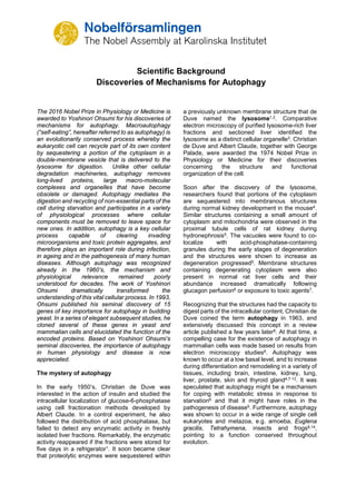
SlideShare a Scribd company logo
1 of 7
Download to read offline
Scientific Background
Discoveries of Mechanisms for Autophagy
The 2016 Nobel Prize in Physiology or Medicine is
awarded to Yoshinori Ohsumi for his discoveries of
mechanisms for autophagy. Macroautophagy
(โ€œself-eatingโ€, hereafter referred to as autophagy) is
an evolutionarily conserved process whereby the
eukaryotic cell can recycle part of its own content
by sequestering a portion of the cytoplasm in a
double-membrane vesicle that is delivered to the
lysosome for digestion. Unlike other cellular
degradation machineries, autophagy removes
long-lived proteins, large macro-molecular
complexes and organelles that have become
obsolete or damaged. Autophagy mediates the
digestion and recycling of non-essential parts of the
cell during starvation and participates in a variety
of physiological processes where cellular
components must be removed to leave space for
new ones. In addition, autophagy is a key cellular
process capable of clearing invading
microorganisms and toxic protein aggregates, and
therefore plays an important role during infection,
in ageing and in the pathogenesis of many human
diseases. Although autophagy was recognized
already in the 1960โ€™s, the mechanism and
physiological relevance remained poorly
understood for decades. The work of Yoshinori
Ohsumi dramatically transformed the
understanding of this vital cellular process. In 1993,
Ohsumi published his seminal discovery of 15
genes of key importance for autophagy in budding
yeast. In a series of elegant subsequent studies, he
cloned several of these genes in yeast and
mammalian cells and elucidated the function of the
encoded proteins. Based on Yoshinori Ohsumiโ€™s
seminal discoveries, the importance of autophagy
in human physiology and disease is now
appreciated.
The mystery of autophagy
In the early 1950โ€™s, Christian de Duve was
interested in the action of insulin and studied the
intracellular localization of glucose-6-phosphatase
using cell fractionation methods developed by
Albert Claude. In a control experiment, he also
followed the distribution of acid phosphatase, but
failed to detect any enzymatic activity in freshly
isolated liver fractions. Remarkably, the enzymatic
activity reappeared if the fractions were stored for
five days in a refrigerator1. It soon became clear
that proteolytic enzymes were sequestered within
a previously unknown membrane structure that de
Duve named the lysosome1,2. Comparative
electron microscopy of purified lysosome-rich liver
fractions and sectioned liver identified the
lysosome as a distinct cellular organelle3. Christian
de Duve and Albert Claude, together with George
Palade, were awarded the 1974 Nobel Prize in
Physiology or Medicine for their discoveries
concerning the structure and functional
organization of the cell.
Soon after the discovery of the lysosome,
researchers found that portions of the cytoplasm
are sequestered into membranous structures
during normal kidney development in the mouse4.
Similar structures containing a small amount of
cytoplasm and mitochondria were observed in the
proximal tubule cells of rat kidney during
hydronephrosis5. The vacuoles were found to co-
localize with acid-phosphatase-containing
granules during the early stages of degeneration
and the structures were shown to increase as
degeneration progressed5. Membrane structures
containing degenerating cytoplasm were also
present in normal rat liver cells and their
abundance increased dramatically following
glucagon perfusion6 or exposure to toxic agents7.
Recognizing that the structures had the capacity to
digest parts of the intracellular content, Christian de
Duve coined the term autophagy in 1963, and
extensively discussed this concept in a review
article published a few years later8. At that time, a
compelling case for the existence of autophagy in
mammalian cells was made based on results from
electron microscopy studies8. Autophagy was
known to occur at a low basal level, and to increase
during differentiation and remodeling in a variety of
tissues, including brain, intestine, kidney, lung,
liver, prostate, skin and thyroid gland4,7-13. It was
speculated that autophagy might be a mechanism
for coping with metabolic stress in response to
starvation6 and that it might have roles in the
pathogenesis of disease5. Furthermore, autophagy
was shown to occur in a wide range of single cell
eukaryotes and metazoa, e.g. amoeba, Euglena
gracilis, Tetrahymena, insects and frogs8,14,
pointing to a function conserved throughout
evolution.
2
During the following decades, advances in the field
were limited. Nutrients and hormones were
reported to influence autophagy; amino acid
deprivation induced15, and insulin-stimulation
suppressed16 autophagy in mammalian tissues. A
small molecule, 3-methyladenine, was shown to
inhibit autophagy17. One study using a combination
of cell fractionation, autoradiography and electron
microscopy provided evidence that the early stage
of autophagy included the formation of a double-
membrane structure, the phagophore, that
extended around a portion of the cytoplasm and
closed into a vesicle lacking hydrolytic enzymes,
the autophagosome18 (Figure 1).
Despite many indications that autophagy could be
an important cellular process, its mechanism and
regulation were not understood. Only a handful of
laboratories were working on the problem, mainly
using correlative or descriptive approaches and
focusing on the late stages of autophagy, i.e. the
steps just before or after fusion with the lysosome.
We now know that the autophagosome is transient
and only exists for ~10-20 minutes before fusing
with the lysosome, making morphological and
biochemical studies very difficult.
Figure 1. Formation of the autophagosome. The
phagophore extends to form a double-membrane
autophagosome that engulfs cytoplasmic material. The
autophagosome fuses with the lysosome, where the
content is degraded.
In the early 1990โ€™s, almost 30 years after de Duve
coined the term autophagy, the process remained
a biological enigma. Molecular markers were not
available and components of the autophagy
machinery were elusive. Many fundamental
questions remained unanswered: How was the
autophagy process initiated? How was the
autophagosome formed? How important was
autophagy for cellular and organismal survival? Did
autophagy have any role in human disease?
Discovery of the autophagy machinery
In the early 1990โ€™s Yoshinori Ohsumi, then an
Assistant Professor at Tokyo University, decided to
study autophagy using the budding yeast
Saccharomyces cerevisae as a model system. The
first question he addressed was whether
autophagy exists in this unicellular organism. The
yeast vacuole is the functional equivalent of the
mammalian lysosome. Ohsumi reasoned that, if
autophagy existed in yeast, inhibition of vacuolar
enzymes would result in the accumulation of
engulfed cytoplasmic components in the vacuole.
To test this hypothesis, he developed yeast strains
that lacked the vacuolar proteases proteinase A,
proteinase B and carboxy-peptidase19. He found
that autophagic bodies accumulated in the vacuole
when the engineered yeast were grown in nutrient-
deprived medium19, producing an abnormal
vacuole that was visible under a light microscope.
He had now identified a unique phenotype that
could be used to discover genes that control the
induction of autophagy. By inducing random
mutations in yeast cells lacking vacuolar proteases,
Ohsumi identified the first mutant that could not
accumulate autophagic bodies in the vacuole20; he
named this gene autophagy 1 (APG1). He then
found that the APG1 mutant lost viability much
quicker than wild-type yeast cells in nitrogen-
deprived medium. As a second screen he used this
more convenient phenotype and additional
characterization to identify 75 recessive mutants
that could be categorized into different
complementation groups. In an article published in
FEBS Letters in 1993, Ohsumi reported his
discovery of as many as 15 genes that are
essential for the activation of autophagy in
eukaryotic cells20. He named the genes APG1-15.
As new autophagy genes were identified in yeast
and other species, a unified system of gene
nomenclature using the ATG abbreviation was
adopted21. This nomenclature will be used
henceforth in the text.
During the following years, Ohsumi cloned several
ATG genes22-24 and characterized the function of
their protein products. Cloning of the ATG1 gene
revealed that it encodes a serine/threonine kinase,
demonstrating a role for protein phosphorylation in
autophagy24. Additional studies showed that Atg1
forms a complex with the product of the ATG13
gene, and that this interaction is regulated by the
target of rapamycin (TOR) kinase23,25. TOR is
active in cells grown under nutrient-rich conditions
and hyper-phosphorylates Atg13, which prevents
the formation of the Atg13:Atg1 complex.
Conversely, when TOR is inactivated by starvation,
dephosphorylated Atg13 binds Atg1 and
autophagy is activated25. Subsequently, the active
kinase was shown to be a pentameric complex26
that includes, in addition to Atg1 and Atg13, Atg17,
Atg29 and Atg31. The assembly of this complex is
a first step in a cascade of events needed for
formation of the autophagosome.
3
Figure 2. Regulation of autophagosome formation. Ohsumi studied the function of the proteins encoded by key
autophagy genes. He delineated how stress signals initiate autophagy and the mechanism by which protein complexes
promote distinct stages of autophagosome formation.
The formation of the autophagosome involves the
integral membrane protein Atg9, as well as a
phosphatidylinositol-3 kinase (PI3K) complex26
composed of vacuolar protein sorting-associated
protein 34 (Vps34), Vps15, Atg6, and Atg14. This
complex generates phosphatidylinositol-3
phosphate and additional Atg proteins are recruited
to the membrane of the phagophore. Extension of
the phagophore to form the mature
autophagosome involves two ubiquitin-like protein
conjugation cascades (Figure 2).
Studies on the localization of Atg8 showed that,
while the protein was evenly distributed throughout
the cytoplasm of growing yeast cells, in starved
cells, Atg8 formed large aggregates that co-
localized with autophagosomes and autophagic
bodies27. Ohsumi made the surprising discovery
that the membrane localization of Atg8 is
dependent on two ubiquitin-like conjugation
systems that act sequentially to promote the
covalent binding of Atg8 to the membrane lipid
phosphatidylethanolamine. The two systems share
the same activating enzyme, Atg7. In the first
conjugation event, Atg12 is activated by forming a
thioester bond with a cysteine residue of Atg7, and
then transferred to the conjugating enzyme Atg10
that catalyzes its covalent binding to the Atg5
protein26,28,29. Further work showed that the
Atg12:Atg5 conjugate recruits Atg16 to form a tri-
molecular complex that plays an essential role in
autophagy by acting as the ligase of the second
ubiquitin-like conjugation system30. In this second
unique reaction, the C-terminal arginine of Atg8 is
removed by Atg4, and mature Atg8 is subsequently
activated by Atg7 for transfer to the Atg3
conjugating enzyme31. Finally, the two conjugation
systems converge as the Atg12:Atg5:Atg16 ligase
promotes the conjugation of Atg8 to
phosphatidylethanolamine26,32. Lipidated Atg8 is a
key driver of autophagosome elongation and
fusion33,34. The two conjugation systems are highly
conserved between yeast and mammals. A
fluorescently tagged version of the mammalian
homologue of yeast Atg8, called light chain 3
(LC3), is extensively used as a marker of
autophagosome formation in mammalian
systems35, 36.
Ohsumi and colleagues were the first to identify
mammalian homologues of the yeast ATG genes,
which allowed studies on the function of autophagy
in higher eukaryotes. Soon after, genetic studies
revealed that mice lacking the Atg5 gene are
apparently normal at birth, but die during the first
day of life due to inability to cope with the starvation
that precedes feeding37. Studies of knockout
mouse models lacking different components of the
autophagy machinery have confirmed the
importance of the process in a variety of
mammalian tissues26,38.
The pioneering studies by Ohsumi generated an
enormous interest in autophagy. The field has
become one of the most intensely studied areas of
biomedical research, with a remarkable increase in
the number of publications since the early 2000โ€™s.
Different types of autophagy
Following the seminal discoveries of Ohsumi,
different subtypes of autophagy can now be
distinguished depending on the cargo that is
degraded. The most extensively studied form of
autophagy, macroautophagy, degrades large
portions of the cytoplasm and cellular organelles.
Non-selective autophagy occurs continuously, and
is efficiently induced in response to stress, e.g.
4
starvation. In addition, the selective autophagy of
specific classes of substrates - protein aggregates,
cytoplasmic organelles or invading viruses and
bacteria - involves specific adaptors that recognize
the cargo and targets it to Atg8/LC3 on the
autophagosomal membrane39. Other forms of
autophagy include microautophagy40, which
involves the direct engulfment of cytoplasmic
material via inward folding of the lysosomal
membrane, and chaperone-mediated autophagy
(CMA). In CMA, proteins with specific recognition
signals are directly translocated into the lysosome
via binding to a chaperone complex41.
Autophagy in health and disease
Insights provided by the molecular characterization
of autophagy have been instrumental in advancing
the understanding of this process and its
involvement in cell physiology and a variety of
pathological states (Figure 3). Autophagy was
initially recognized as a cellular response to stress,
but we now know that the system operates
continuously at basal levels. Unlike the ubiquitin-
proteasome system that preferentially degrades
short-lived proteins, autophagy removes long-lived
proteins and is the only process capable of
destroying whole organelles, such as
mitochondria, peroxisomes and the endoplasmic
reticulum. Thus, autophagy plays an essential role
in the maintenance of cellular homeostasis.
Moreover, autophagy participates in a variety of
physiological processes, such as cell differentiation
and embryogenesis that require the disposal of
large portions of the cytoplasm. The rapid induction
of autophagy in response to different types of
stress underlies its cytoprotective function and the
capacity to counteract cell injury and many
diseases associated with ageing.
Because the deregulation of the autophagic flux is
directly or indirectly involved in a broad spectrum
of human diseases, autophagy is a particularly
interesting target for therapeutic intervention. An
important first insight into the role of autophagy in
disease came from the observation that Beclin-1,
the product of the BECN1 gene, is mutated in a
large proportion of human breast and ovarian
cancers. BECN1 is a homolog of yeast ATG6 that
regulates steps in the initiation of autophagy42. This
finding generated substantial interest in the role of
autophagy in cancer43.
Misfolded proteins tend to form insoluble
aggregates that are toxic to cells. To cope with this
problem the cell depends on autophagy44. In fly and
mouse models of neurodegenerative diseases, the
activation of autophagy by inhibition of TOR kinase
reduces the toxicity of protein aggregates45.
Moreover, loss of autophagy in the mouse brain by
the tissue-specific disruption of Atg5 and Atg7
causes neurodegeneration46,47. Several autosomal
recessive human diseases with impaired
autophagy are characterized by brain
malformations, developmental delay, intellectual
disability, epilepsy, movement disorders and
neurodegeneration48.
Figure 3. Autophagy in health and disease. Autophagy is linked to physiological processes including embryogenesis
and cell differentiation, adaptation to starvation and other types of stress, as well as pathological conditions including
neurodegenerative diseases, cancer and infections.
5
The capacity of autophagy to eliminate invading
microorganisms, a phenomenon called
xenophagy, underlies its key role in the activation
of immune responses and the control of infectious
diseases49,50. Viruses and intracellular bacteria
have developed sophisticated strategies to
circumvent this cellular defense. Additionally,
microorganisms can exploit autophagy to sustain
their own growth.
Conclusion
The discovery of autophagy genes, and the
elucidation of the molecular machinery for
autophagy by Yoshinori Ohsumi have led to a new
paradigm in the understanding of how the cell
recycles its contents. Because of his pioneering
work, autophagy is recognized as a fundamental
process in cell physiology with major implications
for human health and disease.
Nils-Gรถran Larsson and Maria G. Masucci
Karolinska Institutet
References
1. de Duve, C. (2005). The lysosome turns fifty.
Nat Cell Biol 7, 847โ€“849.
2. de Duve, C., Pressman, B.C., Gianetto, R.,
Wattiaux, R., and Appelmans, F. (1955)
Tissue fractionation studies. 6. Intracellular
distribution patterns of enzymes in rat-liver
tissue. Biochem J 60, 604โ€“617.
3. Novikoff, A.B, Beaufay, H., and de Duve, C.
(1956) Electron microscopy of lysosome-rich
fractions from rat liver. Journal Biophys
Biochem Cytol. 2, 179โ€“190.
4. Clark, S.L. (1957) Cellular differentiation in the
kidneys of newborn mice studied with the
electron microscope. J Biophys Biochem
Cytol 3, 349โ€“376.
5. Novikoff, A.B. (1959) The proximal tubule cell
in experimental hydronephrosis. J Biophys
Biochem Cytol 6, 136โ€“138.
6. Ashford, T.P., and Porter, K.R. (1962)
Cytoplasmic components in hepatic cell
lysosomes. J Cell Biol 12, 198โ€“202.
7. Novikoff, A.B., and Essner, E. (1962)
Cytolysomes and mitochondrial degeneration.
J Cell Biol 15, 140โ€“146.
8. de Duve, C., and Wattiaux, R. (1966)
Functions of lysosomes. Annu Rev Physiol 28,
435โ€“492.
9. Behnke, O. (1963) Demonstration of acid
phosphatase-containing granules and
cytoplasmic bodies in the epithelium of foetal
rat duodenum during certain stages of
differentiation. J Cell Biol18, 251โ€“265.
10. Bruni, C., and Porter, K.R. (1965) The fine
structure of the parenchymal cell of the normal
rat liver: I. General observations. Am J Pathol
46, 691โ€“755.
11. Hruban, Z., Spargo, B., Swift, H., Wissler,
R.W., and Kleinfeld, R.G. (1963) Focal
cytoplasmic degradation. Am J Pathol 42,
657โ€“683.
12. Moe, H., and Behnke, O. (1962) Cytoplasmic
bodies containing mitochondria, ribosomes,
and rough surfaced endoplasmic membranes
in the epithelium of the small intestine of
newborn rats. J Cell Biol 13, 168โ€“171.
13. Napolitano, L. (1963) Cytolysomes in
metabolically active cells. J Cell Biol 18, 478โ€“
481.
14. Bonneville, M.A. (1963) Fine structural
changes in the intestinal epithelium of the
bullfrog during metamorphosis. J Cell Biol 18,
579โ€“597.
15. Mortimore, G.E., and Schworer, C.M. (1977)
Induction of autophagy by amino-acid
deprivation in perfused rat liver. Nature 270,
174โ€“176.
16. Pfeifer, U., and Warmuth-Metz, M. (1983)
Inhibition by insulin of cellular autophagy in
proximal tubular cells of rat kidney. Am J
Physiol 244, E109-114.
17. Seglen, P.O., and Gordon, P.B. (1982) 3-
Methyladenine: specific inhibitor of
autophagic/lysosomal protein degradation in
isolated rat hepatocytes. Proc Natl Acad Sci
USA 79, 1889โ€“1892.
18. Arstila, A.U., and Trump, B.F. (1968) Studies
on cellular autophagocytosis. The formation of
autophagic vacuoles in the liver after glucagon
administration. Am J Pathol 53, 687โ€“733.
19. Takeshige, K., Baba, M., Tsuboi, S., Noda, T.,
and Ohsumi, Y. (1992) Autophagy in yeast
demonstrated with proteinase-deficient
mutants and conditions for its induction. J Cell
Biol 119, 301โ€“311.
20. Tsukada, M., and Ohsumi, Y. (1993) Isolation
and characterization of autophagy-defective
mutants of Saccharomyces cerevisiae. FEBS
Lett 333, 169โ€“174.
21. Klionsky, D.J., Cregg, J.M. Dunn, W.A. Jr.,
Emr, S.D., Sakia, J., Sandoval, I.V., Sibirnya,
Y.A., Subramani, S., Thumm, M., Veenhuis,
M., and Ohsumi, Y. (2003) A unified
nomenclature for yeast autophagy-related
genes. Dev Cell 5, 539-545.
22. Kametaka, S., Matsuura, A., Wada Y., and
Ohsumi, Y. (1996) Structural and functional
analyses of APG5, a gene involved in
autophagy in yeast. Gene 178, 139-43.
23. Funakoshi, T., Matsuura, A., Noda, T.,
Ohsumi Y. (1997) Analyses of APG13 gene
involved in autophagy in yeast,
6
Saccharomyces cerevisiae. Gene. 192, 207-
213.
24. Matsuura, A., Tsukada, M., Wada, Y., and
Ohsumi, Y. (1997) Apg1p, a novel protein
kinase required for the autophagic process in
Saccharomyces cerevisiae. Gene 192, 245โ€“
250.
25. Kamada, Y., Funakoshi, T., Shintani, T.,
Nagano, K., Ohsumi, M., and Ohsumi, Y.
(2000) Tor-mediated induction of autophagy
via an Apg1 protein kinase complex. J Cell
Biol 150, 1507โ€“1513.
26. Ohsumi, Y. (2014) Historical landmarks of
autophagy research. Cell Res 24, 9โ€“23.
27. Kirisako, T., Baba, M., Ishihara, N., Miyazawa,
K., Ohsumi, M., Yoshimori, T., Noda, T., and
Ohsumi, Y. (1999) Formation process of
autophagosome is traced with Apg8/Aut7p in
yeast. J Cell Biol 147, 435โ€“446.
28. Mizushima, N., Noda, T., Yoshimori, T.,
Tanaka, Y., Ishii, T., George, M.D., Klionsky,
D.J., Ohsumi, M., and Ohsumi, Y. (1998) A
protein conjugation system essential for
autophagy. Nature 395, 395โ€“398.
29. Shintani, T., Mizushima, N., Ogawa, Y.,
Matsuura, A., Noda, T., and Ohsumi, Y. (1999)
Apg10p, a novel protein-conjugating enzyme
essential for autophagy in yeast. EMBO J 18,
5234โ€“5241.
30. Mizushima, N., Noda, T., and Ohsumi, Y.
(1999) Apg16p is required for the function of
the Apg12p-Apg5p conjugate in the yeast
autophagy pathway. EMBO J 18, 3888โ€“3896.
31. Ichimura, Y., Kirisako, T., Takao, T., Satomi,
Y., Shimonishi, Y., Ishihara, N., Mizushima,
N., Tanida, I., Kominami, E., Ohsumi, M., et al.
(2000) A ubiquitin-like system mediates
protein lipidation. Nature 408, 488โ€“492.
32. Hanada, T., Noda, N.N., Satomi, Y., Ichimura,
Y., Fujioka, Y., Takao, T., Inagaki, F., and
Ohsumi, Y. (2007) The Atg12-Atg5 conjugate
has a novel E3-like activity for protein
lipidation in autophagy. J Biol Chem 282,
37298โ€“37302.
33. Nakatogawa, H., Ichimura, Y., and Ohsumi, Y.
(2007) Atg8, a ubiquitin-like protein required
for autophagosome formation, mediates
membrane tethering and hemifusion. Cell 130,
165โ€“178.
34. Xie Z., Nair U., Klionsky D.J. (2008) ATG8
controls phagophore expansion during
autophagosome formation. Mol Cell Biol 19,
3290-3298.
35. Kabeya, Y., Mizushima, N., Ueno, T.,
Yamamoto, A., Kirisako, T., Noda, T.,
Kominami, E., Ohsumi, Y., and Yoshimori, T.
(2000) LC3, a mammalian homologue of yeast
Apg8p, is localized in autophagosome
membranes after processing. EMBO J 19,
5720โ€“5728.
36. Mizushima, N., Yamamoto, A., Matsui, M.,
Yoshimori, T., and Ohsumi, Y. (2004) In vivo
analysis of autophagy in response to nutrient
starvation using transgenic mice expressing a
fluorescent autophagosome marker. Mol Biol
Cell 15, 1101โ€“1111.
37. Kuma, A., Hatano, M., Matsui, M., Yamamoto,
A., Nakaya, H., Yoshimori, T., Ohsumi, Y.,
Tokuhisa, T., and Mizushima, N. (2004) The
role of autophagy during the early neonatal
starvation period. Nature 432, 1032โ€“1036.
38. Mizushima, N., and Komatsu, M. (2011)
Autophagy: Renovation of cells and tissues.
Cell 147, 728-741.
39. Liu, L., Sakakibara, K., Chen, Q., Okamoto, K.
(2014) Receptor-mediated mitophagy in yeast
and mammalian systems. Cell Res 24, 787-
795.
40. Li, W.W., Li, J., Bao, J.K. (2012)
Microautophagy: lesser-known self-eating.
Cell Mol Life Sci 69, 1125-1136.
41. Cuervo, A.M., and Wong, E. (2014)
Chaperone-mediated autophagy: roles in
disease and aging. Cell Res 24, 92โ€“104.
42. Liang, X.H., Jackson, S., Seaman, M., Brown,
K., Kempkes, B., Hibshoosh, H., and Levine,
B. (1999) Induction of autophagy and
inhibition of tumorigenesis by beclin 1. Nature
402, 672โ€“676.
43. Choi, A.M.K., Ryter, S.W., and Levine, B.
(2013) Autophagy in human health and
disease. N Engl J Med 368, 651โ€“662.
44. Ravikumar, B., Vacher, C., Berger, Z., Davies,
J.E., Luo, S., Oroz, L.G., Scaravilli, F., Easton,
D.F., Duden, R., O'Kane, C.J., et al. (2004)
Inhibition of mTOR induces autophagy and
reduces toxicity of polyglutamine expansions
in fly and mouse models of Huntington
disease. Nat Genet 36, 585โ€“595.
45. Ravikumar, B., Duden, R., and Rubinsztein,
D.C. (2002) Aggregate-prone proteins with
polyglutamine and polyalanine expansions
are degraded by autophagy. Hum Mol Genet
11, 1107โ€“1117.
46. Komatsu, M., Waguri, S., Chiba, T., Murata,
S., Iwata, J.-I., Tanida, I., Ueno, T., Koike, M.,
Uchiyama, Y., Kominami, E., et al. (2006)
Loss of autophagy in the central nervous
system causes neurodegeneration in mice.
Nature 441, 880โ€“884.
47. Hara, T., Nakamura, K., Matsui, M.,
Yamamoto, A., Nakahara, Y., Suzuki-
Migishima, R., Yokoyama, M., Mishima, K.,
Saito, I., Okano, H., et al. (2006) Suppression
of basal autophagy in neural cells causes
neurodegenerative disease in mice. Nature
441, 885โ€“889.
48. Ebrahimi-Fakhari, D., Saffari, A., Wahlster, L.,
Lu, J., Byrne, S., Hoffmann, G.F., Jungbluth,
H., and Sahin, M. (2016) Congenital disorders
7
of autophagy: an emerging novel class of
inborn errors of neuro-metabolism. Brain 139,
317โ€“337.
49. Nakagawa, I., Amano, A., Mizushima, N.,
Yamamoto, A., Yamaguchi, H., Kamimoto, T.,
Nara, A., Funao, J., Nakata, M., Tsuda, K., et
al. (2004) Autophagy defends cells against
invading group A Streptococcus. Science 306,
1037โ€“1040.
50. Gutierrez, M.G., Master, S.S., Singh, S.B.,
Taylor, G.A., Colombo, M.I., and Deretic, V.
(2004) Autophagy is a defense mechanism
inhibiting BCG and Mycobacterium
tuberculosis survival in infected macrophages.
Cell 119, 753โ€“766.
Nils-Gรถran Larsson, MD, PhD
Professor of Mitochondrial Genetics, Karolinska Institutet
Adjunct Member of the Nobel Committee
Member of the Nobel Assembly
Maria G. Masucci, MD, PhD
Professor of Virology, Karolinska Institutet
Adjunct Member of the Nobel Committee
Member of the Nobel Assembly
Illustration: Mattias Karlรฉn
*Footnotes
Additional information on previous Nobel Prize Laureates mentioned in this text can be found at
http://www.nobelprize.org/
The Nobel Prize in Physiology or Medicine 1974 to Albert Claude, Christian de Duve and George E.
Palade โ€œfor their discoveries concerning the structural and functional organization of the cellโ€
http://www.nobelprize.org/nobel_prizes/medicine/laureates/1974/claude-facts.html
http://www.nobelprize.org/nobel_prizes/medicine/laureates/1974/duve-facts.html
http://www.nobelprize.org/nobel_prizes/medicine/laureates/1974/palade-facts.html
Glossary of Terms:
Lysosome: an organelle in the cytoplasm of eukaryotic cells containing degradative enzymes
enclosed in a membrane.
Phagophore: a vesicle that is formed during the initial phases of macroautophagy. The
phagophore is extended by the autophagy machinery to engulf cytoplasmic
components.
Autophagosome: an organelle that encloses parts of the cytoplasm into a double membrane that
fuses to the lysosome where its content is degraded. The autophagosome is the
key structure in macroautophagy.
Selective autophagy: a type of macroautophagy that mediates the degradation of specific cytoplasmic
components. Different forms of selective autophagy are called mitophagy
(degrades mitochondria), ribophagy (degrades ribosomes), lipophagy (degrades
lipid droplets) xenophagy (degrades invading microorganisms) etc.

More Related Content

What's hot

Cells 1 Parts and Functions
Cells 1 Parts and FunctionsCells 1 Parts and Functions
Cells 1 Parts and FunctionsRobin Seamon
ย 
System of a cell
System of a cellSystem of a cell
System of a cellbrenbra15
ย 
Lysosome & Mitochondria- Eukaryotic organless
Lysosome & Mitochondria- Eukaryotic organlessLysosome & Mitochondria- Eukaryotic organless
Lysosome & Mitochondria- Eukaryotic organlessAMIT GAUR
ย 
Single celled eukaryotes
Single celled eukaryotesSingle celled eukaryotes
Single celled eukaryotesHasnahana Chetia
ย 
Lysosomes
LysosomesLysosomes
LysosomesGauri Haval
ย 
X biology full notes chapter 3
X biology full notes chapter 3X biology full notes chapter 3
X biology full notes chapter 3neeraj_enrique
ย 
Lysosome powerpoint
Lysosome powerpointLysosome powerpoint
Lysosome powerpointsquid923
ย 
Lysosomes
LysosomesLysosomes
LysosomesDilip Pandya
ย 
Science p.p.t .ANIMAL CELL's
Science p.p.t .ANIMAL CELL'sScience p.p.t .ANIMAL CELL's
Science p.p.t .ANIMAL CELL'sRahul Jain
ย 
Ribosomes, golgi apparatus, lysosomes
Ribosomes, golgi apparatus, lysosomesRibosomes, golgi apparatus, lysosomes
Ribosomes, golgi apparatus, lysosomesIram Qaiser
ย 
A journey into lysosomes and peroxisomes
A journey into lysosomes and peroxisomesA journey into lysosomes and peroxisomes
A journey into lysosomes and peroxisomessampeel77
ย 
Role of lysosomes as hyrolaytic activity of an enzymes
 Role of lysosomes as hyrolaytic activity of an enzymes Role of lysosomes as hyrolaytic activity of an enzymes
Role of lysosomes as hyrolaytic activity of an enzymesEram Rashid
ย 
Unit1 the cell
Unit1 the cellUnit1 the cell
Unit1 the cellmartamosquera
ย 

What's hot (20)

Sps.final
Sps.finalSps.final
Sps.final
ย 
Cells 1 Parts and Functions
Cells 1 Parts and FunctionsCells 1 Parts and Functions
Cells 1 Parts and Functions
ย 
System of a cell
System of a cellSystem of a cell
System of a cell
ย 
Lysosome & Mitochondria- Eukaryotic organless
Lysosome & Mitochondria- Eukaryotic organlessLysosome & Mitochondria- Eukaryotic organless
Lysosome & Mitochondria- Eukaryotic organless
ย 
Single celled eukaryotes
Single celled eukaryotesSingle celled eukaryotes
Single celled eukaryotes
ย 
Lysosomes
LysosomesLysosomes
Lysosomes
ย 
Lysosome
Lysosome Lysosome
Lysosome
ย 
Lysosome
LysosomeLysosome
Lysosome
ย 
Lysosomes
LysosomesLysosomes
Lysosomes
ย 
X biology full notes chapter 3
X biology full notes chapter 3X biology full notes chapter 3
X biology full notes chapter 3
ย 
Lysosome powerpoint
Lysosome powerpointLysosome powerpoint
Lysosome powerpoint
ย 
lysosomes
lysosomeslysosomes
lysosomes
ย 
2.E Protozoa
2.E Protozoa2.E Protozoa
2.E Protozoa
ย 
Lysosomes
LysosomesLysosomes
Lysosomes
ย 
Science p.p.t .ANIMAL CELL's
Science p.p.t .ANIMAL CELL'sScience p.p.t .ANIMAL CELL's
Science p.p.t .ANIMAL CELL's
ย 
Protozoa
ProtozoaProtozoa
Protozoa
ย 
Ribosomes, golgi apparatus, lysosomes
Ribosomes, golgi apparatus, lysosomesRibosomes, golgi apparatus, lysosomes
Ribosomes, golgi apparatus, lysosomes
ย 
A journey into lysosomes and peroxisomes
A journey into lysosomes and peroxisomesA journey into lysosomes and peroxisomes
A journey into lysosomes and peroxisomes
ย 
Role of lysosomes as hyrolaytic activity of an enzymes
 Role of lysosomes as hyrolaytic activity of an enzymes Role of lysosomes as hyrolaytic activity of an enzymes
Role of lysosomes as hyrolaytic activity of an enzymes
ย 
Unit1 the cell
Unit1 the cellUnit1 the cell
Unit1 the cell
ย 

Viewers also liked

Guerilla Archiving Update 2017
Guerilla Archiving Update 2017Guerilla Archiving Update 2017
Guerilla Archiving Update 2017Civic Tech Toronto
ย 
Informe # 04 del 10 de febrero de 2017
Informe # 04 del 10 de febrero de 2017Informe # 04 del 10 de febrero de 2017
Informe # 04 del 10 de febrero de 2017Hispania Tic
ย 
Dr. Mike Apley - Antibiotics in the Future Tense: How the Last 5 Years Have S...
Dr. Mike Apley - Antibiotics in the Future Tense: How the Last 5 Years Have S...Dr. Mike Apley - Antibiotics in the Future Tense: How the Last 5 Years Have S...
Dr. Mike Apley - Antibiotics in the Future Tense: How the Last 5 Years Have S...John Blue
ย 
nฦกi nร o diฬฃch vuฬฃ giuฬp viรชฬฃc gia ฤ‘iฬ€nh tแป‘t nhแบฅt sร i gรฒn
nฦกi nร o diฬฃch vuฬฃ giuฬp viรชฬฃc gia ฤ‘iฬ€nh tแป‘t nhแบฅt sร i gรฒnnฦกi nร o diฬฃch vuฬฃ giuฬp viรชฬฃc gia ฤ‘iฬ€nh tแป‘t nhแบฅt sร i gรฒn
nฦกi nร o diฬฃch vuฬฃ giuฬp viรชฬฃc gia ฤ‘iฬ€nh tแป‘t nhแบฅt sร i gรฒnadriene730
ย 
้›ฒ็ซฏๅฏต็‰ฉ็›ฃๆŽง็ณป็ตฑ
้›ฒ็ซฏๅฏต็‰ฉ็›ฃๆŽง็ณป็ตฑ้›ฒ็ซฏๅฏต็‰ฉ็›ฃๆŽง็ณป็ตฑ
้›ฒ็ซฏๅฏต็‰ฉ็›ฃๆŽง็ณป็ตฑๆ™ๆฉ ๅผต
ย 
Can i really write
Can i really writeCan i really write
Can i really writeHom Raj Khadka
ย 
Women in Communications Presentation: Marketing Analytics
Women in Communications Presentation: Marketing AnalyticsWomen in Communications Presentation: Marketing Analytics
Women in Communications Presentation: Marketing AnalyticsWebspec Design
ย 

Viewers also liked (9)

Guerilla Archiving Update 2017
Guerilla Archiving Update 2017Guerilla Archiving Update 2017
Guerilla Archiving Update 2017
ย 
Informe # 04 del 10 de febrero de 2017
Informe # 04 del 10 de febrero de 2017Informe # 04 del 10 de febrero de 2017
Informe # 04 del 10 de febrero de 2017
ย 
Dr. Mike Apley - Antibiotics in the Future Tense: How the Last 5 Years Have S...
Dr. Mike Apley - Antibiotics in the Future Tense: How the Last 5 Years Have S...Dr. Mike Apley - Antibiotics in the Future Tense: How the Last 5 Years Have S...
Dr. Mike Apley - Antibiotics in the Future Tense: How the Last 5 Years Have S...
ย 
nฦกi nร o diฬฃch vuฬฃ giuฬp viรชฬฃc gia ฤ‘iฬ€nh tแป‘t nhแบฅt sร i gรฒn
nฦกi nร o diฬฃch vuฬฃ giuฬp viรชฬฃc gia ฤ‘iฬ€nh tแป‘t nhแบฅt sร i gรฒnnฦกi nร o diฬฃch vuฬฃ giuฬp viรชฬฃc gia ฤ‘iฬ€nh tแป‘t nhแบฅt sร i gรฒn
nฦกi nร o diฬฃch vuฬฃ giuฬp viรชฬฃc gia ฤ‘iฬ€nh tแป‘t nhแบฅt sร i gรฒn
ย 
้›ฒ็ซฏๅฏต็‰ฉ็›ฃๆŽง็ณป็ตฑ
้›ฒ็ซฏๅฏต็‰ฉ็›ฃๆŽง็ณป็ตฑ้›ฒ็ซฏๅฏต็‰ฉ็›ฃๆŽง็ณป็ตฑ
้›ฒ็ซฏๅฏต็‰ฉ็›ฃๆŽง็ณป็ตฑ
ย 
Ganoderma
Ganoderma Ganoderma
Ganoderma
ย 
K04
K04K04
K04
ย 
Can i really write
Can i really writeCan i really write
Can i really write
ย 
Women in Communications Presentation: Marketing Analytics
Women in Communications Presentation: Marketing AnalyticsWomen in Communications Presentation: Marketing Analytics
Women in Communications Presentation: Marketing Analytics
ย 

Similar to Advanced medicineprize2016

4.martinez adams 2005
4.martinez adams 20054.martinez adams 2005
4.martinez adams 2005Nicole Rivera
ย 
MICROBODIES
MICROBODIESMICROBODIES
MICROBODIESMadihaAsad5
ย 
Chan & Roth 2008
Chan & Roth 2008Chan & Roth 2008
Chan & Roth 2008Kin Chan
ย 
General Biology 1 - Lesson 1: Cell (structure,function, and theory)
General Biology 1 - Lesson 1: Cell (structure,function, and theory)General Biology 1 - Lesson 1: Cell (structure,function, and theory)
General Biology 1 - Lesson 1: Cell (structure,function, and theory)marvinnbustamante1
ย 
Autophagy the housekeeper in every cell
Autophagy the housekeeper in every cellAutophagy the housekeeper in every cell
Autophagy the housekeeper in every cellfathi neana
ย 
Cellular and Chemical level of Organisation.pptx
Cellular and Chemical level of Organisation.pptxCellular and Chemical level of Organisation.pptx
Cellular and Chemical level of Organisation.pptxSomyaJain603714
ย 
Organelles & Diseases Related
Organelles & Diseases RelatedOrganelles & Diseases Related
Organelles & Diseases Relatedxoxositi
ย 
Legionella Pneumophila Experiment
Legionella Pneumophila ExperimentLegionella Pneumophila Experiment
Legionella Pneumophila ExperimentDiana Turner
ย 
Apoptosis - An Introduction
Apoptosis - An IntroductionApoptosis - An Introduction
Apoptosis - An IntroductionMallikharjuna Palle B
ย 
Seminario 10 Autofagia
Seminario 10 AutofagiaSeminario 10 Autofagia
Seminario 10 AutofagiaMijail JN
ย 
Apoptosis in Bacteria - Programmed cell death in prokaryotic cells
Apoptosis in Bacteria - Programmed cell death in prokaryotic cellsApoptosis in Bacteria - Programmed cell death in prokaryotic cells
Apoptosis in Bacteria - Programmed cell death in prokaryotic cellsRivkah Epstein
ย 
Yeast Respiration Lab Report
Yeast Respiration Lab ReportYeast Respiration Lab Report
Yeast Respiration Lab ReportLindsey Campbell
ย 
Yeast Respiration Lab Report
Yeast Respiration Lab ReportYeast Respiration Lab Report
Yeast Respiration Lab ReportPatty Buckley
ย 
Leeuwenhoek described microorganism as Animalcules. Prokaryotes. Euk.pdf
Leeuwenhoek described microorganism as Animalcules. Prokaryotes. Euk.pdfLeeuwenhoek described microorganism as Animalcules. Prokaryotes. Euk.pdf
Leeuwenhoek described microorganism as Animalcules. Prokaryotes. Euk.pdfrbjain2007
ย 
INTERACTION BETWEEN PROLACTIN AND GONADOTROPINS
INTERACTION BETWEEN PROLACTIN AND GONADOTROPINSINTERACTION BETWEEN PROLACTIN AND GONADOTROPINS
INTERACTION BETWEEN PROLACTIN AND GONADOTROPINSRohit Aggarwal
ย 

Similar to Advanced medicineprize2016 (20)

4.martinez adams 2005
4.martinez adams 20054.martinez adams 2005
4.martinez adams 2005
ย 
MICROBODIES
MICROBODIESMICROBODIES
MICROBODIES
ย 
Chan & Roth 2008
Chan & Roth 2008Chan & Roth 2008
Chan & Roth 2008
ย 
Molecular chaperones
Molecular chaperonesMolecular chaperones
Molecular chaperones
ย 
Pi3P
Pi3PPi3P
Pi3P
ย 
General Biology 1 - Lesson 1: Cell (structure,function, and theory)
General Biology 1 - Lesson 1: Cell (structure,function, and theory)General Biology 1 - Lesson 1: Cell (structure,function, and theory)
General Biology 1 - Lesson 1: Cell (structure,function, and theory)
ย 
cell
cell cell
cell
ย 
cell
cellcell
cell
ย 
Bbc 306
Bbc 306Bbc 306
Bbc 306
ย 
Autophagy the housekeeper in every cell
Autophagy the housekeeper in every cellAutophagy the housekeeper in every cell
Autophagy the housekeeper in every cell
ย 
Cellular and Chemical level of Organisation.pptx
Cellular and Chemical level of Organisation.pptxCellular and Chemical level of Organisation.pptx
Cellular and Chemical level of Organisation.pptx
ย 
Organelles & Diseases Related
Organelles & Diseases RelatedOrganelles & Diseases Related
Organelles & Diseases Related
ย 
Legionella Pneumophila Experiment
Legionella Pneumophila ExperimentLegionella Pneumophila Experiment
Legionella Pneumophila Experiment
ย 
Apoptosis - An Introduction
Apoptosis - An IntroductionApoptosis - An Introduction
Apoptosis - An Introduction
ย 
Seminario 10 Autofagia
Seminario 10 AutofagiaSeminario 10 Autofagia
Seminario 10 Autofagia
ย 
Apoptosis in Bacteria - Programmed cell death in prokaryotic cells
Apoptosis in Bacteria - Programmed cell death in prokaryotic cellsApoptosis in Bacteria - Programmed cell death in prokaryotic cells
Apoptosis in Bacteria - Programmed cell death in prokaryotic cells
ย 
Yeast Respiration Lab Report
Yeast Respiration Lab ReportYeast Respiration Lab Report
Yeast Respiration Lab Report
ย 
Yeast Respiration Lab Report
Yeast Respiration Lab ReportYeast Respiration Lab Report
Yeast Respiration Lab Report
ย 
Leeuwenhoek described microorganism as Animalcules. Prokaryotes. Euk.pdf
Leeuwenhoek described microorganism as Animalcules. Prokaryotes. Euk.pdfLeeuwenhoek described microorganism as Animalcules. Prokaryotes. Euk.pdf
Leeuwenhoek described microorganism as Animalcules. Prokaryotes. Euk.pdf
ย 
INTERACTION BETWEEN PROLACTIN AND GONADOTROPINS
INTERACTION BETWEEN PROLACTIN AND GONADOTROPINSINTERACTION BETWEEN PROLACTIN AND GONADOTROPINS
INTERACTION BETWEEN PROLACTIN AND GONADOTROPINS
ย 

Recently uploaded

Dehradun Call Girls Service 7017441440 Real Russian Girls Looking Models
Dehradun Call Girls Service 7017441440 Real Russian Girls Looking ModelsDehradun Call Girls Service 7017441440 Real Russian Girls Looking Models
Dehradun Call Girls Service 7017441440 Real Russian Girls Looking Modelsindiancallgirl4rent
ย 
Jalandhar Female Call Girls Contact Number 9053900678 ๐Ÿ’šJalandhar Female Call...
Jalandhar  Female Call Girls Contact Number 9053900678 ๐Ÿ’šJalandhar Female Call...Jalandhar  Female Call Girls Contact Number 9053900678 ๐Ÿ’šJalandhar Female Call...
Jalandhar Female Call Girls Contact Number 9053900678 ๐Ÿ’šJalandhar Female Call...Call Girls Service Chandigarh Ayushi
ย 
Dehradun Call Girls Service โค๏ธ๐Ÿ‘ 9675010100 ๐Ÿ‘„๐ŸซฆIndependent Escort Service Dehradun
Dehradun Call Girls Service โค๏ธ๐Ÿ‘ 9675010100 ๐Ÿ‘„๐ŸซฆIndependent Escort Service DehradunDehradun Call Girls Service โค๏ธ๐Ÿ‘ 9675010100 ๐Ÿ‘„๐ŸซฆIndependent Escort Service Dehradun
Dehradun Call Girls Service โค๏ธ๐Ÿ‘ 9675010100 ๐Ÿ‘„๐ŸซฆIndependent Escort Service DehradunNiamh verma
ย 
VIP Call Girls Hyderabad Megha 9907093804 Independent Escort Service Hyderabad
VIP Call Girls Hyderabad Megha 9907093804 Independent Escort Service HyderabadVIP Call Girls Hyderabad Megha 9907093804 Independent Escort Service Hyderabad
VIP Call Girls Hyderabad Megha 9907093804 Independent Escort Service Hyderabaddelhimodelshub1
ย 
Russian Call Girls Hyderabad Indira 9907093804 Independent Escort Service Hyd...
Russian Call Girls Hyderabad Indira 9907093804 Independent Escort Service Hyd...Russian Call Girls Hyderabad Indira 9907093804 Independent Escort Service Hyd...
Russian Call Girls Hyderabad Indira 9907093804 Independent Escort Service Hyd...delhimodelshub1
ย 
Model Call Girl in Subhash Nagar Delhi reach out to us at ๐Ÿ”9953056974๐Ÿ”
Model Call Girl in Subhash Nagar Delhi reach out to us at ๐Ÿ”9953056974๐Ÿ”Model Call Girl in Subhash Nagar Delhi reach out to us at ๐Ÿ”9953056974๐Ÿ”
Model Call Girl in Subhash Nagar Delhi reach out to us at ๐Ÿ”9953056974๐Ÿ”9953056974 Low Rate Call Girls In Saket, Delhi NCR
ย 
Russian Escorts Aishbagh Road * 9548273370 Naughty Call Girls Service in Lucknow
Russian Escorts Aishbagh Road * 9548273370 Naughty Call Girls Service in LucknowRussian Escorts Aishbagh Road * 9548273370 Naughty Call Girls Service in Lucknow
Russian Escorts Aishbagh Road * 9548273370 Naughty Call Girls Service in Lucknowgragteena
ย 
๐Ÿ’š๐Ÿ˜‹Mumbai Escort Service Call Girls, โ‚น5000 To 25K With AC๐Ÿ’š๐Ÿ˜‹
๐Ÿ’š๐Ÿ˜‹Mumbai Escort Service Call Girls, โ‚น5000 To 25K With AC๐Ÿ’š๐Ÿ˜‹๐Ÿ’š๐Ÿ˜‹Mumbai Escort Service Call Girls, โ‚น5000 To 25K With AC๐Ÿ’š๐Ÿ˜‹
๐Ÿ’š๐Ÿ˜‹Mumbai Escort Service Call Girls, โ‚น5000 To 25K With AC๐Ÿ’š๐Ÿ˜‹Sheetaleventcompany
ย 
VIP Call Girls Noida Jhanvi 9711199171 Best VIP Call Girls Near Me
VIP Call Girls Noida Jhanvi 9711199171 Best VIP Call Girls Near MeVIP Call Girls Noida Jhanvi 9711199171 Best VIP Call Girls Near Me
VIP Call Girls Noida Jhanvi 9711199171 Best VIP Call Girls Near Memriyagarg453
ย 
Hot Call Girl In Chandigarh ๐Ÿ‘…๐Ÿฅต 9053'900678 Call Girls Service In Chandigarh
Hot  Call Girl In Chandigarh ๐Ÿ‘…๐Ÿฅต 9053'900678 Call Girls Service In ChandigarhHot  Call Girl In Chandigarh ๐Ÿ‘…๐Ÿฅต 9053'900678 Call Girls Service In Chandigarh
Hot Call Girl In Chandigarh ๐Ÿ‘…๐Ÿฅต 9053'900678 Call Girls Service In ChandigarhVip call girls In Chandigarh
ย 
Basics of Anatomy- Language of Anatomy.pptx
Basics of Anatomy- Language of Anatomy.pptxBasics of Anatomy- Language of Anatomy.pptx
Basics of Anatomy- Language of Anatomy.pptxAyush Gupta
ย 
Leading transformational change: inner and outer skills
Leading transformational change: inner and outer skillsLeading transformational change: inner and outer skills
Leading transformational change: inner and outer skillsHelenBevan4
ย 
VIP Call Girl Sector 25 Gurgaon Just Call Me 9899900591
VIP Call Girl Sector 25 Gurgaon Just Call Me 9899900591VIP Call Girl Sector 25 Gurgaon Just Call Me 9899900591
VIP Call Girl Sector 25 Gurgaon Just Call Me 9899900591adityaroy0215
ย 
๐Ÿ’š๐Ÿ˜‹Chandigarh Escort Service Call Girls, โ‚น5000 To 25K With AC๐Ÿ’š๐Ÿ˜‹
๐Ÿ’š๐Ÿ˜‹Chandigarh Escort Service Call Girls, โ‚น5000 To 25K With AC๐Ÿ’š๐Ÿ˜‹๐Ÿ’š๐Ÿ˜‹Chandigarh Escort Service Call Girls, โ‚น5000 To 25K With AC๐Ÿ’š๐Ÿ˜‹
๐Ÿ’š๐Ÿ˜‹Chandigarh Escort Service Call Girls, โ‚น5000 To 25K With AC๐Ÿ’š๐Ÿ˜‹Sheetaleventcompany
ย 
Call Girls Secunderabad 7001305949 all area service COD available Any Time
Call Girls Secunderabad 7001305949 all area service COD available Any TimeCall Girls Secunderabad 7001305949 all area service COD available Any Time
Call Girls Secunderabad 7001305949 all area service COD available Any Timedelhimodelshub1
ย 
No Advance 9053900678 Chandigarh Call Girls , Indian Call Girls For Full Ni...
No Advance 9053900678 Chandigarh  Call Girls , Indian Call Girls  For Full Ni...No Advance 9053900678 Chandigarh  Call Girls , Indian Call Girls  For Full Ni...
No Advance 9053900678 Chandigarh Call Girls , Indian Call Girls For Full Ni...Vip call girls In Chandigarh
ย 
Vip Kolkata Call Girls Cossipore ๐Ÿ‘‰ 8250192130 โฃ๏ธ๐Ÿ’ฏ Available With Room 24ร—7
Vip Kolkata Call Girls Cossipore ๐Ÿ‘‰ 8250192130 โฃ๏ธ๐Ÿ’ฏ Available With Room 24ร—7Vip Kolkata Call Girls Cossipore ๐Ÿ‘‰ 8250192130 โฃ๏ธ๐Ÿ’ฏ Available With Room 24ร—7
Vip Kolkata Call Girls Cossipore ๐Ÿ‘‰ 8250192130 โฃ๏ธ๐Ÿ’ฏ Available With Room 24ร—7Miss joya
ย 
Call Girls in Mohali Surbhi โค๏ธ๐Ÿ‘ 9907093804 ๐Ÿ‘„๐Ÿซฆ Independent Escort Service Mohali
Call Girls in Mohali Surbhi โค๏ธ๐Ÿ‘ 9907093804 ๐Ÿ‘„๐Ÿซฆ Independent Escort Service MohaliCall Girls in Mohali Surbhi โค๏ธ๐Ÿ‘ 9907093804 ๐Ÿ‘„๐Ÿซฆ Independent Escort Service Mohali
Call Girls in Mohali Surbhi โค๏ธ๐Ÿ‘ 9907093804 ๐Ÿ‘„๐Ÿซฆ Independent Escort Service MohaliHigh Profile Call Girls Chandigarh Aarushi
ย 

Recently uploaded (20)

Dehradun Call Girls Service 7017441440 Real Russian Girls Looking Models
Dehradun Call Girls Service 7017441440 Real Russian Girls Looking ModelsDehradun Call Girls Service 7017441440 Real Russian Girls Looking Models
Dehradun Call Girls Service 7017441440 Real Russian Girls Looking Models
ย 
Jalandhar Female Call Girls Contact Number 9053900678 ๐Ÿ’šJalandhar Female Call...
Jalandhar  Female Call Girls Contact Number 9053900678 ๐Ÿ’šJalandhar Female Call...Jalandhar  Female Call Girls Contact Number 9053900678 ๐Ÿ’šJalandhar Female Call...
Jalandhar Female Call Girls Contact Number 9053900678 ๐Ÿ’šJalandhar Female Call...
ย 
Dehradun Call Girls Service โค๏ธ๐Ÿ‘ 9675010100 ๐Ÿ‘„๐ŸซฆIndependent Escort Service Dehradun
Dehradun Call Girls Service โค๏ธ๐Ÿ‘ 9675010100 ๐Ÿ‘„๐ŸซฆIndependent Escort Service DehradunDehradun Call Girls Service โค๏ธ๐Ÿ‘ 9675010100 ๐Ÿ‘„๐ŸซฆIndependent Escort Service Dehradun
Dehradun Call Girls Service โค๏ธ๐Ÿ‘ 9675010100 ๐Ÿ‘„๐ŸซฆIndependent Escort Service Dehradun
ย 
VIP Call Girls Hyderabad Megha 9907093804 Independent Escort Service Hyderabad
VIP Call Girls Hyderabad Megha 9907093804 Independent Escort Service HyderabadVIP Call Girls Hyderabad Megha 9907093804 Independent Escort Service Hyderabad
VIP Call Girls Hyderabad Megha 9907093804 Independent Escort Service Hyderabad
ย 
Russian Call Girls Hyderabad Indira 9907093804 Independent Escort Service Hyd...
Russian Call Girls Hyderabad Indira 9907093804 Independent Escort Service Hyd...Russian Call Girls Hyderabad Indira 9907093804 Independent Escort Service Hyd...
Russian Call Girls Hyderabad Indira 9907093804 Independent Escort Service Hyd...
ย 
Model Call Girl in Subhash Nagar Delhi reach out to us at ๐Ÿ”9953056974๐Ÿ”
Model Call Girl in Subhash Nagar Delhi reach out to us at ๐Ÿ”9953056974๐Ÿ”Model Call Girl in Subhash Nagar Delhi reach out to us at ๐Ÿ”9953056974๐Ÿ”
Model Call Girl in Subhash Nagar Delhi reach out to us at ๐Ÿ”9953056974๐Ÿ”
ย 
VIP Call Girls Lucknow Isha ๐Ÿ” 9719455033 ๐Ÿ” ๐ŸŽถ Independent Escort Service Lucknow
VIP Call Girls Lucknow Isha ๐Ÿ” 9719455033 ๐Ÿ” ๐ŸŽถ Independent Escort Service LucknowVIP Call Girls Lucknow Isha ๐Ÿ” 9719455033 ๐Ÿ” ๐ŸŽถ Independent Escort Service Lucknow
VIP Call Girls Lucknow Isha ๐Ÿ” 9719455033 ๐Ÿ” ๐ŸŽถ Independent Escort Service Lucknow
ย 
Russian Escorts Aishbagh Road * 9548273370 Naughty Call Girls Service in Lucknow
Russian Escorts Aishbagh Road * 9548273370 Naughty Call Girls Service in LucknowRussian Escorts Aishbagh Road * 9548273370 Naughty Call Girls Service in Lucknow
Russian Escorts Aishbagh Road * 9548273370 Naughty Call Girls Service in Lucknow
ย 
#9711199012# African Student Escorts in Delhi ๐Ÿ˜˜ Call Girls Delhi
#9711199012# African Student Escorts in Delhi ๐Ÿ˜˜ Call Girls Delhi#9711199012# African Student Escorts in Delhi ๐Ÿ˜˜ Call Girls Delhi
#9711199012# African Student Escorts in Delhi ๐Ÿ˜˜ Call Girls Delhi
ย 
๐Ÿ’š๐Ÿ˜‹Mumbai Escort Service Call Girls, โ‚น5000 To 25K With AC๐Ÿ’š๐Ÿ˜‹
๐Ÿ’š๐Ÿ˜‹Mumbai Escort Service Call Girls, โ‚น5000 To 25K With AC๐Ÿ’š๐Ÿ˜‹๐Ÿ’š๐Ÿ˜‹Mumbai Escort Service Call Girls, โ‚น5000 To 25K With AC๐Ÿ’š๐Ÿ˜‹
๐Ÿ’š๐Ÿ˜‹Mumbai Escort Service Call Girls, โ‚น5000 To 25K With AC๐Ÿ’š๐Ÿ˜‹
ย 
VIP Call Girls Noida Jhanvi 9711199171 Best VIP Call Girls Near Me
VIP Call Girls Noida Jhanvi 9711199171 Best VIP Call Girls Near MeVIP Call Girls Noida Jhanvi 9711199171 Best VIP Call Girls Near Me
VIP Call Girls Noida Jhanvi 9711199171 Best VIP Call Girls Near Me
ย 
Hot Call Girl In Chandigarh ๐Ÿ‘…๐Ÿฅต 9053'900678 Call Girls Service In Chandigarh
Hot  Call Girl In Chandigarh ๐Ÿ‘…๐Ÿฅต 9053'900678 Call Girls Service In ChandigarhHot  Call Girl In Chandigarh ๐Ÿ‘…๐Ÿฅต 9053'900678 Call Girls Service In Chandigarh
Hot Call Girl In Chandigarh ๐Ÿ‘…๐Ÿฅต 9053'900678 Call Girls Service In Chandigarh
ย 
Basics of Anatomy- Language of Anatomy.pptx
Basics of Anatomy- Language of Anatomy.pptxBasics of Anatomy- Language of Anatomy.pptx
Basics of Anatomy- Language of Anatomy.pptx
ย 
Leading transformational change: inner and outer skills
Leading transformational change: inner and outer skillsLeading transformational change: inner and outer skills
Leading transformational change: inner and outer skills
ย 
VIP Call Girl Sector 25 Gurgaon Just Call Me 9899900591
VIP Call Girl Sector 25 Gurgaon Just Call Me 9899900591VIP Call Girl Sector 25 Gurgaon Just Call Me 9899900591
VIP Call Girl Sector 25 Gurgaon Just Call Me 9899900591
ย 
๐Ÿ’š๐Ÿ˜‹Chandigarh Escort Service Call Girls, โ‚น5000 To 25K With AC๐Ÿ’š๐Ÿ˜‹
๐Ÿ’š๐Ÿ˜‹Chandigarh Escort Service Call Girls, โ‚น5000 To 25K With AC๐Ÿ’š๐Ÿ˜‹๐Ÿ’š๐Ÿ˜‹Chandigarh Escort Service Call Girls, โ‚น5000 To 25K With AC๐Ÿ’š๐Ÿ˜‹
๐Ÿ’š๐Ÿ˜‹Chandigarh Escort Service Call Girls, โ‚น5000 To 25K With AC๐Ÿ’š๐Ÿ˜‹
ย 
Call Girls Secunderabad 7001305949 all area service COD available Any Time
Call Girls Secunderabad 7001305949 all area service COD available Any TimeCall Girls Secunderabad 7001305949 all area service COD available Any Time
Call Girls Secunderabad 7001305949 all area service COD available Any Time
ย 
No Advance 9053900678 Chandigarh Call Girls , Indian Call Girls For Full Ni...
No Advance 9053900678 Chandigarh  Call Girls , Indian Call Girls  For Full Ni...No Advance 9053900678 Chandigarh  Call Girls , Indian Call Girls  For Full Ni...
No Advance 9053900678 Chandigarh Call Girls , Indian Call Girls For Full Ni...
ย 
Vip Kolkata Call Girls Cossipore ๐Ÿ‘‰ 8250192130 โฃ๏ธ๐Ÿ’ฏ Available With Room 24ร—7
Vip Kolkata Call Girls Cossipore ๐Ÿ‘‰ 8250192130 โฃ๏ธ๐Ÿ’ฏ Available With Room 24ร—7Vip Kolkata Call Girls Cossipore ๐Ÿ‘‰ 8250192130 โฃ๏ธ๐Ÿ’ฏ Available With Room 24ร—7
Vip Kolkata Call Girls Cossipore ๐Ÿ‘‰ 8250192130 โฃ๏ธ๐Ÿ’ฏ Available With Room 24ร—7
ย 
Call Girls in Mohali Surbhi โค๏ธ๐Ÿ‘ 9907093804 ๐Ÿ‘„๐Ÿซฆ Independent Escort Service Mohali
Call Girls in Mohali Surbhi โค๏ธ๐Ÿ‘ 9907093804 ๐Ÿ‘„๐Ÿซฆ Independent Escort Service MohaliCall Girls in Mohali Surbhi โค๏ธ๐Ÿ‘ 9907093804 ๐Ÿ‘„๐Ÿซฆ Independent Escort Service Mohali
Call Girls in Mohali Surbhi โค๏ธ๐Ÿ‘ 9907093804 ๐Ÿ‘„๐Ÿซฆ Independent Escort Service Mohali
ย 

Advanced medicineprize2016

  • 1. Scientific Background Discoveries of Mechanisms for Autophagy The 2016 Nobel Prize in Physiology or Medicine is awarded to Yoshinori Ohsumi for his discoveries of mechanisms for autophagy. Macroautophagy (โ€œself-eatingโ€, hereafter referred to as autophagy) is an evolutionarily conserved process whereby the eukaryotic cell can recycle part of its own content by sequestering a portion of the cytoplasm in a double-membrane vesicle that is delivered to the lysosome for digestion. Unlike other cellular degradation machineries, autophagy removes long-lived proteins, large macro-molecular complexes and organelles that have become obsolete or damaged. Autophagy mediates the digestion and recycling of non-essential parts of the cell during starvation and participates in a variety of physiological processes where cellular components must be removed to leave space for new ones. In addition, autophagy is a key cellular process capable of clearing invading microorganisms and toxic protein aggregates, and therefore plays an important role during infection, in ageing and in the pathogenesis of many human diseases. Although autophagy was recognized already in the 1960โ€™s, the mechanism and physiological relevance remained poorly understood for decades. The work of Yoshinori Ohsumi dramatically transformed the understanding of this vital cellular process. In 1993, Ohsumi published his seminal discovery of 15 genes of key importance for autophagy in budding yeast. In a series of elegant subsequent studies, he cloned several of these genes in yeast and mammalian cells and elucidated the function of the encoded proteins. Based on Yoshinori Ohsumiโ€™s seminal discoveries, the importance of autophagy in human physiology and disease is now appreciated. The mystery of autophagy In the early 1950โ€™s, Christian de Duve was interested in the action of insulin and studied the intracellular localization of glucose-6-phosphatase using cell fractionation methods developed by Albert Claude. In a control experiment, he also followed the distribution of acid phosphatase, but failed to detect any enzymatic activity in freshly isolated liver fractions. Remarkably, the enzymatic activity reappeared if the fractions were stored for five days in a refrigerator1. It soon became clear that proteolytic enzymes were sequestered within a previously unknown membrane structure that de Duve named the lysosome1,2. Comparative electron microscopy of purified lysosome-rich liver fractions and sectioned liver identified the lysosome as a distinct cellular organelle3. Christian de Duve and Albert Claude, together with George Palade, were awarded the 1974 Nobel Prize in Physiology or Medicine for their discoveries concerning the structure and functional organization of the cell. Soon after the discovery of the lysosome, researchers found that portions of the cytoplasm are sequestered into membranous structures during normal kidney development in the mouse4. Similar structures containing a small amount of cytoplasm and mitochondria were observed in the proximal tubule cells of rat kidney during hydronephrosis5. The vacuoles were found to co- localize with acid-phosphatase-containing granules during the early stages of degeneration and the structures were shown to increase as degeneration progressed5. Membrane structures containing degenerating cytoplasm were also present in normal rat liver cells and their abundance increased dramatically following glucagon perfusion6 or exposure to toxic agents7. Recognizing that the structures had the capacity to digest parts of the intracellular content, Christian de Duve coined the term autophagy in 1963, and extensively discussed this concept in a review article published a few years later8. At that time, a compelling case for the existence of autophagy in mammalian cells was made based on results from electron microscopy studies8. Autophagy was known to occur at a low basal level, and to increase during differentiation and remodeling in a variety of tissues, including brain, intestine, kidney, lung, liver, prostate, skin and thyroid gland4,7-13. It was speculated that autophagy might be a mechanism for coping with metabolic stress in response to starvation6 and that it might have roles in the pathogenesis of disease5. Furthermore, autophagy was shown to occur in a wide range of single cell eukaryotes and metazoa, e.g. amoeba, Euglena gracilis, Tetrahymena, insects and frogs8,14, pointing to a function conserved throughout evolution.
  • 2. 2 During the following decades, advances in the field were limited. Nutrients and hormones were reported to influence autophagy; amino acid deprivation induced15, and insulin-stimulation suppressed16 autophagy in mammalian tissues. A small molecule, 3-methyladenine, was shown to inhibit autophagy17. One study using a combination of cell fractionation, autoradiography and electron microscopy provided evidence that the early stage of autophagy included the formation of a double- membrane structure, the phagophore, that extended around a portion of the cytoplasm and closed into a vesicle lacking hydrolytic enzymes, the autophagosome18 (Figure 1). Despite many indications that autophagy could be an important cellular process, its mechanism and regulation were not understood. Only a handful of laboratories were working on the problem, mainly using correlative or descriptive approaches and focusing on the late stages of autophagy, i.e. the steps just before or after fusion with the lysosome. We now know that the autophagosome is transient and only exists for ~10-20 minutes before fusing with the lysosome, making morphological and biochemical studies very difficult. Figure 1. Formation of the autophagosome. The phagophore extends to form a double-membrane autophagosome that engulfs cytoplasmic material. The autophagosome fuses with the lysosome, where the content is degraded. In the early 1990โ€™s, almost 30 years after de Duve coined the term autophagy, the process remained a biological enigma. Molecular markers were not available and components of the autophagy machinery were elusive. Many fundamental questions remained unanswered: How was the autophagy process initiated? How was the autophagosome formed? How important was autophagy for cellular and organismal survival? Did autophagy have any role in human disease? Discovery of the autophagy machinery In the early 1990โ€™s Yoshinori Ohsumi, then an Assistant Professor at Tokyo University, decided to study autophagy using the budding yeast Saccharomyces cerevisae as a model system. The first question he addressed was whether autophagy exists in this unicellular organism. The yeast vacuole is the functional equivalent of the mammalian lysosome. Ohsumi reasoned that, if autophagy existed in yeast, inhibition of vacuolar enzymes would result in the accumulation of engulfed cytoplasmic components in the vacuole. To test this hypothesis, he developed yeast strains that lacked the vacuolar proteases proteinase A, proteinase B and carboxy-peptidase19. He found that autophagic bodies accumulated in the vacuole when the engineered yeast were grown in nutrient- deprived medium19, producing an abnormal vacuole that was visible under a light microscope. He had now identified a unique phenotype that could be used to discover genes that control the induction of autophagy. By inducing random mutations in yeast cells lacking vacuolar proteases, Ohsumi identified the first mutant that could not accumulate autophagic bodies in the vacuole20; he named this gene autophagy 1 (APG1). He then found that the APG1 mutant lost viability much quicker than wild-type yeast cells in nitrogen- deprived medium. As a second screen he used this more convenient phenotype and additional characterization to identify 75 recessive mutants that could be categorized into different complementation groups. In an article published in FEBS Letters in 1993, Ohsumi reported his discovery of as many as 15 genes that are essential for the activation of autophagy in eukaryotic cells20. He named the genes APG1-15. As new autophagy genes were identified in yeast and other species, a unified system of gene nomenclature using the ATG abbreviation was adopted21. This nomenclature will be used henceforth in the text. During the following years, Ohsumi cloned several ATG genes22-24 and characterized the function of their protein products. Cloning of the ATG1 gene revealed that it encodes a serine/threonine kinase, demonstrating a role for protein phosphorylation in autophagy24. Additional studies showed that Atg1 forms a complex with the product of the ATG13 gene, and that this interaction is regulated by the target of rapamycin (TOR) kinase23,25. TOR is active in cells grown under nutrient-rich conditions and hyper-phosphorylates Atg13, which prevents the formation of the Atg13:Atg1 complex. Conversely, when TOR is inactivated by starvation, dephosphorylated Atg13 binds Atg1 and autophagy is activated25. Subsequently, the active kinase was shown to be a pentameric complex26 that includes, in addition to Atg1 and Atg13, Atg17, Atg29 and Atg31. The assembly of this complex is a first step in a cascade of events needed for formation of the autophagosome.
  • 3. 3 Figure 2. Regulation of autophagosome formation. Ohsumi studied the function of the proteins encoded by key autophagy genes. He delineated how stress signals initiate autophagy and the mechanism by which protein complexes promote distinct stages of autophagosome formation. The formation of the autophagosome involves the integral membrane protein Atg9, as well as a phosphatidylinositol-3 kinase (PI3K) complex26 composed of vacuolar protein sorting-associated protein 34 (Vps34), Vps15, Atg6, and Atg14. This complex generates phosphatidylinositol-3 phosphate and additional Atg proteins are recruited to the membrane of the phagophore. Extension of the phagophore to form the mature autophagosome involves two ubiquitin-like protein conjugation cascades (Figure 2). Studies on the localization of Atg8 showed that, while the protein was evenly distributed throughout the cytoplasm of growing yeast cells, in starved cells, Atg8 formed large aggregates that co- localized with autophagosomes and autophagic bodies27. Ohsumi made the surprising discovery that the membrane localization of Atg8 is dependent on two ubiquitin-like conjugation systems that act sequentially to promote the covalent binding of Atg8 to the membrane lipid phosphatidylethanolamine. The two systems share the same activating enzyme, Atg7. In the first conjugation event, Atg12 is activated by forming a thioester bond with a cysteine residue of Atg7, and then transferred to the conjugating enzyme Atg10 that catalyzes its covalent binding to the Atg5 protein26,28,29. Further work showed that the Atg12:Atg5 conjugate recruits Atg16 to form a tri- molecular complex that plays an essential role in autophagy by acting as the ligase of the second ubiquitin-like conjugation system30. In this second unique reaction, the C-terminal arginine of Atg8 is removed by Atg4, and mature Atg8 is subsequently activated by Atg7 for transfer to the Atg3 conjugating enzyme31. Finally, the two conjugation systems converge as the Atg12:Atg5:Atg16 ligase promotes the conjugation of Atg8 to phosphatidylethanolamine26,32. Lipidated Atg8 is a key driver of autophagosome elongation and fusion33,34. The two conjugation systems are highly conserved between yeast and mammals. A fluorescently tagged version of the mammalian homologue of yeast Atg8, called light chain 3 (LC3), is extensively used as a marker of autophagosome formation in mammalian systems35, 36. Ohsumi and colleagues were the first to identify mammalian homologues of the yeast ATG genes, which allowed studies on the function of autophagy in higher eukaryotes. Soon after, genetic studies revealed that mice lacking the Atg5 gene are apparently normal at birth, but die during the first day of life due to inability to cope with the starvation that precedes feeding37. Studies of knockout mouse models lacking different components of the autophagy machinery have confirmed the importance of the process in a variety of mammalian tissues26,38. The pioneering studies by Ohsumi generated an enormous interest in autophagy. The field has become one of the most intensely studied areas of biomedical research, with a remarkable increase in the number of publications since the early 2000โ€™s. Different types of autophagy Following the seminal discoveries of Ohsumi, different subtypes of autophagy can now be distinguished depending on the cargo that is degraded. The most extensively studied form of autophagy, macroautophagy, degrades large portions of the cytoplasm and cellular organelles. Non-selective autophagy occurs continuously, and is efficiently induced in response to stress, e.g.
  • 4. 4 starvation. In addition, the selective autophagy of specific classes of substrates - protein aggregates, cytoplasmic organelles or invading viruses and bacteria - involves specific adaptors that recognize the cargo and targets it to Atg8/LC3 on the autophagosomal membrane39. Other forms of autophagy include microautophagy40, which involves the direct engulfment of cytoplasmic material via inward folding of the lysosomal membrane, and chaperone-mediated autophagy (CMA). In CMA, proteins with specific recognition signals are directly translocated into the lysosome via binding to a chaperone complex41. Autophagy in health and disease Insights provided by the molecular characterization of autophagy have been instrumental in advancing the understanding of this process and its involvement in cell physiology and a variety of pathological states (Figure 3). Autophagy was initially recognized as a cellular response to stress, but we now know that the system operates continuously at basal levels. Unlike the ubiquitin- proteasome system that preferentially degrades short-lived proteins, autophagy removes long-lived proteins and is the only process capable of destroying whole organelles, such as mitochondria, peroxisomes and the endoplasmic reticulum. Thus, autophagy plays an essential role in the maintenance of cellular homeostasis. Moreover, autophagy participates in a variety of physiological processes, such as cell differentiation and embryogenesis that require the disposal of large portions of the cytoplasm. The rapid induction of autophagy in response to different types of stress underlies its cytoprotective function and the capacity to counteract cell injury and many diseases associated with ageing. Because the deregulation of the autophagic flux is directly or indirectly involved in a broad spectrum of human diseases, autophagy is a particularly interesting target for therapeutic intervention. An important first insight into the role of autophagy in disease came from the observation that Beclin-1, the product of the BECN1 gene, is mutated in a large proportion of human breast and ovarian cancers. BECN1 is a homolog of yeast ATG6 that regulates steps in the initiation of autophagy42. This finding generated substantial interest in the role of autophagy in cancer43. Misfolded proteins tend to form insoluble aggregates that are toxic to cells. To cope with this problem the cell depends on autophagy44. In fly and mouse models of neurodegenerative diseases, the activation of autophagy by inhibition of TOR kinase reduces the toxicity of protein aggregates45. Moreover, loss of autophagy in the mouse brain by the tissue-specific disruption of Atg5 and Atg7 causes neurodegeneration46,47. Several autosomal recessive human diseases with impaired autophagy are characterized by brain malformations, developmental delay, intellectual disability, epilepsy, movement disorders and neurodegeneration48. Figure 3. Autophagy in health and disease. Autophagy is linked to physiological processes including embryogenesis and cell differentiation, adaptation to starvation and other types of stress, as well as pathological conditions including neurodegenerative diseases, cancer and infections.
  • 5. 5 The capacity of autophagy to eliminate invading microorganisms, a phenomenon called xenophagy, underlies its key role in the activation of immune responses and the control of infectious diseases49,50. Viruses and intracellular bacteria have developed sophisticated strategies to circumvent this cellular defense. Additionally, microorganisms can exploit autophagy to sustain their own growth. Conclusion The discovery of autophagy genes, and the elucidation of the molecular machinery for autophagy by Yoshinori Ohsumi have led to a new paradigm in the understanding of how the cell recycles its contents. Because of his pioneering work, autophagy is recognized as a fundamental process in cell physiology with major implications for human health and disease. Nils-Gรถran Larsson and Maria G. Masucci Karolinska Institutet References 1. de Duve, C. (2005). The lysosome turns fifty. Nat Cell Biol 7, 847โ€“849. 2. de Duve, C., Pressman, B.C., Gianetto, R., Wattiaux, R., and Appelmans, F. (1955) Tissue fractionation studies. 6. Intracellular distribution patterns of enzymes in rat-liver tissue. Biochem J 60, 604โ€“617. 3. Novikoff, A.B, Beaufay, H., and de Duve, C. (1956) Electron microscopy of lysosome-rich fractions from rat liver. Journal Biophys Biochem Cytol. 2, 179โ€“190. 4. Clark, S.L. (1957) Cellular differentiation in the kidneys of newborn mice studied with the electron microscope. J Biophys Biochem Cytol 3, 349โ€“376. 5. Novikoff, A.B. (1959) The proximal tubule cell in experimental hydronephrosis. J Biophys Biochem Cytol 6, 136โ€“138. 6. Ashford, T.P., and Porter, K.R. (1962) Cytoplasmic components in hepatic cell lysosomes. J Cell Biol 12, 198โ€“202. 7. Novikoff, A.B., and Essner, E. (1962) Cytolysomes and mitochondrial degeneration. J Cell Biol 15, 140โ€“146. 8. de Duve, C., and Wattiaux, R. (1966) Functions of lysosomes. Annu Rev Physiol 28, 435โ€“492. 9. Behnke, O. (1963) Demonstration of acid phosphatase-containing granules and cytoplasmic bodies in the epithelium of foetal rat duodenum during certain stages of differentiation. J Cell Biol18, 251โ€“265. 10. Bruni, C., and Porter, K.R. (1965) The fine structure of the parenchymal cell of the normal rat liver: I. General observations. Am J Pathol 46, 691โ€“755. 11. Hruban, Z., Spargo, B., Swift, H., Wissler, R.W., and Kleinfeld, R.G. (1963) Focal cytoplasmic degradation. Am J Pathol 42, 657โ€“683. 12. Moe, H., and Behnke, O. (1962) Cytoplasmic bodies containing mitochondria, ribosomes, and rough surfaced endoplasmic membranes in the epithelium of the small intestine of newborn rats. J Cell Biol 13, 168โ€“171. 13. Napolitano, L. (1963) Cytolysomes in metabolically active cells. J Cell Biol 18, 478โ€“ 481. 14. Bonneville, M.A. (1963) Fine structural changes in the intestinal epithelium of the bullfrog during metamorphosis. J Cell Biol 18, 579โ€“597. 15. Mortimore, G.E., and Schworer, C.M. (1977) Induction of autophagy by amino-acid deprivation in perfused rat liver. Nature 270, 174โ€“176. 16. Pfeifer, U., and Warmuth-Metz, M. (1983) Inhibition by insulin of cellular autophagy in proximal tubular cells of rat kidney. Am J Physiol 244, E109-114. 17. Seglen, P.O., and Gordon, P.B. (1982) 3- Methyladenine: specific inhibitor of autophagic/lysosomal protein degradation in isolated rat hepatocytes. Proc Natl Acad Sci USA 79, 1889โ€“1892. 18. Arstila, A.U., and Trump, B.F. (1968) Studies on cellular autophagocytosis. The formation of autophagic vacuoles in the liver after glucagon administration. Am J Pathol 53, 687โ€“733. 19. Takeshige, K., Baba, M., Tsuboi, S., Noda, T., and Ohsumi, Y. (1992) Autophagy in yeast demonstrated with proteinase-deficient mutants and conditions for its induction. J Cell Biol 119, 301โ€“311. 20. Tsukada, M., and Ohsumi, Y. (1993) Isolation and characterization of autophagy-defective mutants of Saccharomyces cerevisiae. FEBS Lett 333, 169โ€“174. 21. Klionsky, D.J., Cregg, J.M. Dunn, W.A. Jr., Emr, S.D., Sakia, J., Sandoval, I.V., Sibirnya, Y.A., Subramani, S., Thumm, M., Veenhuis, M., and Ohsumi, Y. (2003) A unified nomenclature for yeast autophagy-related genes. Dev Cell 5, 539-545. 22. Kametaka, S., Matsuura, A., Wada Y., and Ohsumi, Y. (1996) Structural and functional analyses of APG5, a gene involved in autophagy in yeast. Gene 178, 139-43. 23. Funakoshi, T., Matsuura, A., Noda, T., Ohsumi Y. (1997) Analyses of APG13 gene involved in autophagy in yeast,
  • 6. 6 Saccharomyces cerevisiae. Gene. 192, 207- 213. 24. Matsuura, A., Tsukada, M., Wada, Y., and Ohsumi, Y. (1997) Apg1p, a novel protein kinase required for the autophagic process in Saccharomyces cerevisiae. Gene 192, 245โ€“ 250. 25. Kamada, Y., Funakoshi, T., Shintani, T., Nagano, K., Ohsumi, M., and Ohsumi, Y. (2000) Tor-mediated induction of autophagy via an Apg1 protein kinase complex. J Cell Biol 150, 1507โ€“1513. 26. Ohsumi, Y. (2014) Historical landmarks of autophagy research. Cell Res 24, 9โ€“23. 27. Kirisako, T., Baba, M., Ishihara, N., Miyazawa, K., Ohsumi, M., Yoshimori, T., Noda, T., and Ohsumi, Y. (1999) Formation process of autophagosome is traced with Apg8/Aut7p in yeast. J Cell Biol 147, 435โ€“446. 28. Mizushima, N., Noda, T., Yoshimori, T., Tanaka, Y., Ishii, T., George, M.D., Klionsky, D.J., Ohsumi, M., and Ohsumi, Y. (1998) A protein conjugation system essential for autophagy. Nature 395, 395โ€“398. 29. Shintani, T., Mizushima, N., Ogawa, Y., Matsuura, A., Noda, T., and Ohsumi, Y. (1999) Apg10p, a novel protein-conjugating enzyme essential for autophagy in yeast. EMBO J 18, 5234โ€“5241. 30. Mizushima, N., Noda, T., and Ohsumi, Y. (1999) Apg16p is required for the function of the Apg12p-Apg5p conjugate in the yeast autophagy pathway. EMBO J 18, 3888โ€“3896. 31. Ichimura, Y., Kirisako, T., Takao, T., Satomi, Y., Shimonishi, Y., Ishihara, N., Mizushima, N., Tanida, I., Kominami, E., Ohsumi, M., et al. (2000) A ubiquitin-like system mediates protein lipidation. Nature 408, 488โ€“492. 32. Hanada, T., Noda, N.N., Satomi, Y., Ichimura, Y., Fujioka, Y., Takao, T., Inagaki, F., and Ohsumi, Y. (2007) The Atg12-Atg5 conjugate has a novel E3-like activity for protein lipidation in autophagy. J Biol Chem 282, 37298โ€“37302. 33. Nakatogawa, H., Ichimura, Y., and Ohsumi, Y. (2007) Atg8, a ubiquitin-like protein required for autophagosome formation, mediates membrane tethering and hemifusion. Cell 130, 165โ€“178. 34. Xie Z., Nair U., Klionsky D.J. (2008) ATG8 controls phagophore expansion during autophagosome formation. Mol Cell Biol 19, 3290-3298. 35. Kabeya, Y., Mizushima, N., Ueno, T., Yamamoto, A., Kirisako, T., Noda, T., Kominami, E., Ohsumi, Y., and Yoshimori, T. (2000) LC3, a mammalian homologue of yeast Apg8p, is localized in autophagosome membranes after processing. EMBO J 19, 5720โ€“5728. 36. Mizushima, N., Yamamoto, A., Matsui, M., Yoshimori, T., and Ohsumi, Y. (2004) In vivo analysis of autophagy in response to nutrient starvation using transgenic mice expressing a fluorescent autophagosome marker. Mol Biol Cell 15, 1101โ€“1111. 37. Kuma, A., Hatano, M., Matsui, M., Yamamoto, A., Nakaya, H., Yoshimori, T., Ohsumi, Y., Tokuhisa, T., and Mizushima, N. (2004) The role of autophagy during the early neonatal starvation period. Nature 432, 1032โ€“1036. 38. Mizushima, N., and Komatsu, M. (2011) Autophagy: Renovation of cells and tissues. Cell 147, 728-741. 39. Liu, L., Sakakibara, K., Chen, Q., Okamoto, K. (2014) Receptor-mediated mitophagy in yeast and mammalian systems. Cell Res 24, 787- 795. 40. Li, W.W., Li, J., Bao, J.K. (2012) Microautophagy: lesser-known self-eating. Cell Mol Life Sci 69, 1125-1136. 41. Cuervo, A.M., and Wong, E. (2014) Chaperone-mediated autophagy: roles in disease and aging. Cell Res 24, 92โ€“104. 42. Liang, X.H., Jackson, S., Seaman, M., Brown, K., Kempkes, B., Hibshoosh, H., and Levine, B. (1999) Induction of autophagy and inhibition of tumorigenesis by beclin 1. Nature 402, 672โ€“676. 43. Choi, A.M.K., Ryter, S.W., and Levine, B. (2013) Autophagy in human health and disease. N Engl J Med 368, 651โ€“662. 44. Ravikumar, B., Vacher, C., Berger, Z., Davies, J.E., Luo, S., Oroz, L.G., Scaravilli, F., Easton, D.F., Duden, R., O'Kane, C.J., et al. (2004) Inhibition of mTOR induces autophagy and reduces toxicity of polyglutamine expansions in fly and mouse models of Huntington disease. Nat Genet 36, 585โ€“595. 45. Ravikumar, B., Duden, R., and Rubinsztein, D.C. (2002) Aggregate-prone proteins with polyglutamine and polyalanine expansions are degraded by autophagy. Hum Mol Genet 11, 1107โ€“1117. 46. Komatsu, M., Waguri, S., Chiba, T., Murata, S., Iwata, J.-I., Tanida, I., Ueno, T., Koike, M., Uchiyama, Y., Kominami, E., et al. (2006) Loss of autophagy in the central nervous system causes neurodegeneration in mice. Nature 441, 880โ€“884. 47. Hara, T., Nakamura, K., Matsui, M., Yamamoto, A., Nakahara, Y., Suzuki- Migishima, R., Yokoyama, M., Mishima, K., Saito, I., Okano, H., et al. (2006) Suppression of basal autophagy in neural cells causes neurodegenerative disease in mice. Nature 441, 885โ€“889. 48. Ebrahimi-Fakhari, D., Saffari, A., Wahlster, L., Lu, J., Byrne, S., Hoffmann, G.F., Jungbluth, H., and Sahin, M. (2016) Congenital disorders
  • 7. 7 of autophagy: an emerging novel class of inborn errors of neuro-metabolism. Brain 139, 317โ€“337. 49. Nakagawa, I., Amano, A., Mizushima, N., Yamamoto, A., Yamaguchi, H., Kamimoto, T., Nara, A., Funao, J., Nakata, M., Tsuda, K., et al. (2004) Autophagy defends cells against invading group A Streptococcus. Science 306, 1037โ€“1040. 50. Gutierrez, M.G., Master, S.S., Singh, S.B., Taylor, G.A., Colombo, M.I., and Deretic, V. (2004) Autophagy is a defense mechanism inhibiting BCG and Mycobacterium tuberculosis survival in infected macrophages. Cell 119, 753โ€“766. Nils-Gรถran Larsson, MD, PhD Professor of Mitochondrial Genetics, Karolinska Institutet Adjunct Member of the Nobel Committee Member of the Nobel Assembly Maria G. Masucci, MD, PhD Professor of Virology, Karolinska Institutet Adjunct Member of the Nobel Committee Member of the Nobel Assembly Illustration: Mattias Karlรฉn *Footnotes Additional information on previous Nobel Prize Laureates mentioned in this text can be found at http://www.nobelprize.org/ The Nobel Prize in Physiology or Medicine 1974 to Albert Claude, Christian de Duve and George E. Palade โ€œfor their discoveries concerning the structural and functional organization of the cellโ€ http://www.nobelprize.org/nobel_prizes/medicine/laureates/1974/claude-facts.html http://www.nobelprize.org/nobel_prizes/medicine/laureates/1974/duve-facts.html http://www.nobelprize.org/nobel_prizes/medicine/laureates/1974/palade-facts.html Glossary of Terms: Lysosome: an organelle in the cytoplasm of eukaryotic cells containing degradative enzymes enclosed in a membrane. Phagophore: a vesicle that is formed during the initial phases of macroautophagy. The phagophore is extended by the autophagy machinery to engulf cytoplasmic components. Autophagosome: an organelle that encloses parts of the cytoplasm into a double membrane that fuses to the lysosome where its content is degraded. The autophagosome is the key structure in macroautophagy. Selective autophagy: a type of macroautophagy that mediates the degradation of specific cytoplasmic components. Different forms of selective autophagy are called mitophagy (degrades mitochondria), ribophagy (degrades ribosomes), lipophagy (degrades lipid droplets) xenophagy (degrades invading microorganisms) etc.