BEST Call Girls In Old Faridabad ✨ 9773824855 ✨ Escorts Service In Delhi Ncr,
1 s2.0-s0378874113003917-main
1. Topical anti-inflammatory and analgesic activities of standardized
pomegranate rind extract in comparison with its marker compound
ellagic acid in vivo
Jiao Mo a
, Pharkphoom Panichayupakaranant b
, Nattha Kaewnopparat c
,
Anupong Nitiruangjaras d
, Wantana Reanmongkol a,n
a
Department of Clinical Pharmacy, Faculty of Pharmaceutical Sciences, Prince of Songkla University, Hat Yai, Songkhla 90110 Thailand
b
Department of Pharmacognosy and Pharmaceutical Botany, Faculty of Pharmaceutical Sciences, Prince of Songkla University, Hat Yai,
Songkhla 90110 Thailand
c
Department of Pharmaceutical Technology, Faculty of Pharmaceutical Sciences, Prince of Songkla University, Hat Yai, Songkhla 90110 Thailand
d
Department of Pathology, Faculty of Medicine, Prince of Songkla University, Hat Yai, Songkhla 90110 Thailand
a r t i c l e i n f o
Article history:
Received 25 April 2013
Received in revised form
13 May 2013
Accepted 23 May 2013
Available online 3 June 2013
Keywords:
Punica granatum
Topical administration
Inflammation
Pain
Ear edema
Arthritis
a b s t r a c t
Ethnopharmacological relevance: In Chinese traditional medicine, the peels of Punica granatum L. have
been used to treat traumatic hemorrhage, burn, and ulcers.
Aims of the study: This study aimed to assess the topical anti-inflammatory and analgesic activities of a
standardized pomegranate rind extract (SPRE) of which ellagic acid (EA) was the major antioxidant
constituent and the marker compound.
Material and methods: The topical anti-inflammatory effects of SPRE were evaluated against acute
models (croton oil-induced mouse ear edema and carrageenan-induced rat paw edema) and chronic
model (complete Freund's adjuvant (CFA)-induced polyarthritis). The topical analgesic activities of SPRE
were investigated in the rat punctuate mechanical hyperalgesia test and in the mouse formalin test. All
studies of SPRE were carried out in parallel with its marker compound EA.
Results: SPRE (5%, 2.5%, and 1%, w/w) and the equivalent EA (0.65%, 0.325%, and 0.13%, w/w) dose-
dependently reduced the croton oil-induced mouse ear edema with a maximal inhibition of 86.30% and
80.82%, respectively. SPRE dose-dependently attenuated the inflammatory responses in the carrageenan-
induced rat paw edema and in the CFA-induced polyarthritis but the equivalent EA were effective only at
the doses of 0.65% and 0.325%. Both SPRE (5%) and EA (0.65%) showed significant topical analgesic
activities in the rat punctuate mechanical hyperalgesia test and in the mouse formalin test.
Conclusions: SPRE was more active as an anti-inflammatory agent than EA. The anti-inflammatory and
analgesic effects of SPRE were achieved through inhibiting the leukocyte infiltration and modulating the
pro-inflammatory cytokines IL-β and TNF-α. These results clearly demonstrated that SPRE is a promising
phytomedicine that could find use in the treatment of inflammatory diseases.
& 2013 Elsevier Ireland Ltd. All rights reserved.
1. Introduction
Though non-steroidal anti-inflammatory drugs (NSAIDs) and
corticosteroids remain the mainstream treatments for inflammatory
diseases, their benefits are compromised by the side effects and their
limited capacity to relieve inflammatory symptoms (Price et al.,
1996). Chronic use of NSAIDs is restricted by the gastrointestinal-
related toxicities. Even the better tolerated NSAIDs, such as the
cyclooxygenase-2 (COX-2) inhibitors are criticized for their cardio-
vascular risks (Lenzer, 2005). Corticosteroids provide effective alle-
viation for a broad spectrum of inflammatory symptoms but the
long-term use is prohibited because of their immunosuppressive
effects and substantial toxicities (Saag et al., 1994). Topical delivery is
an alternative route to administer NSAIDs without systemic side
Contents lists available at SciVerse ScienceDirect
journal homepage: www.elsevier.com/locate/jep
Journal of Ethnopharmacology
0378-8741/$ - see front matter & 2013 Elsevier Ireland Ltd. All rights reserved.
http://dx.doi.org/10.1016/j.jep.2013.05.040
Abbreviations: CIA, collagen-induced arthritis; CFA, complete Freund's adjuvant;
COX, cyclooxygenase; ctrl, control; DF, diclofenac; EA, ellagic acid; HTAB, hexade-
cyltrimethylammonium bromide; IL-1β, interleukin-1 beta; iNOS, inducible NO
synthase; LPS, lipopolysaccharide; MPO, myeloperoxidase; NF-κB, nuclear factor
kappa B; NGF, nerve growth factor; nml, normal control; NO, nitric oxide; NSAIDs,
non-steroidal anti-inflammatory drugs; PBS, phosphate-buffered saline; PEG,
polyethylene glycol; PGE2, prostaglandin E2; SPRE, standardized pomegranate rind
extract; TA, triamcinolone; TNF-α, tumor necrosis factor-alpha; TMB, tetramethyl-
benzidine HCl.
n
Corresponding author. Tel.: +66 74 288871; fax: +66 74 428222.
E-mail address: wantana.r@psu.ac.th (W. Reanmongkol).
Journal of Ethnopharmacology 148 (2013) 901–908
2. effects but such delivery systems are hindered by the low availability
of the drug at the sites of inflammation (Subramanian et al., 2005).
Topical formulations of corticosteroids are available and are potent in
treating cutaneous inflammation; however, chronic use of these
formulations is not recommended because of the known risk of
systemic adverse reactions due to cumulative skin absorption (Saag
et al., 1994). Therefore it is necessary to develop novel effective
topical pharmaceuticals for the treatment of inflammatory diseases.
Punica granatum L. (Punicaceae), commonly known as pome-
granate, has a long history of involvement in ethnopharmacy.
In the traditional Chinese medicine, the peels are considered as a
powerful astringent and anti-inflammatory agent and are applied
in the treatment for traumatic hemorrhage, ulcers and infections,
and disorders of the digestive tract such as diarrhea and dysentery
(National Pharmacopoeia Committee, 2010). In India, Tunisia, and
Guatemala, water decoction of dried pomegranate peels is
employed as cure for aphthae and ulcers (Lansky and Newman,
2007). In the past two decades, it has been revealed that the
experimentally or clinically proven therapeutic effects of pome-
granate products encompass but are not limited to attenuating the
chemotherapy-induced nephrotoxicity and hepatotoxicity (Cayir
et al., 2011), alleviating allergic symptoms (Lansky and Newman,
2007), protecting the cardiovascular system (Aviram et al., 2004),
and improving skin wound healing (Hayouni et al., 2011). As most
previous studies of pomegranate products were performed under
the condition of systemic administration, the topical anti-
inflammatory efficacy of pomegranate products and the mechan-
isms have not been exploited. In addition, the methodologies used
for fractioning or extracting pomegranate in previous studies are
different, leading to the need of an investigation that is carried out
with a standardized pomegranate product.
Recently, we established an antioxidant assay-guided extrac-
tion of pomegranate peels. In this procedure, ellagic acid (EA) was
found to be the major antioxidant constituent and thus was
selected as the marker compound for the standardization and
quality control of the extraction method (Panichayupakaranant
et al., 2010a). The content of EA in this standardized pomegranate
rind extract (SPRE) must be not less than 13%. SPRE was stable
for at least two years when kept in a well-closed container and
stored at room temperature as indicated by its EA content
(Panichayupakaranant et al., 2010b). Based on the ethnopharma-
cological profile of pomegranate and the physiochemical proper-
ties of SPRE, we have been encouraged to investigate the effects of
topical application of SPRE to treat inflammatory disorders. In the
present research, we thoroughly evaluated the anti-inflammatory
and analgesic activities of a topical formulation of SPRE in several
animal models. As the marker compound as well as the major
antioxidant of SPRE, EA alone was also formulated at doses
equivalent to its content in SPRE and was studied in parallel in
order to compare the efficacy of a standardized extract as a
mixture to that of its marker compound.
2. Materials and methods
2.1. Reagents
Standard ellagic acid, complete Freund's adjuvant (CFA), carragee-
nan, croton oil, hexadecyltrimethylammonium bromide (HTAB), tetra-
methylbenzidine HCl (TMB), bovine serum albumin, benzethonium
chloride, and protease inhibitor cocktail were from Sigma-Aldrich (St.
Louis, MO, USA). 0.1% triamcinolone cream was from Seven Stars
Pharmaceutical Co. Ltd. (Nakornpathom, Thailand). One percent diclo-
fenac gel was from the Government Pharmaceutical Organization
(Bangkok, Thailand). Solvents for HPLC were from Labscan (Bangkok,
Thailand). Other chemicals were of analysis grade and were from Ajax
Finechem Pty Ltd. (Auckland, New Zealand). Solvents for extraction
were of commercial grade and were from local corporations.
2.2. Preparation of SPRE
Pomegranate fruits were collected from Mengzi pomegranate
garden, Yunnan, China, in August 2011. A voucher specimen (No.
SKP 158 16 07 01) was identified by Assoc. Prof. Dr. Pharkphoom
Panichayupakaranant, (Department of Pharmacognosy and Phar-
maceutical Botany, Faculty of Pharmaceutical Sciences, Prince of
Songkla University, Thailand), and deposited at the herbarium of
the Faculty of Pharmaceutical Sciences, Prince of Songkla Univer-
sity, Thailand. The fruit peels were dried at 50 1C for 24 h in a hot
air oven then reduced to powder using a grinder and a no. 45
sieve. SPRE dry powder was prepared and standardized to contain
13% w/w EA according to the previously described methods
(Panichayupakaranant et al., 2010b). Briefly, the dried powder of
pomegranate rind (0.5 kg) was extracted twice with 90% methanol
in water (v/v) (2 L) under reflux conditions for 1 h. The pooled
extracts were evaporated to dryness. The crude extract (10 g) was
then suspended in 2% aqueous acetic acid and partitioned with
ethyl acetate (400 mL Â 4). The pooled ethyl acetate fractions were
then evaporated to dry powder.
2.3. HPLC analysis of SPRE
SPRE powder was accurately weighed to 5 mg and diluted to
10 mL in a volumetric flask with methanol. The solutions were
filtered through a 0.45 μm membrane filter and subjected to HPLC
analysis. HPLC analysis was carried out using Agilent 1100 series
equipped with photodiode-array detector (PDA) and autosampler.
Data were analyzed with Agilent 3D ChemStation software (Agi-
lent, Santa Clara, CA, USA). Separation was achieved at 25 1C on a
150 mm  4.6 mm TSK-gel ODS-80Tm column. The mobile phase
consisted of methanol and 2% aqueous acetic acid with gradient
mode elution (0–15 min, 40–60% v/v methanol and 15–25 min, 60%
v/v methanol) at a flow rate of 1 mL/min. The injection volume
was 20 μL. The wavelength was set at 254 nm. The calibration
curve was established with the standard EA at concentrations
between 3–50 μg/mL (Panichayupakaranant et al., 2010a).
2.4. Preparation of topical formulations containing SPRE or EA
Polyethylene glycol 4000 (PEG 4000) (40 g) was melted in a
water bath and the liquid PEG 400 (60 g) was added. The mixture
was cooled while being stirred until it congealed. SPRE powder
(5%, 2.5% and 1%, w/w) or EA (0.65%, 0.325%, and 0.13%, w/w,
equivalent to the EA content in SPRE) was levigated with the blank
base and was serially diluted using a mortar and pestle.
2.5. Animals
Male ICR mice with weights of between 25–35 g and male
Wistar rats with weights between 180–220 g were used. All
animals were obtained from the Southern Laboratory Animal
Facility, Prince of Songkla University, Hat Yai, Songkhla, Thailand.
The animals were housed for at least 1 week in the laboratory
prior to testing. Animals were allowed free access to food and
water. All experimental protocols were approved by the Animal
Ethics Committee, Prince of Songkla University (MOE 0521.11/173,
Ref. 03/2012).
2.6. Croton oil-induced ear edema
The experiment was carried out as described previously
(Morteza-Semnani et al., 2004; Tubaro et al., 1986). Mice were
J. Mo et al. / Journal of Ethnopharmacology 148 (2013) 901–908902
3. divided randomly into several groups. Ear edema was induced by
topical application of 2.5% croton oil in acetone (0.5 mg per ear,
20 μL) to the outer and inner surfaces of the right ear of each
mouse. Five minutes after the application of the irritant agent,
0.1 g of the topical formulation was applied to the outer and inner
surfaces of the right ear of each mouse by gently rubbing for 10 s
with the index finger. The control group received the formulation
base in the same way. 0.1% triamcinolone cream and 1% diclofenac
gel were used as the reference drugs. Edema was quantified as the
difference (μm) in the thickness of the right ear before and 4 h
after the application of irritant agent by applying an electronic
caliper (Insize, 1137-150, China) near the tip of the ear just distal to
the cartilaginous ridge. The inhibition percentage was calculated
as (1−increase in the ear thickness of the treated group/increase in
the ear thickness of the control group)100%. Ear samples (6 mm
plugs) were collected 24 h after induction of inflammation and
were assayed for myeloperoxidase (MPO) activity.
2.7. Tissue myeloperoxidase (MPO) activity assay
MPO activity was evaluated according to the techniques pro-
posed by Bradley et al. (1982) and modified by De Young et al.
(1989). Each ear plug sample was placed in 1 mL of 80 mM
phosphate buffered saline (pH 5.4) containing 0.5% HTAB and
homogenized at 0 1C. The homogenate was centrifuged at 12000g,
4 1C for 15 min. The resulting supernatant was added into 96-well
plate in triplicate at a volume of 20 μL. Then 200 μL of a mixture
containing 100 μL of 80 mM PBS pH 5.4, 85 μL of 0.22 M PBS pH
5.4 and 15 μL of 0.017% hydrogen peroxide was added to the well.
The reaction was started by addition of 20 μL of 18.4 mM TMB in
dimethylformamide. The plate was incubated at 37 1C for 4 min
and then placed on ice. The reaction was stopped by adding 30 μL
of 1.46 M sodium acetate pH 3.0. The absorbance (OD) at 620 nM
was measured using a plate reader to determine the enzyme
activity. The inhibition percentage was calculated as (1−MPO
activity of the treated group/ MPO activity of the control group)
100%.
2.8. Carrageenan-induced rat paw edema
The experiment was carried out as described previously
(Winter et al., 1962). Briefly, male Wistar rats were divided
randomly into several groups. Hundred microlitres of 1% suspen-
sion of carrageenan in saline was injected into the plantar area of
the rat right hind paw. One hour before the injection of carragee-
nan, 0.3 g of the topical formulation was applied to the plantar
surface of the paw by gently rubbing with the index finger for
1 min. The control group received the formulation base in the
same way. 0.1% triamcinolone cream and 1% diclofenac gel were
used as the reference drugs. The paw volume was measured before
(0 h) and at 1, 2, 3, and 4 h after the injection of carrageenan using
a plethysmometer (Ugo Basile, Milan, Italy).
2.9. Complete Freund's adjuvant-induced polyarthritis
Polyarthritis was induced using complete Freund's adjuvant
(CFA) in rats as previously described (Newbould, 1963; Stein et al.,
1988). Male Wistar rats were divided randomly into several
groups. Fifty microlitres of 10% CFA in corn oil was injected into
the plantar area of the rat right hind foot. From one day before the
injection of CFA (day 0), 0.3 g of the topical formulation was
applied to the plantar surface of the paw by gently rubbing with
the index finger for 1 min on a daily basis until 12 days after the
CFA injection (day 13). The control group received the formulation
base in the same way. 0.1% triamcinolone cream and 1% diclofenac
gel were used as the reference drugs. A swollen paw was the
indication of polyarthritis and was measured in the same method
as for the carrageenan-induced rat paw edema on day 4, 7, 10,
and 13.
2.10. Mechanical hyperalgesia test
Punctuate mechanical hyperalgesia (Randall Selitto) test (Ugo
Basile, Milan, Italy) was performed on the CFA-injected animals
from all treatment groups on day 3, 6, 9, and 12 as described
below. Animals were allowed to acclimatize for at least 30 min
before assessment of their pain behavior. The rat right hind paw
was placed on a small plinth under a cone-shaped pusher with a
rounded tip, through which a force that increased at a constant
rate was applied to the rat. This force was continuously monitored
by a pointer moving along a linear scale and was recorded when
the animal withdrew paw upon feeling pain. Three readings were
taken for each rat at each test session (at least 1 min elapsed
between each test), and the average was determined.
2.11. Pro-inflammatory cytokines and histopathological examination
Twelve days after the CFA injection (day 13), animals from all
treatment groups were sacrificed by an overdose of pentobarbital
and the subcutaneous plantar tissues of the injected paws were
removed. All tissue samples were weighed and stored at −20 1C.
For cytokine assays, 0.1 g of sample was homogenized in 1 mL of
phosphate-buffered saline (PBS, pH 7.4) containing 0.4 mol/L NaCl,
0.05% Tween-20, 0.5% bovine serum albumin, 0.1 mmol/L ben-
zethonium chloride, 10 mmol/L EDTA, and 0.1% protease inhibitor
cocktail. The homogenate was centrifuged at 12000g for 30 min at
4 1C. The supernatant was removed and the levels of the pro-
inflammatory cytokines interleukin-1 beta (IL-1β) and tumor
necrosis factor-alpha (TNF-α) were determined using commer-
cially available rat cytokine ELISA kits (R&D Systems, Minneapolis,
MN, USA) according to the manufacturer's instructions. Their
concentrations were calculated from the calibration curve of the
standards. Histopathological examination was performed on the
animals having received the following treatments: the formulation
base, 0.1% triamcinolone cream, 1% diclofenac gel, 5% SPRE topical
formulation, and 0.65% EA topical formulation. Biopsies of the paw
were taken, fixed in 10% neutral buffered formaldehyde for 7 days,
embedded in paraffin, and sectioned (4 μm) using an Olympus
microtome. The sections were stained with hematoxylin and eosin.
2.12. Formalin test
The formalin test was performed according to the method
previously described with slight modifications (Hunskaar and
Hole, 1987). 0.1 g of the topical formulation was applied to the
mouse right hind paw by gently rubbing for 1 min with the index
finger. The control group received the formulation base in the
same way. One percent diclofenac gel was used as the reference
drug. Fifteen minutes after application of the drug, 20 μL of 2.5%
formalin was injected subcutaneously into the mouse right hind
paw. The time (in seconds) that the animal spent on licking the
injected paw served as an indicator of the pain response. The time
spent licking the injected paw was recorded and expressed as the
total licking time in the early phase (0–5 min) and in the late phase
(15–30 min) after formalin injection. Inhibition of the pain (%) was
calculated as (1−licking time of the treated group/licking time of
the control group)100%.
2.13. Statistical analysis
The data obtained were expressed as mean 7SD. Data from the
croton-oil induced edema, MPO assay, and the formalin test were
J. Mo et al. / Journal of Ethnopharmacology 148 (2013) 901–908 903
4. analyzed by one-way ANOVA followed by the post-hoc Tukey's
test. Data from the carrageenan-induced rat paw edema, complete
Freund's adjuvant-induced polyarthritis, and the Randall Selitto
test were analyzed by repeated measures ANOVA followed by
post-hoc Tukey's test. A significant difference was considered at
po0.05.
3. Results
3.1. HPLC analysis of SPRE
EA was the major constituent in SPRE and its content was not
less than 13%. The SPRE used in this study contained 13.41% EA.
3.2. Croton oil-induced mouse ear edema and tissue MPO activity
Topical application of croton oil elicited an inflammatory
response in mice as determined by the increase in the thickness
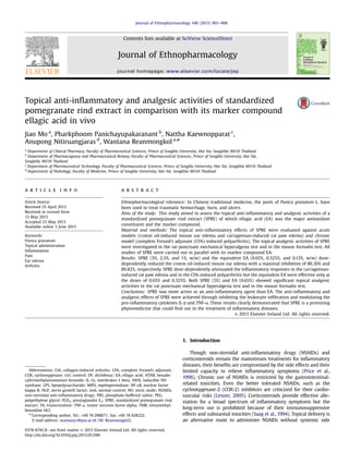
of the ear and tissue MPO activity. As shown in Fig. 1A, topical
application of both SPRE formulations (5%, 2.5% and 1%) and the
equivalent EA formulations (0.65%, 0.325% and 0.13%) resulted in a
dose-dependent reduction in the ear edema with a maximal
inhibition percentage of 86.30% and 80.82% (po0.001), respec-
tively. 0.1% triamcinolone cream and 1% diclofenac gel as reference
drugs reduced the ear edema by 87.67% and 58.90%, respectively.
MPO is an enzyme present in the intracellular granules of
neutrophils, and is used as a marker for polymorphonuclear
leukocyte infiltration into the inflamed tissues, which is an
indicator of an inflammatory reaction. As shown in Fig. 1B, SPRE
and EA dose-dependently inhibited the increase of tissue MPO
activity upon croton oil challenge by 70.25% and 74.13%, maxi-
mally. The reference drugs 0.1% triamcinolone cream and 1%
diclofenac gel caused inhibition of 93.04% and 54.60%, respectively.
3.3. Carrageenan-induced rat paw edema
As determined by repeated measures ANOVA, the rat paw
volume after carrageenan injection showed a statistically signifi-
cant difference between treatments. A mean rat paw swelling of
1.5 ml was observed in the formulation base group (Table 1).
Triamcinolone and diclofenac as positive controls prominently
reduced paw edema compared to the formulation base
(po0.001 vs. the control, repeated measures ANOVA). In the
similar way to the reference drugs, 5% and 2.5% SPRE remarkably
inhibited paw edema (po0.001 vs. the control, repeated measures
ANOVA). 1% SPRE had a moderate but statistically significant effect
(po0.05 vs. the control, repeated measures ANOVA). 0.65% and
0.325% EA were effective in reducing paw edema (p¼0.001 and
po0.01 vs. the control, respectively, repeated measures ANOVA),
while 0.13% EA failed to inhibit paw edema though a mild
reduction was observed (p¼0.15).
3.4. CFA-induced polyarthritis
Polyarthritis of the toe and ankle joint was induced by plantar
injection of CFA and persisted over 13 days as indicated by the
joint swelling. As shown in Table 2, SPRE (5%, 2.5%, and 1%) dose-
dependently inhibited the joint swelling (po0.001, po0.01, and
po0.05 vs. the control, respectively, repeated measures ANOVA),
as did the reference drugs. 0.65% and 0.325% EA similarly reduced
the joint volume (po0.001, po0.01 vs. the control, respectively,
repeated measures ANOVA), but 0.13% EA showed no significant
effect.
3.5. Pro-inflammatory cytokines
In order to elucidate the mechanisms underlying the anti-
inflammatory action of these topical formulations, the levels of the
pro-inflammatory cytokines TNF-α and IL1-β were assessed by
ELISA. As shown in Fig. 2A, TNF-α level in the CFA-injected paw
was higher than that of the normal paw (po0.001 vs. the normal
control). Both SPRE and EA suppressed the elevation of TNF-α in a
dose-dependent manner, as did the reference drugs triamcinolone
and diclofenac (po0.001 vs. the control). Just like TNF-α, the IL1-β
level increased dramatically after CFA injection (po0.001 vs. the
normal control) (Fig. 2B). Triamcinolone and diclofenac exerted a
remarkable inhibition on IL1-β production (po0.001 vs. the
control). SPRE also dose-dependently suppressed the production
of IL1-β (5%: po0.001, 2.5%: po0.05, vs. the control, respectively).
The suppression of EA on IL1-β was significant only at the dose of
0.65% (po0.001 vs. the control).
3.6. Histopathological examination
Histopathologic analysis was performed on normal paws and
affected paws from the control group, groups receiving reference
drugs, 5% SPRE and 0.65% EA. Representative images of paw tissue
sections stained with hematoxylin and eosin from these groups
are shown in Fig. 3. Chronic granulomatous inflammation in
the dermis and subcutaneous tissue were present in all groups.
Severe focal acute inflammation was observed in the dermis and
ctrl 0.1 1 1 2.5 5 0.13 0.325 0.65
0
25
50
75
100
***
***
***
***
***
***
***
***
87.67%
60.27%
80.82%
52.05%
70.55%
86.30%
58.90%
48.63%
TA SPRE EA
####
DF
(%)
Treatment
Increaseinearthickness
(μm)
ctrl 0.1 1 1 2.5 5 0.13 0.325 0.65
0.00
0.05
0.10
0.15
***
***
***
***
***
***
93.04%
34.13%
53.44%
74.13%
31.98%
50.87%
70.25%
54.60%
SPRE EADFTA
(%)
Treatment
MPOactivity(OD/Biopsy)
Fig. 1. Effects of SPRE and EA on the croton oil-induced mouse ear edema (A) and
tissue MPO activity (B). Ear edema was measured in 4 h after application of croton
oil and MPO activity was determined in 24 h after croton oil application. Data are
expressed as mean7SD, n¼8. The number above each column denotes the
inhibition percentage. nnn
po0.001 vs. the control group, ## po0.01 vs. diclofenac.
ctrl: control; TA: triamcinolone; DF: diclofenac.
J. Mo et al. / Journal of Ethnopharmacology 148 (2013) 901–908904
5. subcutaneous tissue in the control group as indicated by the dense
neutrophilic infiltrates (Fig. 3B). Groups receiving drug treatment also
exhibited acute inflammation but it was less severe (1% diclofenac,
Fig. 3D) or mild (0.1% triamcinolone, Fig. 3C; 5% SPRE, Fig. 3E; and
0.65% EA, Fig. 3F) compared to the control group. The inflammation-
induced morphological changes (mainly tissue destruction) in all
treatment groups were less severe than in the control group.
3.7. Mechanical hyperalgesia test
Mechanical hyperalgesia is an indication of inflammatory pain.
Table 3 shows that there was a decrease in the pain threshold
upon CFA injection from the first test session (day 2), it reached its
maximum at day 5 and persisted until the end day. One percent
diclofenac, 5% SPRE and 0.65% EA reversed the pain threshold
decrease (po0.001, po0.001, and po0.05 vs. the control, respec-
tively, repeated measures ANOVA). 0.1% triamcinolone attenuated
the hyperalgesia on day 5 (po0.01 vs. the control, one-way
ANOVA on day 5) but when it was assessed through the whole
test sessions, the analgesic effect of 0.1% triamcinolone was not
significant (repeated measures ANOVA).
3.8. Formalin test
Neither the reference drug 1% diclofenac nor the test formula-
tions inhibited the early phase of the formalin-induced pain
(Fig. 4). For the late phase, 1% diclofenac showed the strongest
analgesic effect (53.81% inhibition, po0.001 vs. the control),
followed by 5% SPRE (35.63% inhibition, po0.01 vs. the control)
and 0.65% EA (33.76% inhibition, po0.01 vs. the control). There
was no statistical significance between the effects of 1% diclofenac,
5% SPRE and 0.65% EA observed.
4. Discussion
The present study has evaluated the topical anti-inflammatory
and analgesic activities of SPRE in parallel with its marker
compound EA in several classical animal models and investigated
the possible mechanisms.
Croton oil-induced ear edema is a widely used model for
identifying potential anti-inflammatory agents for the treatment
of skin disorders (De Young et al., 1989). Topical application of
SPRE inhibited two characteristic inflammatory responses induced
by croton oil: edema formation and neutrophilic infiltration, as
demonstrated by the reduction of the mouse ear thickness
(Fig. 1A) and the suppression on ear tissue MPO activity (Fig. 1B).
Corticoids and COX inhibitors are highly effective against croton
oil-induced inflammation, although their mechanisms are differ-
ent. Both 5% SPRE and 0.65% EA showed a strong inhibitory effect
which is similar to that of triamcinolone but more pronounced
than diclofenac (Fig. 1A, po0.01 vs. diclofenac). This indicated that
the anti-inflammatory mechanism of SPRE was analogous to that
of the corticoids. SPRE did not show superiority over its EA
equivalent either in reducing edema or suppressing MPO activity,
Table 1
Effects of SPRE and EA on carrageenan-induced rat paw edema.
Treatment Volume of the injected paw (mL) (Inhibition percentage (%))
0 h 1 h 2 h 3 h 4 h
Control 1.0670.09 1.5570.16 2.2070.26 2.4870.16 2.4870.20
0.1% TA 1.0370.10 1.3070.19 (44.67) 1.4070.13 (67.32)nnn
1.4570.12 (70.56)nnn
1.5470.16 (63.86)nnn
1% DF 1.0070.06 1.2670.11 (47.21)n
1.3270.11 (72.35)nnn
1.4670.19 (68.10)nnn
1.6270.27 (56.75)nnn
5% SPRE 1.0370.06 1.2670.08 (52.28)n
1.4970.15 (59.23)nnn
1.7970.21 (46.49)nnn
1.8270.12 (44.47)nnn
2.5% SPRE 1.0070.06 1.3470.17 (31.47) 1.6770.23 (41.53)nnn
1.9570.21 (33.22)nnn
2.0070.21 (30.18)nn
1% SPRE 1.0470.05 1.4570.22 (16.75) 1.7970.28 (34.54)nn
2.0970.29 (25.83)n
2.1670.28 (21.14)
0.65% EA 1.0370.10 1.3870.20 (30.46) 1.7370.25 (39.02)nnn
1.9570.23 (35.50)nnn
1.9370.25 (37.11)nnn
0.325% EA 1.0470.10 1.4270.15 (23.10) 1.7470.26 (39.02)nn
2.0170.25 (31.81)nn
2.1170.21 (25.35)nn
0.13% EA 1.0170.06 1.4670.13 (9.39) 1.9270.16 (20.77) 2.1870.16 (17.57) 2.1570.12 (19.74)
One-way ANOVA at each time point followed by post-hoc Tukey's test. TA: triamcinolone; DF: diclofenac. Data are expressed as mean7SD, n¼8.
n
po0.05.
nn
po0.01.
nnn
po0.001 vs. the control
Table 2
Effects of SPRE and EA on CFA-induced rat polyarthritis.
Treatment Volume of the injected paw (mL) (Inhibition percentage (%))
Day 0 Day 4 Day 7 Day 10 Day 13
Control 1.1670.03 1.7170.18 1.747 0.09 1.7270.08 1.7670.10
0.1% TA 1.1470.05 1.3770.11 (58.31)nnn
1.3370.14 (67.80)nnn
1.4370.18 (47.79)nnn
1.3570.11 (66.05)nnn
1% DF 1.1570.04 1.6070.10 (16.63) 1.3470.11 (67.19)nnn
1.3370.10 (67.49)nnn
1.4770.06 (46.30)nnn
5% SPRE 1.1470.02 1.4770.06 (41.00)nn
1.3070.09 (73.99)nnn
1.3870.11 (58.41)nnn
1.3770.07 (63.17)nnn
2.5% SPRE 1.1770.05 1.6170.12 (20.50) 1.4870.09 (43.97)nnn
1.5670.08 (31.64) 1.5670.08 (36.42)nn
1% SPRE 1.1570.03 1.6970.11 (2.05) 1.5270.08 (36.25)nnn
1.6070.08 (21.02) 1.5770.12 (30.86)nn
0.65% EA 1.1270.04 1.4870.14 (33.71)n
1.3970.05 (54.58)nnn
1.4170.08 (48.45)nnn
1.3870.10 (56.58)nnn
0.325% EA 1.1370.02 1.6070.14 (14.58) 1.5170.07 (20.95)nnn
1.5670.05 (23.67) 1.5870.12 (27.16)n
0.13% EA 1.1570.05 1.7470.13 (-7.06) 1.6570.09 (18.79) 1.6470.13 (12.39) 1.6370.12 (20.16)
One-way ANOVA at each test session. TA: triamcinolone; DF: diclofenac. Data are expressed as mean7SD, n¼8.
n
po0.05.
nn
po0.01.
nnn
po0.001 vs. the control.
J. Mo et al. / Journal of Ethnopharmacology 148 (2013) 901–908 905
6. indicating that EA is the major if not the only active constituent of
SPRE responsible for the anti-inflammatory effect in the croton oil-
induced mouse ear edema test.
Carrageenan-induced paw edema is an in vivo model of
inflammation that has been commonly employed to assess the
anti-edematous effect of natural products (Kang et al., 2010). The
edematous response that occurs 0–2.5 h after carrageenan injec-
tion has been correlated with the exudative stage of inflammation
which is featured with the release of histamine, serotonin, and
bradykinin (Antonisamy and Ignacimuthu, 2011). In the late phase
when the edema reaches its highest volume in about 4 h after
carrageenan exposure, it is characterized by the presence of
prostaglandins (Spector and Willoughby, 1963). The significant
inhibition of SPRE on each phase demonstrated that the extract
had a non-selective inhibitory effect on the release or actions of
these mediators (Table 1). A finding in this model that could not be
overlooked is that SPRE at the lowest dose (1%) was effective in
reducing paw edema but its equivalent EA counterpart was not.
This indicated that although EA is the major active constituent of
SPRE which is responsible for its anti-inflammatory activity, SPRE
possesses better activity than EA in terms of controlling
carrageenan-induced acute inflammation. Synergy, or potentia-
tion, is an important concept in the field of herbal medicine and
also a commonly found phenomenon in phytopharmaceuticals
(Marquele-Oliveira et al., 2007; Wagner and Ulrich-Merzenich,
2009). As we have pointed out before, SPRE is a complex mixture
of chemicals some of which might have pharmacological activities,
thus it cannot be ruled out that constituents other than EA might
affect different targets in the inflammatory process and the
synergetic action of these constituents with EA would result in
greater effects of SPRE than by EA alone.
The test of CFA-induced rat polyarthritis has been well char-
acterized and has been frequently used for discovering and
evaluating agents for arthritis and chronic inflammation, with a
proven track record of predictability (Bendele et al., 1999; Stein
et al., 1988; Walz et al., 1971). CFA-induced polyarthritis is largely
driven by the recruitment of T-lymphocytes and macrophages into
the rat paw, progressing with paw swelling and massive infiltra-
tion of leukocytes into the synovium, and resulting in chronic
inflammation and osteolytic lesions (Butler et al., 1992; Romas
et al., 2002). The activation of macrophages leads to increased
levels of lysosomal enzymes, production of pro-inflammatory
cytokines like TNF-α and IL-1β, growth factors and other mediators
nml ctrl 0.1 1 1 2.5 5 0.13 0.325 0.65
0
5
10
15
20
*** ***
***
***
***
***
###
##
###
EASPREDFTA
(%)
Treatment
ConcentrationofTNF-α
(pg/0.1gtissue)
nml ctrl 0.1 1 1 2.5 5 0.13 0.325 0.65
0
500
1000
1500
**
***
*
***
***
###
######
###
#
###
EASPREDFTA
(%)
Treatment
ConcentrationofIL1-β
(pg/0.1gtissue)
Fig. 2. Effects of SPRE and EA on TNF-α (A) and IL1-β (B) production 12 days after
CFA injection. Data are expressed as mean7SD, n¼8, nnn
po0.001, nn
po0.01,
n
po0.05, vs. the control, ### po0.001, ## po0.01, # po0.05 vs. the normal
control, nml: normal control, ctrl: control, TA: triamcinolone; DF: diclofenac.
Fig. 3. Histological appearance of the rat hind footpad 12 days after injection of CFA (40 Â ). (A): Normal paw, (B): control, (C): 0.1% triamcinolone, (D): 1% diclofenac, (E): 5%
SPRE and (F): 0.65% EA.
J. Mo et al. / Journal of Ethnopharmacology 148 (2013) 901–908906
7. of inflammation (Kumar et al., 2010). Over-expressed pro-inflam-
matory cytokines by these activated cells contribute to the
irreversible joint and tissue destruction. Furthermore, studies with
animal models of inflammatory arthritis and in patients with
rheumatoid arthritis have demonstrated an important role for
the high levels of nitric oxide (NO) in the pathogenesis of the
disease (Stichtenoth and Frolich, 1998). In addition, TNF-α induces
NO synthesis by activating the inducible NO synthase (iNOS) and
augments the responses of neutrophils to inflammatory stimuli
(Kumar et al., 2010). In the present study, SPRE showed a strong
inhibition on neutrophilic infiltration (Fig. 1B) as well as suppres-
sion on TNF-α and IL-1β (Fig. 2A and B). SPRE also has been found
to be an efficient NO scavenger with an IC50 of 10.7 μg/mL in
RAW264.7 cells (Panichayupakaranant et al., 2010c). Therefore the
therapeutic effects of SPRE on CFA-induced polyarthritis probably
lie in its inhibition on leukocytes recruitment, down-regulation of
pro-inflammatory cytokines and NO neutralizing property. This is
substantiated by a previous report that pomegranate extract
reduced collagen-induced arthritis (CIA) in mice through inhibit-
ing lipopolysaccharide (LPS)-induced NO production in mouse
macrophages (Shukla et al., 2008). The anti-inflammatory activity
of SPRE was corroborated by the results of histological assay that
the histological changes in the SPRE treated groups were less
severe (Fig. 3). In a similar way to the test of carrageenan-induced
paw edema, 1% SPRE was again found to be superior to its EA
counterpart (Tables 1 and 2), lending support to the idea that the
anti-inflammatory activity of SPRE is better than EA. The anti-
inflammatory efficacies of SPRE and EA are consistent with their
capabilities to block the pro-inflammatory cytokine, as revealed by
the result that SPRE at 2.5% was able to block IL-1β but the
equivalent 0.325% EA was not. This suggested that the superiority
of SPRE might probably lie in its better inhibition on IL-1β.
Peripheral inflammation is symptomized by heightened pain
sensitivity. In the CFA-induced arthritis model, the pain behavior
of animal is characterized by profound referred hyperalgesia and
that can be measured as a decrease in the pain threshold to
mechanical stimulus (Table 3). In animals the mechanisms under-
lying hyperalgesia have been related to increased afferent activity
and a lowered threshold of the spinal nerves to innocuous
stimulation (Schaible et al., 2002). Sensitization of nociceptors is
known to be responsible for the decrease of the pain threshold.
Among the biochemical agents released in the process of inflam-
mation, bradykinin is a potent pain stimulator and prostaglandin
E2 (PGE2) sensitizes the nociceptors to the stimulating influence of
bradykinin, thus causes the nociceptors' threshold for pain to be
lowered. TNF-α contributes to inflammatory sensory hypersensi-
tivity by inducing IL-1β and nerve growth factor (NGF), and the
administration of anti-TNF-α antiserum delayed the onset of CFA-
induced mechanical hyperalgesia in rats (Woolf et al., 1997). In our
study, SPRE eliminated the elevation in the levels of both TNF-α
and IL-1β (Fig. 2A and B), thus indicating that the analgesic activity
of SPRE was mediated via blocking these two cytokines.
In addition, there is evidence that the activation of the nuclear
factor kappa B (NF-κB), which is essential for the induction and
expression of many pro-inflammatory genes, also contributes to
hyperalgesia (Chan et al., 2000). Pomegranate extracts have been
found to suppress the LPS-induced activation of NF-κB in mouse
macrophages in vitro (Shukla et al., 2008) and to inhibit PGE2 in
human intestinal Caco-2 cells (Romier-Crouzet et al., 2009). Taken
together, the analgesic effects of SPRE in CFA-induced polyarthritis
could be attributed to the suppression of the NF-κB pathways and
PGE2 production in addition to its inhibition on TNF-α and IL-1β.
The formalin test produced a distinct biphasic response con-
sisting of the early phase which measures the direct chemical
stimulation of the nociceptors and the late phase which is
dependent on peripheral inflammation and discharge of pain-
producing substances surrounding the nociceptors (Hunskaar and
Hole, 1987). Central analgesics such as opioids inhibit both phases
equally, but peripheral analgesics like COX inhibitors relieve only
the late-phase pain. Our results showed that SPRE produced a
significant relief of pain only in the late phase and mimicked the
effect of COX inhibitor diclofenac (Fig. 4). This is evidence that the
analgesic effect of SPRE is peripheral, does not act through the
Table 3
Effects of SPRE and EA on CFA-induced rat mechanical hyperalgesia.
Treatment Threshold of pain (g) (Inhibition of pain threshold decrease (%))
Day 0 Day 3 Day 6 Day 9 Day 12
Control 87.7576.43 68.5075.93 53.887 8.68 64.88710.96 60.63713.55
0.1% TA 86.88712.46 76.38710.04 (45.45) 74.3873.50 (63.10)nn
72.2579.27 (36.07) 67.3877.95 (28.11)
1% DF 87.88712.08 85.50712.25 (87.66)n
85.75 711.15 (93.73)nnn
77.25711.39 (53.55) 82.3379.51 (79.57)nn
5% SPRE 87.3879.59 86.00712.05 (92.86)n
76.3879.15 (67.53)nnn
81.13710.86 (72.68)n
84.1379.51 (88.02)nnn
2.5% SPRE 84.2577.34 72.0076.52 (36.36) 69.5079.01 (56.46) 65.63710.94 (18.58) 68.3875.66 (41.47)
1% SPRE 83.2577.05 77.1379.01 (68.18) 66.1377.14 (49.45) 65.8877.04 (24.04) 66.63710.39 (38.71)
0.65% EA 82.8875.28 84.6377.95 (109.09)n
75.13712.11 (77.12)nn
72.75710.05 (55.74) 79.88710.67 (88.94)nn
0.325% EA 83.88710.97 74.25710.39 (50.00) 61.6376.61 (34.32) 61.3877.21 (1.64) 65.2579.63 (31.34)
0.13% EA 85.75713.01 74.38713.68 (40.91) 56.88711.33 (14.76) 63.2575.42 (1.64) 58.6378.53 (0.00)
One-way ANOVA at each test session. TA: triamcinolone; DF: diclofenac.
n
po0.05.
nn
po0.01.
nnn
po0.001 vs. the control.
Control 1% DF 5% SPRE 0.65% EA
0
100
200
300 Early phase
Late phase
33.76%35.63%
53.81%
-14.31%
8.07%3.73%
****
***
Treatment
Lickingtime(s)
Fig. 4. Analgesic effects of topical formulations of SPRE and EA in the formalin test.
Data are expressed as mean7SD, n¼7. The number above each column denotes the
inhibition percentages. nnn
po0.001, nn
po0.01 vs. the control group. DF: diclofenac.
J. Mo et al. / Journal of Ethnopharmacology 148 (2013) 901–908 907
8. central pathway, and is dependent on its anti-inflammatory
activities. In neither the rat punctuate mechanical hyperalgesia
test nor the mouse formalin test, did SPRE exhibit any superiority
over EA, indicating that EA is fully responsible for the analgesic
activity of SPRE.
5. Conclusion
Our research has for the first time provided firm evidence that
topical application of SPRE has excellent anti-inflammatory and
analgesic activities. Its marker compound EA is the major active
constituent that is responsible for the pharmacological effects of
SPRE, but SPRE is superior over EA in terms of anti-inflammation
and IL-1β modulation. The analgesic activity of SPRE is significant
and is better than the steroid anti-inflammatory drug triamcino-
lone in chronic inflammation though this effect is milder than
diclofenac in acute inflammatory pain. These therapeutic benefits
of SPRE are probably due to its ability to inhibit the leukocyte
infiltration and to block the pro-inflammatory cytokines besides
its NO scavenging property. Therefore, topical SPRE formulations
are promising phytopharmaceutical candidates and have the
potential to be applied in the clinical treatment of inflammatory
diseases such as cutaneous inflammation and arthritis as a
complementary and alternative therapy.
Acknowledgment
This research is supported by a Grant from Prince of Songkla
University (Grant no. PHA550382S). Thanks to Dr. Brian Hodgson
for assistance with the English.
References
Antonisamy, P., Ignacimuthu, S., 2011. Immunomodulatory, analgesic and antipyre-
tic effects of violacein isolated from Chromobacterium violaceum. Phytomedi-
cine 17, 300–304.
Aviram, M., Rosenblat, M., Gaitini, D., Nitecki, S., Hoffman, A., Dornfeld, L., Volkova,
N., Presser, D., Attias, J., Liker, H., Hayek, T., 2004. Pomegranate juice consump-
tion for 3 years by patients with carotid artery stenosis reduces common
carotid intima-media thickness, blood pressure and LDL oxidation. Clinical
Nutrition 23, 423–433.
Bendele, A., McComb, J., Gould, T., McAbee, T., Sennello, G., Chlipala, E., Guy, M.,
1999. Animal models of arthritis: relevance to human disease. Toxicologic
Pathology 27, 134–142.
Bradley, P.P., Priebat, D.A., Christensen, R.D., Rothstein, G., 1982. Measurement of
cutaneous inflammation: estimation of neutrophil content with an enzyme
marker. Journal of Investigative Dermatology 78, 206–209.
Butler, S.H., Godefroy, F., Besson, J.M., Weil-Fugazza, J., 1992. A limited arthritic
model for chronic pain studies in the rat. Pain 48, 73–81.
Cayir, K., Karadeniz, A., Simsek, N., Yildirim, S., Karakus, E., Kara, A., Akkoyun, H.T.,
Sengul, E., 2011. Pomegranate seed extract attenuates chemotherapy-induced
acute nephrotoxicity and hepatotoxicity in rats. Journal of Medicinal Food 14,
1254–1262.
Chan, C.F., Sun, W.Z., Lin, J.K., Lin-Shiau, S.Y., 2000. Activation of transcription
factors of nuclear factor kappa B, activator protein-1 and octamer factors in
hyperalgesia. European Journal of Pharmacology 402, 61–68.
De Young, L.M., Kheifets, J.B., Ballaron, S.J., Young, J.M., 1989. Edema and cell
infiltration in the phorbol ester-treated mouse ear are temporally separate and
can be differentially modulated by pharmacologic agents. Agents Actions 26,
335–341.
Hayouni, E.A., Miled, K., Boubaker, S., Bellasfar, Z., Abedrabba, M., Iwaski, H., Oku,
H., Matsui, T., Limam, F., Hamdi, M., 2011. Hydroalcoholic extract based-
ointment from Punica granatum L. peels with enhanced in vivo healing
potential on dermal wounds. Phytomedicine 18, 976–984.
Hunskaar, S., Hole, K., 1987. The formalin test in mice: dissociation between
inflammatory and non-inflammatory pain. Pain 30, 103–114.
Kang, M., Jung, I., Hur, J., Kim, S.H., Lee, J.H., Kang, J.Y., Jung, K.C., Kim, K.S., Yoo, M.C.,
Park, D.S., Lee, J.D., Cho, Y.B., 2010. The analgesic and anti-inflammatory effect
of WIN-34B, a new herbal formula for osteoarthritis composed of Lonicera
japonica Thunb and Anemarrhena asphodeloides BUNGE in vivo. Journal of
Ethnopharmacology 131, 485–496.
Kumar, V., Abbas, A.K., Fausto, N., Aster, J.C., 2010. Acute and chronic inflammation,
Robbins and Cotran Pathologic Basis of Disease. Saunders, Philadelphia, pp.
43–77.
Lansky, E.P., Newman, R.A., 2007. Punica granatum (pomegranate) and its potential
for prevention and treatment of inflammation and cancer. Journal of Ethno-
pharmacology 109, 177–206.
Lenzer, J., 2005. FDA advisers warn: COX 2 inhibitors increase risk of heart attack
and stroke. British Medical Journal 330, 440.
Marquele-Oliveira, F., Fonseca, Y.M., de Freitas, O., Fonseca, M.J.V., 2007. Develop-
ment of topical functionalized formulations added with propolis extract:
stability, cutaneous absorption and in vivo studies. International Journal of
Pharmaceutics 342, 40–48.
Morteza-Semnani, K., Saeedi, M., Hamidian, M., 2004. Anti-inflammatory and
analgesic activity of the topical preparation of Glaucium grandiflorum. Fitoter-
apia 75, 123–129.
National Pharmacopoeia Committee, 2010. Pharmacopoeia of People's Republic of
China, Part 1, Chemical Industry Press, Beijing, p. 87.
Newbould, B.B., 1963. Chemotherapy of arthritis induced in rats by mycobacterial
adjuvant. British Journal of Pharmacology and Chemotherapy 21, 127–136.
Panichayupakaranant, P., Issuriya, A., Sirikatitham, A., Wang, W., 2010a. Antioxidant
assay-guided purification and LC determination of ellagic acid in pomegranate
peel. Journal of Chromatographic Science 48, 456–459.
Panichayupakaranant, P., Itsuriya, A., Sirikatitham, A., 2010b. Preparation method
and stability of ellagic acid-rich pomegranate fruit peel extract. Pharmaceutical
Biology 48, 201–205.
Panichayupakaranant, P., Tewtrakul, S., Yuenyongsawad, S., 2010c. Antibacterial,
anti-inflammatory and anti-allergic activities of standardised pomegranate rind
extract. Food Chemistry 123, 400–403.
Price, D.D., Mao, J., Lu, J., Caruso, F.S., Frenk, H., Mayer, D.J., 1996. Effects of the
combined oral administration of NSAIDs and dextromethorphan on behavioral
symptoms indicative of arthritic pain in rats. Pain 68, 119–127.
Romas, E., Gillespie, M.T., Martin, T.J., 2002. Involvement of receptor activator of NF-
kappa-B ligand and tumor necrosis factor-alpha in bone destruction in
rheumatoid arthritis. Bone 30, 340–346.
Romier-Crouzet, B., Van De Walle, J., During, A., Joly, A., Rousseau, C., Henry, O.,
Larondelle, Y., Schneider, Y.-J., 2009. Inhibition of inflammatory mediators by
polyphenolic plant extracts in human intestinal Caco-2 cells. Food and
Chemical Toxicology 47, 1221–1230.
Saag, K.G., Koehnke, R., Caldwell, J.R., Brasington, R., Burmeister, L.F., Zimmerman,
B., Kohler, J.A., Furst, D.E., 1994. Low dose long-term corticosteroid therapy in
rheumatoid arthritis: an analysis of serious adverse events. American Journal of
Medicine 96, 115–123.
Schaible, H.G., Ebersberger, A., Von Banchet, G.S., 2002. Mechanisms of pain in
arthritis. Annals of the New York Academy of Sciences 966, 343–354.
Shukla, M., Gupta, K., Rasheed, Z., Khan, K.A., Haqqi, T.M., 2008. Consumption of
hydrolyzable tannins-rich pomegranate extract suppresses inflammation and
joint damage in rheumatoid arthritis. Nutrition 24, 733–743.
Spector, W.G., Willoughby, D.A., 1963. The inflammatory response. Bacteriological
Reviews 27, 117–154.
Stein, C., Millan, M.J., Herz, A., 1988. Unilateral inflammation of the hindpaw in rats
as a model of prolonged noxious stimulation: alterations in behavior and
nociceptive thresholds. Pharmacology Biochemistry Behavior 31, 445–451.
Stichtenoth, D.O., Frolich, J.C., 1998. Nitric oxide and inflammatory joint diseases.
British Journal of Rheumatology 37, 246–257.
Subramanian, N., Ghosal, S.K., Moulik, S.P., 2005. Enhanced in vitro percutaneous
absorption and in vivo anti-inflammatory effect of a selective cyclooxygenase
inhibitor using microemulsion. Drug Development and Industrial Pharmacy 31,
405–416.
Tubaro, A., Dri, P., Delbello, G., Zilli, C., Della Loggia, R., 1986. The croton oil ear test
revisited. Agents Actions 17, 347–349.
Wagner, H., Ulrich-Merzenich, G., 2009. Synergy research: approaching a new
generation of phytopharmaceuticals. Phytomedicine 16, 97–110.
Walz, D.T., DiMartino, M.J., Misher, A., 1971. Adjuvant-induced arthritis in rats. II.
Drug effects on physiologic, biochemical and immunologic parameters. Journal
of Pharmacology and Experimental Therapeutics 178, 223–231.
Winter, C.A., Risley, E.A., Nuss, G.W., 1962. Carrageenin-induced edema in hind paw
of the rat as an assay for anti-inflammatory drugs. In: Proceedings of the
Society for Experimental Biology and Medicine 111, pp. 544–547.
Woolf, C.J., Allchorne, A., Safieh-Garabedian, B., Poole, S., 1997. Cytokines, nerve
growth factor and inflammatory hyperalgesia: the contribution of tumour
necrosis factor alpha. British Journal of Pharmacology 121, 417–424.
J. Mo et al. / Journal of Ethnopharmacology 148 (2013) 901–908908