ijoi_vol_46_updated.pdf
•
0 likes•274 views

The document discusses using temporary anchorage devices (TADs) placed in the infrazygomatic crest (IZC) region to retract the maxillary arch. It describes the relevant anatomy of the IZC region and recommends placing TADs at the IZC 7 site buccal to the second molar, as there is thicker bone in this area compared to the IZC 6 site by the first molar. CBCT images show examples of TAD placement interfering with retraction when placed too close to tooth roots. Proper placement of TADs in an extra-radicular position is important for reliable maxillary arch retraction.

Recommended
Recommended
More Related Content
What's hot
What's hot (20)
Similar to ijoi_vol_46_updated.pdf
Similar to ijoi_vol_46_updated.pdf (20)
Recently uploaded
Recently uploaded (20)
ijoi_vol_46_updated.pdf
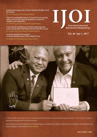
- 1. Guided Infra-Zygomatic Screws: Reliable Maxillary Arch Retraction Drs. John Jin-Jong Lin & W. Eugene Roberts Class II Crowded Malocclusion Treated Conservatively with a Passive Self Ligating Appliance: Expansion, Stability and Adaptation Drs. Shih-Yung Lin, Chris Chang & W. Eugene Roberts Trans-Alveolar Uprighting of a Horizontally Impacted Lower Canine with a Mandibular Buccal Shelf Bone Screw Drs. Szu Rou Yeh, Chris Chang & W. Eugene Roberts Archwire Sequence for Insignia® : a Custom Bracket System with a Bright Future Drs. Angle Lee, Chris Chang & W. Eugene Roberts IJOI International Journal of Orthodontics & Implantology Vol. 46 Apr 1, 2017 CC (left) and DD (right) with the new Trumpology book at the Damon Forum Speakers Dinner, Orlando, FL, USA. They are smiling in agreement having decided to drain the swamp! International Journal of Orthodontics & Implantology is an experience sharing magazine for worldwide orthodontists and Implantologists. Download it at http://iaoi.pro. 《僅供牙科專業人士參閱》
- 2. 10/2 11/ 7/ /10 /2 /21 23 / / 10/1 11/22 12/13 / /20 /11 / /13 7/11 2/1 3/1 12/21 1/ 2/1 /22-2 /1 -1 11/2 -12/1 11/27-30
- 3. IAOI welcomes general submissions and inquiries to be sent to contact@iaoi.pro BE PE T E I F RMATI A I EAS Trumpology p. 72 Featured on the cover of this issue is a photo of Dr. Dwight Damon (DD) and myself at the Damon Forum 2017 Speakers Dinner. I was not sure whether or not DD would be so welcoming after my Keynote speech this year as I tackled a rather controversial, almost taboo, subject for the Damon Forum: extraction vs. non-extraction. However, as those of you who have read my new Trumpology book know: “Be willing to take on new challenges.” (Trumpology p. 73) I was very hesitant to take on this subject, as for the last 10 years in which I have participated in the Damon Forums, I have never heard anyone talk about extraction issues and DD has an extraction rate of 1% (compared to my 65%). He is considered to be a non-extraction Dr and I have also been repeatedly labelled so (I have been asked “You don’t extract, do you?” for 6 consecutive years at a prestigious organisation’s meeting), as have probably most Orthodontists who use Damon braces. Extraction is very common in Asia, due to profile and the desired results of our patients. If Asian doctors tried to replicate DD’s 1% extraction rate, it would only end in disaster!! My speech could well have been a disaster, I almost didn’t have the guts to present it, but……. At the Speakers Dinner I was cornered by DD and for a whole hour we spoke about my speech. He told me he considered it to be the best speech of all the Damon Forums and that somebody should have talked about this important subject a long time ago, to make it clear to the participants and worldwide audience that extraction is OK. DD fully understood that Caucasians and Asians are different and although he advocates minimally invasive treatments, he also has to extract when necessary (as do I!). How we interpret information is as important as the information itself and DD proved to me that he is very open-minded to new information. Therefore, I also presented him with a copy of my new Trumpology book and discussed with him how we can “drain the swamp” (Trumpology p.166) of our profession to allow all Orthodontists to practise to their fullest potential without a perceived opinion being pushed onto them. I sincerely hope that all of you will help to “drain the swamp” and break down perceived opinions in our profession and continue to march with us along the path to glory. Wishing you every success in your practice and a Happy Easter, s n DDS, PhD, Publisher of IJOI. Dr. Baldwin W. Marchack Dr. Thomas Han Dr. Fernando Rojas-Vizcaya Dr. Homa Zadeh Dr. Larry White Dr. J. Michael Steffen Dr. W. Eugene Roberts Dr. Tucker Haltom Consultants Dr. Frank Chang Dr. Johnny Liaw Examiners Dr. John J. J. Lin Dr. Hong Po Chang Dr. Yu Lin Hsu Dr. Yu-Hsin Huang Dr. Bill Su Dr. Ming-Jen Chang Associate editors Dr. Chris Lin Dr. Ariel Chang Editorial Board Editor-in-chief Dr. W. Eugene Roberts ublisher Dr. Chris Chang Research Dr. Chi Huang Case Report Dr. Angle Lee Surgery Dr. Shih-Yung Lin Editors Desk editor Bella Chu English editing Paul Head Proofreader Tzu Han Huang Illustration Dr. Rungsi Thavarungkul Editorial LI E FROM T E MASTER Guided Infra-Zygomatic Screws: Reliable Maxillary Arch Retraction iAOI CASE RE ORT Class II Crowded Malocclusion Treated Conservatively with a Passive Self Ligating Appliance: Expansion, Stability and Adaptation Trans-Alveolar Uprighting of a Horizontally Impacted Lower Canine with a Mandibular Buccal Shelf Bone Screw Clinical Tips Archwire Sequence for Insignia®: a Custom Bracket System with a Bright Future Damon Workbook Class I Anterior Crossbite Crowded Class II Division 2 Malocclusion with Class I Molars Due to Blocked In Lower Second Premolars iAOI OT ERS In memory of Charles Burstone: He has been missing almost two years, but the void is still fresh. FEEDBACK FROM T E WORLD Feedback from the world EDITORIAL IJOI 46
- 4. 4 IJOI 46 LI E RO T E A TER █ Fig. 1A: The skeletal anatomy (osteology) of the Infrazygomatic Crest (IZC) is marked with an ovoid magenta circle. █ Fig. 1B: A practical position for an IZC bone screw is anterior to the anatomic ridge and buccal to the mesiobuccal root of the maxillary first molar (MBR of U6). Distal to the anatomic ridge, the preferred site is buccal to the MBR of U7. These TAD sites are designated as IZC 6 and IZC 7 (yellow ovoid circles), respectively. i n ra o atic cr ia a i ar rc traction Introduction The infra-zygomatic crest (IZC) is a buccal process on the maxilla, connecting to the zygoma. Intraorally it is a crest of bone emanating from the buccal plate of the alveolar process, lateral to the roots of the rst and second maxillary molars (Fig. 1A). The ridge of bone extends 2cm or more superiorly to the zygomatic-maxillary suture, and the inferior portion can be subdivided into the IZC and IZC 7 areas, respectively (Fig. 1B). The IZC is a common site for insertion of temporary anchorage devices (TADs). Melsen1 and Uribe2 placed routine TADs along the intraoral anatomical ridge of the IZC, and illegas3 used a 2 mm long screw to engage the superior aspects of the IZC, approximating the zygoma. The amount of alveolar bone buccal to the maxillary molars is the critical factor for placing OrthoBoneScrew® (OBS, Newton’s A Ltd, Hsinchu, Taiwan) in an Extra-Radicular (E-R) position. Inter- radicular (I-R) TADs are also effective for maxillary retraction, if the screws avoid the path of distal tooth movement. This article reviews the relevant anatomy and clinical procedures for routinely achieving maxillary retraction with TADs, inserted directly into alveolar bone of the posterior maxilla. (Int J Orthod Implantol 2017;46:4-16)
- 5. 5 I IJOI 46 █ Fig. 2: An IZC anchorage screw penetrates about a 3mm thickness of attached gingiva and cortical bone. The clearance of the screw head to the soft tissue should be ~1.5mm, so there is a distance about 4.5mm between the base of the screw and the inner surface of the cortical bone. Thus, screws of 8-12mm length will extend into the non-cortical bone space (medullary bone or sinus) about 3.5-7.5mm. r ohn in- ong Lin, Examiner of IJOI, Director of Jin-Jong Lin Orthodontic Clinic (Left) r Eugene Roberts, Editor-in-chief, International Journal of Orthodontics & Implantology (Right) Anatomical onsiderations Soft tissue irritation is a common problem if the inferior aspect of the screw platform is contacting or near the mucosa. To control this problem the IZC TADs are placed in attached gingiva with 1. mm of clearance from soft tissue to the base of the TAD platform. , It is important to carefully consider the anatomy of the IZC site to select an appropriate screw length. The average thickness of the attached gingiva in the maxillary first molar is about 1.0mm, and the cortical bone thickness is about 1.1-1. mm.7 The screw threads must engage cortical bone to insure primary stability. Generalizing the widths, for soft tissue clearance, attached gingiva and cortical bone at 1. mm each (Fig. 2), reveals that -12mm IZC screws penetrate the medullary bone or sinus from . -7. mm. Under most clinical conditions, an mm screw is adequate to engage the cortical plate and secure primary stability (Fig. 2). Liou suggested orienting screws about -70 degrees inferior to the horizontal plane to achieve maximal buccal bone engagement, but it was not clear whether IZC or 7 was the preferred site from an anatomic perspective (Figs. 1-3). Because the alveolar bone is thicker on the buccal surface of the second molar (Figs. 4 and 5), the IZC 7 site is usually preferable for TADs. In Taiwan, most orthodontists utilize the IZC 7 site (Fig. 6A),10,11 because the buccal plate covering the first molar roots is too thin. For clinical convenience and the advantage of attached gingiva, the preferred IZC bone screw sites are considerably inferior (Fig. 6B) to the anatomic zygomatic crest (Figs. 1A & B). Initially, it was thought that all OBSs (OrthoBoneScrews, Newton’s A Ltd, Hsinchu, Taiwan) placed buccal to the MBR of U7 were routinely achieving E-R (Extra- Radicular) placement for arch retraction. However,
- 6. 6 IJOI 46 LI E RO T E A TER █ Fig. 5B: A similar series of CBCT axial cuts (3-8) from the CEJ apically (series of upper views), is shaded in the series of lower views to show available buccal bone for extra-radicular placement of IZC screws: IZC 7 (green) and IZC 6 (red). See text for details. █ Fig. 4: The coronal view of a CBCT (red rectangle) documents alveolar bone thickness on the buccal (B) and palatal (P) aspects of molar roots. Extra-radicular placement of a TAD is more predictable for the IZC 7 compared to the IZC 6 sites, because the bone is thicker over the distobuccal (7DB) and mesiobuccal (7MB) roots of the second molar (U7), compared to the corresponding 6DB, 6MB sites for the first molar (U6). █ Fig. 3: The alveolar bone is too thin to place a TAD buccal to the MBR of 6, even with an increased angulation of 55-70º (Liou’s IZC 6). The senior author (JJ-JL) proposes placing an IZC screw lateral to the MBR of 7 (Lin’s IZC 7) to more reliably achieve an E-R position for maxillary arch retraction. █ Fig. 5A: Five CBCT axial cuts (1-5) in the area demarcated by the red rectangle show the alveolar bone anatomy for ~1mm apical to the cementoemamel junction (Cut 1). The teeth are labeled with Palmer notation 4-8. 5-6mm above CEJ is the level where IZC screws are usually inserted. Note that the alveolar bone is much thicker buccal to the U7 compared to the U6.
- 7. 7 I IJOI 46 █ Fig. 6A: IZC 7 interference with maxillary retraction is documented for four months (4ms) of treatment from 19y4m to 19y8m of age. Canine cusps are marked with yellow lines in the upper arch and blue lines in the lower arch. Four months (4ms) of retraction with IZC 7 bone screws produced ~2mm of maxillary arch retraction on the patient’s right side (upper views), but little or no retraction on the left side (lower views). See text for details. █ Fig. 6B: 3D CBCT images of molars (white) and TADs (yellow) are superimposed on a panoramic radiograph of the patient illustrated in Fig. 6A. The screw on the patient’s left side overlaps the root apex and may be interfering with maxillary arch retraction. See text for details █ Fig. 6C: A horizontal maxillary cut of the CBCT show the IZC 7 screws are inserted in bone, buccal to the roots of the molars (red shading). █ Fig. 6D: A similar CBCT cut near the apices of the second molars shows the screw on the right side penetrated the sinus, but it is extra-radicular and still provided effective anchorage to retract the right buccal segment, and the patient had no symptoms or complaints. The left IZC 7 impinged on the distal surface of the MBR of the U7, preventing retraction of the left buccal segment in the direction of the yellow arrows. CBCT analysis of consecutive patients revealed some TADs were I-R (Inter-Radicular) (Figs. 6A-D), and mostly they were E-R (Fig. 7). Consistently placing IZC bone screws in an E-R, or carefully selected I-R position, is critical for reliable maxillary arch retraction.
- 8. 8 IJOI 46 LI E RO T E A TER █ Fig. 7A: IZC 6 interference with maxillary retraction is documented. A patient 14y1m of age was treated with nine months of maxillary retraction, that was anchored with IZC 6 and IZC 7 screws. Note there is a further deviation of the upper midline to the right, which is consistent with more retraction on the right than the left sides. Little or no progress was noted in correcting the Class II buccal segment on the left side (lower views). See text for details. █ Fig. 7B: The right IZC 7 is buccal to the roots, and has provided effective anchorage for maxillary retraction (yellow arrows). The MBR of the U6 on the left side interfered with retraction (Stop!). See text for details. Anc orage equirements Originally, the main purpose for TADs in the posterior maxillary arch was for maximal retraction of the anterior segment following extraction of premolars. Ideal I-R placement was right in the middle of the roots of upper 2nd premolar and 1st molar to avoid hitting the roots. In effect, the TADs provided solid anchorage for the extraction space closure. Similar I-R placement of TADs did not work to retract maxillary molars, paradoxically for the same reason: they blocked the path of distalizing tooth movement. Thus, substantial retraction of the entire maxillary arch was not much or not possible with routine I-R TADs. Clinical experience with OBSs placed in the IZC and 7 areas revealed that maxillary arch retraction was accomplished more often with TADs in the IZC 7, rather than the IZC position. CBCT evaluation demonstrated that the TADs, successfully anchoring maxillary arch retraction, were placed in an E-R position of the relatively thick buccal bone (Figs. 3-5). A truly reliable (“fail-proof”) method for maxillary retraction must evaluate bone quality as well as quantity at the TAD site. Chang et al.12 recently found an IZC screw failure rate of 7%, compared to 20%, as reported by Uribe.2 The improved performance of the Chang et al.12 method may relate to favorable soft tissue on the more inferior sites like IZC 7 (Fig. 1B), compared to the more superior crestal positions of the TADs used by
- 9. 9 I IJOI 46 █ Fig. 8A: Maximal anchorage for routine space closure after premolars extractions is inadequate for correction of a severe malocclusion in a 16y4m old patient. Retraction of the maxillary buccal segments is required. Uribe2 and Melsen1 (Fig. 1A). From the evidence reviewed, it is clear that an E-R bone screw is more reliable for maxillary retraction compared to high IZC1,2 or routine I-R TADs.1 The current challenge is to develop a clinical approach that consistently achieves TAD anchorage that reliably supports posterior retraction of the molars. ouble ilm et od Dr. Leslie Chen13 suggested that screws can be placed mesial to the MBR of either U or U7 to achieve maxillary arch retraction. This generalization worked well for the thick buccal bone of the IZC 7, but the IZC site was problematic. It was more di cult to achieve E-R anchorage due to thin buccal bone, so the IZC TADs usually produced I-R interference that blocked the path of distal tooth movement. A CBCT scan is valuable for confirming that an IZC TAD is unlikely to interfere with retraction of the maxillary molars, but D imaging is not necessary for placing the TAD. The Chen13 double lm method is a 2D radiographic guide for screw placement in patients requiring IZC anchorage (Fig. 8A). Two Screw Insertion Procedure A radiograph after the initial, preliminary positioning of the TAD serves as baseline for planning its final position, which is confirmed with a follow-up radiograph. Either periapical, preferably with the long cone technique, or panoramic radiographs are suitable for the double lm procedure: 1. Initial insertion of the TAD with 1mm of bone engagement (Fig. 8B). 2. Check the position with an initial radiograph (Fig. 8C). . The screw position is adjusted according to the radiographic image. . The preferred position for the screw is immediately mesial to the MBR, for either the IZC or IZC 7 sites. . Take a second periapical film to confirm that the position is over the mesial of MBR (Fig. 8C). . De ning the D position of the screw relative to the roots of adjacent teeth requires a CBCT (Fig. 8D). If necessary, the TAD can be repositioned.
- 10. 10 IJOI 46 LI E RO T E A TER █ Fig. 8D: Axial view of a CBCT shows the right IZC 6 is between the roots of the U5 and U6, positioned so the right maxillary segment can be retracted. █ Fig. 8E: Axial view of a CBCT shows that the left IZC 6 is also in a favorable position between the roots of the first molar and second premolar (shaded in red), so the left maxillary segment can also be retracted. █ Fig. 8B: The double film method is effective with periapical radiographs. For the initial film (lower left) the screw is engaged into the bone about 1mm (upper left). The clinical view after the TAD was placed (upper right) and the final film (lower right) shows that the screw (arrow) was reinserted in a more gingival and axial position. █ Fig. 8C: The double film method can also be used with panoramic radiography. On the initial film (upper), space for screw placement is noted between the U5 and U6 roots. The final film shows the screw positioned mesial to the MBR of the U6s. This configuration is suitable for maxillary arch retraction.
- 11. 11 I IJOI 46 Advantages: 1. There is usually enough I-R space between the U root and the MBR of the U for an IZC TAD. Placing the screw in this area is suitable for maxillary arch retraction if it’s mesial to the MBR of the U and there is adequate clearance for distal movement of the root of the U . 2. If it is desirable to position the TAD over the mesial of MBR of U7 (IZC 7), the chance of extra radicular is much higher. . Local infiltration analgesia is recommended to control pain, and fortunately this form of anesthesia does not interfere with the patient s perception of a screw contacting the root. If the patient feels the screw touching the root of a tooth, the TAD can be repositioned. Disadvantages: 1. Additional wounds occur if screw positioning is changed. 2. Repositioning of the screw is associated with saliva contamination. Pin Head Soft Tissue Penetration Method Dr. Mala Ram Manohar1 presented this innovative method at the th World Implant Orthodontics Congress in Goa in 201 . The distinct advantage over the Chen13 double film method is the lack of saliva contamination associated with reinserting the TAD. The procedure is as follows: 1. Use stainless steel dot pins (Fig. 9-1) cut off the heads (Fig. 9-2) leaving about a 1mm piece of the shank (Fig. 9-3). 2. Canker sore patches (Fig. 9-4) used to cover aphthous ulcers (Fig. 9-5) are thin, opaque strips or circular patches and position a pin head in the center of the patch, with the point up (Fig. 9-6). . Following topical and then local infiltration anesthesia, the circular patch covering the pin-head point is pressed firmly into place, penetrating the soft tissue in the anticipated position of a TAD. . Image the area with a 2D radiograph, and reinsert the TAD if needed into a more desirable position use the soft tissue mark left by the pin head as a landmark. . Take a follow-up radiograph to check the nal position of the screw, and adjust the TAD position as needed. Advantages: 1. o saliva contamination of the screw, unless it must be repositioned after the follow-up radiograph. 2. Avoids multiple screw placement wounds.
- 12. 12 IJOI 46 LI E RO T E A TER Transparent Adhesive Patch for Double Film Technique 1. Source a transparent, adhesive canker sore patch for oral use. It is important that the patch remains rmly attached to soft tissue in the presence of saliva (Fig. 10A-1,2). 2. Cut mm rectangular patches, remove the protective lm, and press the back side of the pin head into the center of the adhesive side of the patch (Fig. 10A-3). . Under local anesthesia, press the pin head into the soft tissue as before (Fig. 10A-5). . The transparent patch allows the clinician to see the exact position of the pin head (Fig. 10A-4,6). █ Fig. 9: The double film method employs a soft tissue marker that is secured to the soft tissue with an opaque canker sore patch. 1. Source standard steel dot pins (SS is preferred for sterilization). 2. Cut off the pin head. 3. Leave about 1mm of the pin shaft to serve as a soft tissue indentation marker. 4. Choose a canker sore patch. 5. Large patches are inappropriate because they are easily distorted by movable mucosa. 6.Select the round canker sore patch and place a pin head in the center of the circle. 7. Position the patch with a pin head on the tip of a gloved finger. 8. Under local anesthesia, press the pin head into the soft tissue; the patch should maintain the position of the pin head on the soft tissue during the initial radiograph. 9. When the patch is removed, the soft tissue indentation of the pin head serves as the mark to reposition the TAD. 10. The initial film (x-ray with pin head) has pin head dots marking the soft tissue penetration points. 11. The final film shows the “Correct TADs” as placed based on the positions of the initial pin heads.
- 13. 13 I IJOI 46 █ Fig. 10A: Testing of multiple canker sore patches revealed that ComfortBrace® strips are a better material for the double film method initial film (1). 1. Protective strips to cover braces have the advantage of soft tissue adhesion up to 24 hours. 2. The transparent material is packed in strips with a protective layer that is peeled off prior to application. 3. Place the pin head on the adhesive side, with the pin shaft pointed out. 4. The very sticky material is best positioned with two sets of forceps. 5. After anesthetizing the soft tissue, press the pin head attached to the strip firmly into the mucosa to produce a mark to serve as reference for planning the penetration point for the screw. 6. The pin head is held securely by the adhesive strip in preparation for the initial radiograph. After an extensive search, ComfortBrace® strips (Lighthouse Point FL), originally designed for preventing xed appliance irritation, were selected as the ideal product because they: 1. Are clear, so the position of the pin head is readily apparent (Fig. 10A-1). 2. Can be cut into small mm rectangular pieces to be easily positioned on the attached gingiva, and do not move (Fig. 10A-2,3). . Are made with FDA approved ingredients for use in the oral cavity: polyvinylpyrrolidone, acrylates copolymer, sorbitol, water, hydroxyethyl cellulose, carbomer 0, and mint (www.comfortbracestrip.com/ safety.asp).
- 14. 14 IJOI 46 LI E RO T E A TER Indication or I 1. At least a mm gap is required between the roots of the U and the MBR of U to avoid root contact with an I-R TAD. 2. Small oral cavities are often more convenient to place IZC rather than placing the IZC 7. . A buccal frenum on or near the site can induce laceration, in ammation and screw failure fortunately, there is usually no buccal frenum at the IZC site. . The mm width of attached gingiva is adequate for most IZC TADs. . Avoid placing TADs between the roots of teeth where the sinus oor is low because these areas usually have low density bone and a thin cortical plate. █ Fig. 10B: Double film method using ComfortBrace® strips in preparation for TAD placement (2). 1. Initial film shows the dot image of the radio-opaque pin-head. 2. Peel off the clear strip of ComfortBrace® covering the pin head. 3. Remove the pin head, and the soft tissue indentation mark is apparent. 4. Based on the pin head reference, derived from the first film, the TAD is positioned more superior and posterior. 5. The self-drilling screw is installed in the final position. 6. Final film shows the screw positioned mesial to the MBR, as intended.
- 15. 15 I IJOI 46 Indication o I 1. At least mm width of attached gingiva is advantageous. 2. Access to the IZC 7 area requires a large oral cavity, as well as lip reflection for adequate access to the screw insertion site. . Avoid placing between the roots of teeth where the sinus oor is low because this area has a thin cortical plate. ailure rate According to a recent study by Chang et al.12 the IZC screw failure rate is 7%. Most of the failures are due to: 1. Poor bone quality: Unfortunately there is no reliable method for evaluating bone quality. The sensation for poor bone quality, beneath a sound layer of cortical bone, is like punching through an egg shell, followed by a lack of screw stability. Unless the TAD can be stabilized by deeper penetration, it is best to remove it and try another site. 2. Immediate loading: SS screws are excellent TADs because they do not osseointegrate and are easily repositioned to another site, if necessary. 3. Sinus floor: A low sinus between the roots of teeth is undesirable for an IZC TAD site. 4. Movable mucosa: Unattached alveolar mucosa at the TAD site is usually undesirable. , However, Chang et al.12 found no signi cant difference in the failure rate between movable mucosa and attached gingiva if the platform of the screw is at least mm away from the soft tissue surface. The disadvantages of the latter approach are a longer screw is required (~12mm) and it must be carefully positioned for patient comfort. uccal el one crews The senior author (JJ-JL) previously introduced mandibular buccal shelf bone screws, which were usually placed by periodontists or oral surgeons, using the apically positioned ap to provide attached gingiva at the TAD site.1 When the mandibular buccal shelf is steep and if patients require an extra- radicular placement, with an apically repositioned ap of attached gingiva, an experienced surgeon is needed. Flap surgery is more expensive and tends to be painful postoperatively, particularly if a TAD must be repositioned. Currently, a skillful orthodontist can produce a good result with a self drilling screw by using the double lm method to place the I-R buccal shelf screw (right mesial to the MBR of L7). Reliable retraction of the mandibular arch can be done. onclusions 1. The double film method is advantageous for installing TADs in the three most common sites: IZC , IZC 7, and mandibular buccal shelf. 2. ComfortBrace® strips have proven superior for maintaining a pin head in a stable position relative to the soft tissue.
- 16. 16 IJOI 46 LI E RO T E A TER . The double film method is indicated for selecting the appropriate point of entry for IZC or buccal shelf screws. . Extensive experience with the double film method has demonstrated it is an advantageous approach for reliably placing IZC IZC 7 bone screws to retract the dentition. . The double film method has significant advantages for both the clinician and the patient. Ac nowledgement 1. Thanks to Dr. Leslie en-Peng Chen for the innovative idea leading to the current double film method for accurately placing TADs for orthodontic anchorage. 2. Thanks to Dr. Mala Ram Manohar for the practical idea of using pin heads with a 1mm shaft to mark the initial point of tissue penetration for a TAD. . Thanks to Dr. Po-Jung Chen for the CBCT cross- sectional evaluation of the IZC vs. IZC 7 sites as shown in Fig. . . Thanks to Dr. Po-Jan Kuo for the CBCT information in Fig. which illustrates the buccal anatomy of IZC 7 sites. e erences 1. Melsen B. Overview of mini-implants: where are we? J Clin Ortho 2005;39(9):539-47. 2. Uribe F, Mehr R, Mother A, Janakiraman N, Allareddy V. Failure rates of mini-implants placed in the infrazygomatic region. Prog Orthod 2015;16:31. 3. Villegas C. 100% successful TAD system. Presentation at the 2016 AAO Meeting, Orlando FL. 4. Chang MJ, Lin JJ, Roberts WE. Probable airway etiology for a severe Class III openbite malocclusion: conservative treatment with extra-alveolar bone screws and intermaxillary elastics. Int J Orthod Implantol 2017;45:4-20. 5. Shih YH, Lin JJ, Roberts WE. Conservative management of Class I crowded malocclusion complicated by severe maxillary protrusion, facial convexity and deepbite. Int J Orthod Implantol 2016;44:4-16. 6. Goaslind GD, et al. Thickness of facial gingiva. J Perio 1977;12:768-71. 7. Chen MY, et al. Eastern and western studies of interdental cortical bone thickness for temporary anchorage devices. J Taiwan Periodontal 2013;18(1):33-43. 8. Liou EJ, Chen PH, Wang YC, Lin JC. A computed tomographic image study on the thickness of the infrazygomatic crest of the maxilla and its clinical implications for mini-screw insertion. Am J Orthod Dentofacial Orthop 2007;131:352-6. 9. Chen PJ, et al. Bone thickness of the infrazygomatic crest for orthodontic implant placement. Poster, Annual Meeting of Taiwan Association of Orthodontists, 2008. 10. Lin JJ. Mini-screw or mini-plate, which is better for whole upper arch distalization. News and Trends in Orthodontics 2007;5:1-2. 11. Lin JJ. Creative orthodontics: Blending the Damon System and TADs to Manage Difficult Malocclusions. Taipei: Yong Chien Ltd; 2010. 12. Chris CH, Hsu E, Lin JSY, Yeh HY, Roberts WE. Comparison of the failure rate for infrazygomatic bone screws placed in movable mucosa or attached gingiva. Eur J Orthod 2017 (In Preparation). 13. Personal communication with Dr. Leslie Chen, 2012. 14. Manohar MR. Versatility of miniscrews-implants & implants guides. 8th Goa, India: 8th WIOC; 2016. 15. Lin JJ, et al. Treatment of Severe Class III with Buccal Shelf Mini-Screws. News and Trends in Orthodontics 2010;18:4-15.
- 17. 專題演講 Int ucing T th ntics in the a n ste Dr. Elizabeth Menzel 國立交通大學-浩然圖書館B1國際會議廳 ( n) Dr. Diego Peydro Herrero 金牛頓藝術科技 ( h ) 合報價 e n th ntics with lea ligne s The a 匯款資訊: 03-5711377 蔡小姐 o a iaoi.pro 台灣顱顎障礙症學會 中華民國兒童牙科醫學會 iaoi.pro 好康雙響炮 元 000 日盛國際商業銀行815光復分行0347 帳號:105-27376210-000 戶名:國際矯正植牙學會 專題演講 報名2017兩場大會 專題演講 Int ucing T th ntics in the a n ste Dr. en el 來自德國,於法蘭克福大學完成矯正專科訓練,專長在以 矯正方式處理下顎周圍疼痛與疾病,具有 方式處理下顎周圍疼痛與疾病,具有 年以上的臨床治療經驗。 年以上的臨床治療經驗。 自 年以來經常在國際上發表相關演講,使用 以來經常在國際上發表相關演講,使用 Damon system 已超 已超 過 年的時間,講學和臨床治療的經驗都非常豐富。 講學和臨床治療的經驗都非常豐富。 年來台演講, 大獲好評。 應邀再次來台,內容豐富,精彩可期。 Dr. Elizabeth Menzel 國立交通大學-浩然圖書館B1國際會議廳(新竹市大學路1001號) ( n) Dr. Diego Peydro Herrero 金牛頓藝術科技 (新竹市建中一路25號2樓) ( h ) 合報價 e n th ntics with lea ligne s The a uccess ul t ategies t lve the st i icult ases with Invisalign Technique Dr. Diego Peydro Herrero 為西班牙的矯正專科醫師,並於馬德里和瓦倫 西亞大學開設矯正專業課程。此外,他也是隱適美系統的專家,在歐洲 和中東地區開設隱適美系統的國際繼續教育課程-Clear Ortho Inter- national Program(COIP)。本次的演講他將著重利用 。本次的演講他將著重利用 aligners aligners 和骨釘來 和骨釘來 處理複雜 處理複雜的案例,透過分解步驟的解說,來闡述他的治療原理和方式。 匯款資訊: 03-5711377 蔡小姐 台灣顱顎障礙症學會 中華民國兒童牙科醫學會 iaoi.pro 好康雙響炮 元 000 *4/21前因故退費需扣除一成行政費用、4/21後因故退費需扣除三成行政費用。 日盛國際商業銀行815光復分行0347 帳號:105-27376210-000 戶名:國際矯正植牙學會 專題演講 報名2017兩場大會 (特價至2017/4/21前 額滿為止) (特價至2017/4/21前 額滿為止)
- 20. 20 IJOI 46 AOI A E RE ORT istor and tiolog A 10yr female presented with her parents for orthodontic consultation. The chief complaint was excessive overjet. Facial evaluation showed a convex pro le, hypermentalis activity, mm of lip incompetence, and a retrusive mandible (Fig. 1). Intra-oral examination revealed retained maxillary primary second molars, relatively narrow arches, and an 7mm overjet (Fig. 2). Except for the Class I molars, due to the retained maxillary deciduous molars, the casts were consistent with an end-on Class II, division 1 malocclusion (Fig. 3). There was no additional contributing medical or dental history. Conservative orthodontic treatment produced an excellent alignment and a pleasing smile (Figs. 4-6). Panoramic and cephalometric radiographs before and after treatment are shown in Figs. 7 and , respectively. Fig. documents the dentofacial treatment and the unfavorable vertical growth response with superimposed cephalometric tracings. Cephalometric measurements are presented in Table 1. a ro a occ ion r at on r ati it a a i i atin pp ianc pan ion ta i it an aptation tract A 10-year-old female presented with a retrusive mandible (SNB 76°), Class I molars and Class II canines due to the delayed eruption of the maxillary second premolars. There was 7mm overjet, 5mm overbite, 7mm of lower arch crowding, steep mandibular plane angle (FMA 32°), and increased axial inclination of the lower incisors to the mandibular plane (102°). The Discrepancy Index (DI) was 21. Despite the indication for extraction of premolars, the patient and her parents preferred conservative (noninvasive) treatment with a simple, fixed appliance. The revised treatment plan was to open the bite with posterior bite turbos on lower first molars, expand the arches with a passive self-ligating (PSL) appliance, and correct the sagittal discrepancy with Class II elastics. During 30 months of active treatment there was an unfavorable vertical growth response, resulting in a posterior rotation of the mandible, which was associated with less natural development of arch length. Thus, increased expansion was required to resolve crowding and produce an excellent alignment, documented by a cast-radiograph evaluation (CRE) of 20, with a Pink & White dental esthetics score of 4. Despite the desirable result, there were stability concerns because the lower and upper canines, as well as the molars, were expanded 3-5 and 11-12mm, respectively. Both arches were retained with 3-3 fixed retainers, bonded to each tooth, and overlay appliances. The pleasing result was stable 6 years later indicating that arch expansion to correct crowding is a viable option if there is a commitment to permanent retention. (Int J Orthod Implantol 2017;46:20-37) Key words: Arch expansion, posterior and anterior bite turbos, lower facial height, inter-canine and inter-molar widths, fixed retention, passive self-ligating brackets, vertical facial growth, Class II elastics
- 21. 21 T IJOI 46 █ Fig. 2: Pre-treatment intraoral photographs █ Fig. 1: Pre-treatment facial photographs, 10yr female █ Fig. 3: Pre-treatment study models █ Fig. 4: Post-treatment facial photographs, after 30 months of active treatment █ Fig. 5: Post-treatment intraoral photographs █ Fig. 6: Post-treatment study models r Shih- ung Lin, Editor, International Journal of Orthodontics & Implantology (Left) r Chris Chang, Founder, Beethoven Orthodontic Center Publisher, International Journal of Orthodontics & Implantology (Center) r Eugene Roberts, Editor-in-chief, International Journal of Orthodontics & Implantology (Right)
- 22. 22 IJOI 46 AOI A E RE ORT █ Fig. 7: Pre-treatment cephalometric and panoramic radiographs document the original dentofacial morphology. The panoramic film reveals that the upper second premolars are erupting. █ Fig. 8: Post-treatment cephalometric and panoramic radiographs reveal the dentofacial morphology immediately after fixed appliances are removed. █ Fig. Pre- and post-treatment cephalometric tracings are superimposed on the anterior cranial base (left), the maxilla (upper right), and the stable internal structures of the mandible (lower right). Principal changes during treatment were posterior rotation of the mandible, retraction of the maxillary incisors, and decreased lip protrusion. Note the unfavorable vertical growth response does not appear to be associated with excessive lower molar extrusion, due to the Class II elastics.
- 23. 23 T IJOI 46 iagnosis Skeletal Retrusive mandible (SNA 81˚, SNB 77˚, ANB 4˚) High mandibular plane angle (SN-MP 40˚, FMA 32˚) Dental Class II canine bilaterally (Class II molar is expected when upper 2nd premolars erupt) Excessive overjet: 7mm Deep overbite: 5mm Moderate crowding in the upper and lower arches: 4 and 7mm respectively Facial Convex pro le with protrusive lips to the A-Pg’ line Mentalis strain The ABO Discrepancy Index (DI) was 21 as shown in the subsequent worksheet. eci ic Ob ecti es o reatment Maxilla (all three planes) A - P: Maintain ertical: Maintain Transverse: Expand to correct crowding and occlude with the lower arch Mandible (all three planes) A - P: Maintain ertical: Maintain Transverse: Maintain Maxillary Dentition A - P: Retract the maxillary anterior segment ertical: Maintain Inter-molar Inter-canine Width: Expand to occlude with expanded lower dentition Mandibular Dentition A - P: Maintain ertical: Maintain Inter-molar Inter-canine Width: Expand Facial Esthetics Retract protrusive lips A O I A A A I PRE-Tx POST-Tx DIFF. SNA° (82°) 1 0 1 SNB° (80°) 77 7 1 ANB° (2°) 0 SN-MP° (32°) 0 2 2 FMA° (25°) 2 2 A A A I U1 TO NA mm (4 mm) mm 2 mm mm U1 TO SN° (110°) 107 L1 TO NB mm (4 mm) 10 mm mm 1 mm L1 TO MP° (90°) 102 102 0 A IA A A I E-LINE UL (2-3 mm) mm 1 mm mm E-LINE LL (1-2 mm) mm mm 2 mm Convexity: G-Sn-Pg' (13°) . % % 1. %FH: Na-ANS-Gn (53%) 1 1 █ Table 1: Cephalometric summary
- 24. 24 IJOI 46 AOI A E RE ORT reatment lan The initial treatment plan was to extract upper first and lower second premolars (Fig. 10), and use infrazygomatic crest miniscrews as anchorage to correct the large overjet (Fig. 11) as needed. However, the parents refused to have any teeth removed except the remaining primary teeth, and also declined the use of miniscrews. A conservative, non-extraction approach without any temporary anchorage devices was formulated to open the bite with bite turbos, expand the arches with a PSL appliance, and correct the overjet with Class II elastics. The parents and patient were warned that an ideal result with the conservative treatment plan they desired was dependent of a favorable (forward) growth response of the mandible. An unfavorable (vertical) growth response may result in opening the bite and excessive expansion of the arches, requiring permanent retention. They accepted these limitations and treatment proceeded. A liances and reatment rogress An 0.022-in slot passive self-ligating (PSL) bracket system (Damon D3MX®, Ormco, Glendora, CA) was bonded on the lower arch with standard torque brackets in the anterior segment. Bite turbos were constructed by bonding light cured glass ionomer cement on the occlusal surface on both mandibular first molars. The bite was opened mm at the incisors to prevent occlusal contact with brackets. The initial archwire was 0.01 -in Cu iTi fitted with resin balls that were bonded on the ends of the archwires to avoid mucosal irritation. The patient was then scheduled to have the upper primary second molars extracted to facilitate the eruption of the permanent second premolars. █ Fig. 10: The original treatment plan was to extract upper first premolars and lower second premolars. However, the parents desired nonextraction, conservative treatment without miniscrews. █ Fig. 11: An inferior view of the large overjet shows that it was ~7mm.
- 25. 25 T IJOI 46 for Class II elastics (Fox 1/4”, 3.5-oz) to correct the sagittal discrepancy as the arches were expanding (Fig. 13). In the 1 th month, an upper 0.01 x0.02 -in stainless steel archwire was placed with vertical hooks mesial to the canines, to continue the Class II elastics (Fox 1/4”, 3.5-oz). Two months later, both mandibular second molars erupted with a lingual inclination. Each second molar was bonded with a buccal bracket and two lingual buttons, and a 0.01 -in Cu iT lower archwire was placed, that extended to the tubes of the second molars. The height of the bite turbos on the lower first molars was increased to accommodate posterior cross elastics (Chipmunk 1/8”, 3.5-oz), which were applied from the buccal hooks of the upper rst molars to the lingual buttons of the lower second molars (Fig. 14). After one month, the lingually tipped mandibular second molars were corrected, so the bite turbos were removed and a 0.01 x0.02 -in Cu iTi lower archwire was placed. In the 21st month of treatment, the maxillary second molars erupted. They were bonded and █ Fig. 12: PSL brackets were bonded on the upper arch after the permanent second premolars eruption. The lower arch was previously under treatment for 4 months with an 0.014- in CuNiTi wire and bite turbos on the first molars (below). At this appointment upper 0.014-in and expanded lower 0.014x0.025-in CuNiTi archwires were placed. In the th month of active treatment, the maxillary permanent second premolars were erupted and the upper arch was bonded using standard torque brackets in the anterior segment. The initial archwire was 0.01 -in Cu iTi, with resin balls light cured on the ends of the wire. The lower archwire was changed to 0.01 x0.02 -in Cu iTi with an expanded arch form to correct crowding (Fig. 12). Three months later, both archwires were changed to 0.017x0.02 -in TMA and the anterior segments were ligated with stainless steel in a gure-eight pattern, to maintain firm contact. Two drop-in hooks were tted into the vertical slots of the maxillary canines █ Fig. 13: Light short Class II elastics (3.5-oz) were applied from the maxillary canine to the mandibular first molar. 4M 7M
- 26. 26 IJOI 46 AOI A E RE ORT The progression of the arch expansion (development) process from 1-2 months is shown in Fig. 1 . In the last month of active treatment, up and down (vertical) elastics (Ostrich 3/4”, 2-oz) were used to improve occlusal contacts. After 0 months of active treatment, all xed appliances were removed (Table 2 & Fig. 17). esults Ac ie ed Maxilla (all three planes) A - P : Maintained ertical : Maintained Transverse : Maintained Mandible (all three planes) A - P : Decreased ertical : Increased with posterior rotation Transverse : Maintained Maxillary Dentition A - P : Maxillary anterior segment was retracted ertical : Extruded slightly Inter-molar and Inter-canine Width : Expanded Mandibular Dentition A - P : Maintained ertical : Extruded slighlty Inter-molar and Inter-canine Width : Expanded Facial Esthetics Protrusive lips retracted Superimpositions The upper incisors and the protrusive lips were retracted. █ Fig.14: Posterior cross elastics (Chipmunk ⅛”, 3.5-oz) which were activated from the buccal hooks of the upper first molars to the lingual buttons of the lower second molars. █ Fig.15: Class II elastics (Fox ¼”, 3.5-oz) were supplemented with bilateral triangular elastics: maxillary central incisor and canine and mandibular canine. a 0.01 x0.02 -in Cu iTi lower archwire was engaged for the entire upper arch. In the 2 th month, additional bite turbos were placed on the palatal surface of the maxillary central incisors to help correct the deep bite. Both archwires were replaced with 0.017x0.02 -in TMA. Additional drop- in hooks were inserted into the vertical slots of the maxillary central incisors and mandibular canines to accommodate bilateral triangular elastics (Fox 1/4”, 3.5-oz): maxillary central incisor and canine to the mandibular canine (Fig. 15). 17M 24M
- 27. 27 T IJOI 46 █ Fig. 16: The arch expansion process is shown at the following time intervals: 1, 4, 14 and 24 month(s). Note that at about 14m the negative influence of the poor growth response was becoming evident. Retraction of the upper incisors to correct the increasing overjet deepened the bite, requiring bite turbos and more Class II elastics. The side effects of these mechanics required more expansion to achieve ideal buccolingual alignment, as documented in the CRE score. █ Fig. 17: Occlusal views of the arch forms is shown immediately prior to maxillary arch treatment (0m), but the lower arch had been in treatment for 4 months. The center images (30m) are arch forms at the end of active treatment and start of retention. Three and six years of post-treatment follow-up (3yr and 6yr) document the stability of the ovoid shaped arches. 24M 14M 4M 1M 6yr 3yr 30M 0M
- 28. 28 IJOI 46 AOI A E RE ORT 11mm 5mm 12mm 3mm etention Anterior xed retainers were bonded on both arches from canine to canine (3-3). Removable clear overlay retainers were delivered for both arches, and the patient was instructed to wear them full time for the rst months and nights only thereafter. Instructions were provided for home hygiene and maintenance of the retainers (Fig. 17). inal aluation o reatment For this challenging malocclusion (DI=21), an ABO Cast-Radiograph Evaluation (CRE) score of 20 points was achieved, which indicates an excellent intermaxillary alignment. The major residual CRE discrepancy was slight overjet of the anterior teeth (6 points). arrow arches (Figs. 2 and 3) were resolved by expanding the upper (Fig. 18) and lower dentition (Fig. 19). To correct crowding and achieve near ideal buccolingual relationships, it was necessary to expand both arches as documented in Figs. 1 and 1 , as the Class II relationship and overjet were resolved (Fig. 16). iscussion As the parents requested, the treatment approach was a simple appliance that was noninvasive and required minimal cooperation. However, treatment outcomes were mixed. The dentition was well aligned (CRE 20) for this challenging malocclusion (DI 21), but the mandible rotated posteriorly, apparently due to the unfavorable growth response. If the opening of the mandibular plane angle were due to the effect of the Class II elastics, more lower molar extrusion would be expected than is documented in Fig. . Bimaxillary arch expansion (development) was achieved from 10-12. years of age with light wires and PSL brackets (Damon 3MX®). Although there was a substantial increase maxillary and mandibular arch widths (Figs. 18 and 19), the result was the full, broad █ Fig. 18: The pre-treatment maxillary arch form (left) is compared to the post-treatment result (right). The inter-canine width increased ~5mm (green line) and the inter-molar width increased ~11mm (blue line) at the mesiobuccal cusp. █ Fig. 19: The pre-treatment mandibular arch form (left) is compared to the post-treatment result (right). The inter-canine width was increased ~3mm (green line) and the inter-molar width was increased ~12mm (blue line) at the mesiobuccal cusp.
- 29. 29 T IJOI 46 smile which was the objective of the patient and her parents. They realized that it would be necessary to permanently retain both arches with - fixed and clear overlay retainers at night. Three years after the completion of treatment, the result is stable because it is well retained (Fig. 17). Is it possible for the facial musculoskeletal system of a preadolescent to adapt to this degree of expansion or is retention required inde nitely Although the arches were narrow at the onset (Figs. 2 and 3), resolving 7mm of lower crowding resulted in mm of mandibular canine expansion, which is usually a stability concern. Regarding the stability of arch expansion, many studies have reported that there is a strong tendency for the arch to return to its original shape after appliances have been removed.1,2 Lee and Kirschen3 concluded that there is no evidence for longterm stability when the upper first molars are expanded more than mm. The results for current patient were quite stable after years (Fig. 17), but the desired outcome was permanently retained. Previous stability studies1-3 used retention for a limited period of time or not at all. It is not physiologically valid to compare stability between patients who are retained and not retained. The important stability issues for extensive arch development (expansion) are the mechanism of expansion, retention, as well as the long- term satisfaction and cooperation of the patient, particularly if removable retainers are involved. The objectives of the patient and parents are important considerations, but all concerned must understand the consequences of their choices. Aligning teeth over the apical base of bone is the best choice for long-term stability if there is no commitment to permanent retention.1-3 The degree of arch development shown in Figs. , 1 -1 is the expectation for conservative alignment, when there are no extractions or interproximal enamel reduction (IPR). For the present patient (Figs. 4-6), the desired result is expected to be stable as long as the permanent retention scheme is maintained. Tongue Cheek Cheek █ Fig. 20: Damon1 ,1 philosophy is summarized for arch expansion to correct crowding. Superelastic CuNiTi archwires in passive self- ligating brackets move teeth to the “position of least resistance.” The arch expansion is viewed as “posterior transverse functional adaptation.”
- 30. 30 IJOI 46 AOI A E RE ORT So from the initial consultation, it is very important that the patient and her parents understand that reality of their choice. It is possible over time that the facial musculoskeletal system (tongue, lips and cheeks) will adapt to the expanded arch form (Fig. 20), but that mechanism has not been established with randomized clinical trails. Arch expansion is only one of the nonextraction possibilities for delivering a pleasing smile. There are conservative treatment alternatives for correcting the malocclusion with little or no arch expansion. First, interproximal enamel reduction (IPR) is an excellent option because 0.2 mm reduction of each approximating surface could have produced over 7mm of arch length in both arches. This is more than enough to allow ideal alignment with light wires in a PSL appliance, without producing any arch expansion. Even in the absence of arch expansion, a - xed retainer bonded to all of the lower teeth is still indicated to prevent incisor crowding and arch collapse. However, a removable retainer such as a Hawley at night is all that would be needed to retain the upper arch. Avoiding xed retention in the maxillary anterior region is advantageous because retainer debonding is common on the palatal surfaces of maxillary incisors. Second, retraction of the upper and or lower molars to create arch length is readily accomplished with extra-alveolar (E-A) OrthoBoneScrews® (OBS) (Newton’s A Ltd., Hsinchu, Taiwan). Seven millimeters of arch length can be easily achieved with OBS anchorage, particularly if there is IPR simultaneously. For the present patient, some expansion was indicated, but it was not necessary to expand to the degree shown in Fig. 1 to correct the crowding. Although IPR was an attractive alternative for treating the present patient, it is an invasive procedure which was undesirable to the patient and her parents. When marked arch expansion is the outcome, whether it was planned to or not, some degree of permanent retention is indicated. on entional ansion A liances For the present patient, PSL brackets with light wires was an effective and relatively comfortable arch expansion appliance (Figs. 16-19). There are many different types of expansion appliances: Hyrax, Haas, bonded rapid palatal expander, Schwarz appliance, lingual arches, quadhelix, W arch, pendex (pendulum) appliance, lip bumper, and conventional fixed appliances with arch wires. The rapid palatal or maxillary expanders (RPE or RME) have long been among the most popular expansion appliances. Table compares some of the most popular expansion appliances. a id a illar ansion A liances om ared to ig t ires wit rac ets The most common undesirable effects of rapid expansion are pain and discomfort, from the time of activation up to several days later. The size of the appliance is uncomfortable and soft tissue impingement may result in irritation and or ulceration. The most significant longterm complication is compromised periodontal health and gingival recession. , If rapid expansion is performed after the mid-palatal suture begins to fuse (~14-16 years of age), there may be a delayed risk of recession of the buccal gingival tissue in the maxillary buccal segments. Garib reported that RME exerts a high level of force (up to 20-40 lb.), reducing the buccal bone plate thickness from 0. to 0. mm, and it may even result in dehiscence.
- 31. 31 T IJOI 46 Upper/ Lower Fixed/ Removable Working method ra e ander Upper Fixed Activate the expansion screw (One turn ~ 0.25mm/day) aas e ander Upper Fixed Activate the expansion screw (One turn ~ 0.7mm/day) onded ra id alatal e ander Upper Fixed Activate the expansion screw per day ower c war a liance Lower Removable Activate the expansion screw per week i ed mandibular e ander Lower Fixed Activate the expansion screw per week uad eli Upper Fixed Activate the helical loops by compressing it arc Upper Fixed Activate by compressing it ende Upper Fixed Activate the helical loops of the appliance by compressing it i bum er Either Fixed By removing the buccal pressure █ Table : The comparison of different arch expansion appliances. █ Table 2: Archwire sequence chart
- 32. 32 IJOI 46 AOI A E RE ORT bone screws were a viable option for preventing the mesial molar drift, that is a natural consequence of arch expansion (Fig. 9). Bone screws would help correct the crowding with less expansion, and reduce the overjet without as much tipping of the maxillary incisors. This option was not available for the present patient because her parents had rejected the use of miniscrews. ongterm tabilit o Arc ansion In 1 , Riedel2 reviewed stability studies of arch form without retention, concluding that changes in inter-canine and inter-molar width during orthodontic treatment tend to return to their pre- treatment position. In 1 , Sandstrom et al.12 observed that the average amount of increased lower inter-canine width was about 1.1mm and the inter-molar width was 2. mm these small changes result in a negligible increase in arch Expansion complications are rare when the arches are expanded with light wires in PSL brackets associated with minimal friction and binding (Damon philosophy).7 Light lateral loads achieve substantial arch expansion without pain, discomfort and periodontal problems.7, Lin compared expansion philosophies and organized them into a table which has been modi ed for the current report (Table 4). Arc ircum erence elati e to ansion Many studies report a relationship between arch width increase and the creation of space to resolve crowding. -11 Intermolar expansion of 1mm creates 0. mm arch length space because the molars tend to drift mesially as the arches expand (Fig. 9), due to the pull of the supracrestal fibers. For the present patient, 11-12mm of expansion was necessary to create the necessary arch length to resolve 7mm of crowding. However, infrazygomatic crest (IZC) Light Force, Archwire Expansion Rapid Maxillary Expansion A liances Simple Complicated orce Light continue Heavy short (20-40 lb) om liance o es reate diastema o es Age limit o es, unpredictable suture splitting o ing teet wit bone es o o ing teet t oug bone o es ide e ects o Thinning of buccal plate █ Table : Light force, archwire vs. rapid maxillary expansion (Revision of Dr. John Lin’s table).
- 33. 33 T IJOI 46 of treatment is an essential aspect of informed consent. The present patient and her parents were well pleased with both the treatment and the outcome. Ac nowledgement Thanks to Mr. Paul Head for proofreading this article. e erences 1. Felton MJ, Sinclair PM, Jones DL, Alexander RG, A computerized analysis of the shape and stability of mandibular arch form. Am J Orthod Dentofacial Orthop 1987;92:478-483. 2. Riedel RA 1969 In: Graber TM (ed) Current orthodontic concepts and techniques, Saunders, Philadelphia. 3. Lee R, Kirschen R. Space planning for the dentition (space analysis). Orthodontics Principle and Pracice 2011;10:88-98. 4. Bayirli B, Riolo CS, Thornberg M, Riolo ML. Treatment tactics for problems related to dentofacial discrepancies in three planes of space. In: Mosby’s Orthodontic Review, eds: English J, Peltomaki T, Pham-Litschel K; 2009. Chapter 10. p.128-136. 5. Graber TM, Vanarsdall RL. Orthodontics: current principles and techniques, 2nd ed. St. Louis: Mosby Year Book; 1994. p. 719-749. 6. Garib DG, Henriques JF, Janson G, de Freitas MR, Fernandes AY. Periodontal effects of rapid maxillary expansion with tooth-tissue-borne and tooth-borne expanders: A computed tomography evaluation. Am J Orthod Dentofacial Orthop 2006;129:749-58. 7. Bagden AA. Conversation the Damon system: questions and answers. Clinical Impression 2005;14:4-13. 8. Lin JJ. Creative Orthodontics: Blending the Damon system & TADs to manage difficult malocclusion. 2nd ed. Taipei: Yong Chieh Enterprise Co, Ltd; 2010. 9. O’Higgins EA, Lee RT. How much space is created from expansion or premolar extracion? J Orthod 2000;27:11-13. 10. Adkins MD, Nanda RS, Currier GF. Arch perimeter changes on rapid palatal expansion. Am J Orthod Dentofacial Orthod 1990;97:194-199. 11. Akkaya S, Lorenzon S, Ucem T. Comparison of dental arch and arch perimeter changes between bonded rapid and slow maxillary expansion procedures. Eur J Orthod 1998;20:255- 261. length to correct crowding. Haas13 studied the long- term outcomes of RME and reported a few cases with an increase in inter-canine width of - mm. The predictability of the latter result is unclear, particularly for archwire expansion. In contrast to expansion of the mid palatal suture (RPE or RME), numerous authors7, ,1 ,1 have proposed that expansion with very light wires in PSL brackets results in a more physiologically determined tooth positions (Fig. 20). The present case study confirms that a major malocclusion (DI=21) can be treated to a very good functional (CRE 20) and esthetic (P&W Esthetic Score 4) result, that is stable for at least three years, with permanent retention: fixed - in both arches and clear overlay retainers at night. However, there are no longterm studies indicating that large increases in dental arch width, achieved with light wires and PSL brackets, are stable without retention. onclusion A challenging malocclusion (DI 21), was treated non-invasively to an excellent alignment (CRE 20) with simple mechanics. Unfortunately there was an unfavorable (vertical) growth response and the mandible rotated posteriorly. Because of failure to grow anteriorly and develop arch length with natural expansion, it was necessary to over-expand the arches to conservatively correct the crowding. Patients may choose “simple treatment” options that produce good functional and esthetic results, but they should be informed of the potential for adverse outcomes, particularly with respect to unpredictable growth patterns and stability. Accepting the limitations for a desired course
- 34. 34 IJOI 46 AOI A E RE ORT 12. Sandstrom RA, Klapper L, Papaconstantinou S Expansion of the lower arch concurrent with rapid maxillary expansion. Am J Orthod Dentofacial Orthop 1988;94:296-302. 13. Haas AJ. Long-term post-treatment evaluation of rapid palatal expansion. Angle Orthod 1980 Jul;50:189-217. 14. Pollard AP. Capturing the essence of the Damon approach. Clinical Impression 2003;12:4-11. 15. Damon D. Damon system. The workbooks. Ormco Corporation; 2004. 16. Chang CH. Advanced Damon Course No. 1: Crowding: Ext. vs. Non-ext., Beethoven Podcast Encyclopedia in Orthodontics [podcast]. Hsinchu: Newton’s A Ltd; 2011.
- 35. 35 T IJOI 46 OVERJET 0 mm. (edge-to-edge) = 1 pt. 1 Ð 3 mm. = 0 pts. 3.1 Ð 5 mm. = 2 pts. 5.1 Ð 7 mm. = 3 pts. 7.1 Ð 9 mm. = 4 pts. > 9 mm. = 5 pts. Negative OJ (x-bite) 1 pt. per mm. per tooth = OVERBITE 0 Ð 3 mm. = 0 pts. 3.1 Ð 5 mm. = 2 pts. 5.1 Ð 7 mm. = 3 pts. Impinging (100%) = 5 pts. ANTERIOR OPEN BITE 0 mm. (edge-to-edge), 1 pt. per tooth then 1 pt. per additional full mm. per tooth LATERAL OPEN BITE 2 pts. per mm. per tooth CROWDING (only one arch) 1 Ð 3 mm. = 1 pt. 3.1 Ð 5 mm. = 2 pts. 5.1 Ð 7 mm. = 4 pts. > 7 mm. = 7 pts. OCCLUSION Class I to end on = 0 pts. End on Class II or III = 2 pts. per side pts. Full Class II or III = 4 pts. per side pts. Beyond Class II or III = 1 pt. per mm. pts. pts. additional Total Total T = Total Total T = Total Total T = Total Total T = Total Total T = Total = TOTAL D.I. D.I. SCORE CORE LINGUAL POSTERIOR X-BITE 1 pt. per tooth Total = BUCCAL POSTERIOR X-BITE 2 pts. per tooth Total = CEPHALOMETRICS (See Instructions) ANB ≥ 6¡ or ≤ -2¡ = 4 pts. SN-MP ≥ 38¡ = 2 pts. Each degree > 38¡ Each degree > 38¡ x 2 pts. = x 2 pts. = ≤ 26¡ = 1 pt. Each degree < 26¡ Each degree < 26¡ x 1 pt. = x 1 pt. = 1 to MP ≥ 99¡ = 1 pt. Each degree > 99¡ Each degree > 99¡ x 1 pt. = x 1 pt. = OTHER (See Instructions) Supernumerary teeth x 1 pt. = Ankylosis of perm. teeth x 2 pts. = Anomalous morphology x 2 pts. = Impaction (except 3rd molars) rd molars) rd x 2 pts. = Midline discrepancy (≥3mm) @ 2 pts. = Missing teeth (except 3rd molars) rd molars) rd x 1 pts. = Missing teeth, congenital x 2 pts. = Spacing (4 or more, per arch) x 2 pts. = Spacing (Mx cent. diastema ≥ 2mm) @ 2 pts. = Tooth transposition x 2 pts. = Skeletal asymmetry (nonsurgical tx) @ 3 pts. = Addl. treatment complexities Addl. treatment complexities x 2 pts. = x 2 pts. = Identify: Each degree > 6¡ x 1 pt. = Each degree < -2¡ Each degree < -2¡ x 1 pt. = x 1 pt. = Total = Total = 21 21 4 3 3 0 0 0 0 4 4 0 0 0 10 10 0 0 = 1 pt. = 1 pt. = 1 pt. = 1 pt. 2 3 3 38¡ = 2 pts. 38¡ = 2 pts. 38¡ = 2 pts. 38¡ = 2 pts. 4 3 3 0 i cr panc n or t
- 36. 36 IJOI 46 AOI A E RE ORT Total Score: Case # Patient 2 2 1 1 1 11 1 0 6 0 1 1 4 5 1 Alignment/Rotations Marginal Ridges Buccolingual Inclination Overjet Occlusal Contacts Occlusal Relationships Interproximal Contacts INSTRUCTIONS: Place score beside each deficient tooth and enter total score for each parameter in the white box. Mark extracted teeth with ÒXÓ. Second molars should be in occlusion. 20 Root Angulation 2 1 1 1 1 1 1 1 1 1 1 1 1 1 1 1 1 1 1 1 a t a io rap a ation
- 37. 37 T IJOI 46 1 2 3 4 4 1 2 3 1 2 3 4 1 2 3 4 1 2 3 4 4 1 2 3 1 2 3 4 1 2 3 4 1 2 3 4 4 1 2 3 1 2 3 4 1 2 3 4 1. Pink Esthetic Score in it t tic cor or r ica ro n n t nin Total Score: = 4 2. White Esthetic Score ( for Micro-esthetics ) 1 2 3 4 4 1 2 3 1 2 3 4 1 2 3 4 1. M & D Papilla 0 1 2 2. Keratinized Gingiva 0 1 2 3. Curvature of Gingival Margin 0 1 2 4. Level of Gingival Margin 0 1 2 5. Root Convexity ( Torque ) 0 1 2 6. Scar Formation 0 1 2 1. Midline 0 1 2 2. Incisor Curve 0 1 2 3. Axial Inclination (5°, 8°, 10°) 0 1 2 4. Contact Area (50%, 40%, 30%) 0 1 2 5. Tooth Proportion (1:0.8) 0 1 2 6. Tooth to Tooth Proportion 0 1 2 1. M & D Papilla 0 1 2 2. Keratinized Gingiva 0 1 2 3. Curvature of Gingival Margin 0 1 2 4. Level of Gingival Margin 0 1 2 5. Root Convexity ( Torque ) 0 1 2 6. Scar Formation 0 1 2 1. Midline 0 1 2 2. Incisor Curve 0 1 2 3. Axial Inclination (5°, 8°, 10°) 0 1 2 4. Contact Area (50%, 40%, 30%) 0 1 2 5. Tooth Proportion (1:0.8) 0 1 2 6. Tooth to Tooth Proportion 0 1 2 Total = 2 Total = 2
- 38. International Workshop D a m o n , O B S & VISTA Dr. Chris Chang Dr. John Lin Day 123 work-shop hands-on lecture OBS Damon surgeries Class III mini screws chair-side observation in e eetho en International Damon O I or sho has re ei ed o er arti i ants rom more than o ntries. his three-day ad an ed hands-on o rse om ines ra ti al le t res and in-o i e lini al learning and has attra ted orthodonti ra titioners orld ide to strengthen their s ills and no ledge in the Damon ystem Ds and minimally in asi e s rgeries. In addition to lini al de elo ment arti i ants ill e e osed to lini management and sta training o a orld- lass orthodonti entre. he o tional eynote or sho ill hel im ro e yo r a ility to master ro essional digital omm ni ation. Come oin s and e art o a omm nity o e ellen e. Day 4 USD 600 USD 2,700 Early bird rate: $200 off by 2017/3/16 (A), 9/28 (B); 2018/3/22 (A), 9/27 (B) Early bird rate: $100 off by 2017/3/16 (A), 9/28 (B); 2018/3/22 (A), 9/27 (B) http://iworkshop.beethoven.tw course@newtonsa.com.tw For more information and registration, visit @Taiwan 05/16-19 (Full) (Full) 11/28-12/1 Session A: Session B: +886-3-5735676 #218 Bella 2017 05/22-25 11/27-30 Session A: Session B: 2018 o r o o i a p a ro p. r i i i o p io o a r i i a i r o o i ro ia a i r i i . p i r o International Journal of Orthodontics & Implantology, a a i i o i i a app i a io o or o o i o r . r i o i o i r o o i i i . r. i r i i . ro ar i r i a i a i r a io a r o r r. a o a or o r a i r o o i a o a o International Journal of Orthodontics & Implantology. KEYNOTE VISTA Day 1 C rs h Chair-side observation Day 2 Lecture, Model workshop, Chair-side observation Day 3 VISTA Lecture, Lecture by Dr. John Lin, VISTA workshop Day 4 Keynote workshop i o r a o r i a or op a r i i a i a i pro r or i pa io a o i a . opi i . or r a . or o i i ra . r i o i i i a o p r o r a i o o p p o o app i a io p a r o i r. o a o i pro o i a io i o r pra i a i pa i i o r o r i i i or i . r. i a ar ai a r r a o or op
- 39. B) B) .tw ella . y . s f Advanced Keynote Workshop 1 2 Design illustration in Keynote. Animation & Illustration Workshop 12/2-4 (Sat-Mon) 09:00-17:00 Topics: Requirements: Effective dental presentation in today’s digital world requires not only clear clinical photos but also diagrams and animation to engage the audience. Moreover, these visual tools are excellent aids to make your presentation unique and memorable. In this workshop Dr. Rungsi will share his dental illustration experiences and demonstrate step by step how to create an illustration from an initial sketch to a finished piece. Active participation and completion completion of workshop assignments are required for workshop participants. Lecturer: Dr. Rungsi Thavarungkul, Thailand course@newtonsa.com.tw by Showcase your own drawing with stunning animation in Keynote. 3 Animation composition. Mac computer with OSX 10.11.X; Keynote 6.X (the latest)
- 40. 40 IJOI 46 AOI A E RE ORT ran o ar pri tin o a ori onta pact o r anin it a an i ar cca on cr tract A 10yr 6m female presented with an unerupted mandibular left canine and crowding of the maxillary incisors. Cone-beam computer tomography (CBCT) revealed the unerupted cuspid was a deep transalveolar impaction, positioned lingual to the roots of the left mandibular incisors and buccal to the root of the adjacent first premolar. Extraction posed serious surgical risks to the mental nerve, sublingual artery, and periodontium. So a carefully sequenced treatment plan was devised to reverse the etiology of the aberrant development, and recover the cuspid by uprighting it in an oblique plane corresponding to the long axis of the impaction. Two stages of conservative surgery exposed and progressively bonded the impaction as it was uprighted. To help avoid root resorption, the adjacent lateral incisor was not bonded and engaged on the archwire. The precise mechanics to upright the cuspid in the prepared oblique plane was provided by a rectangular lever arm anchored by a mandibular buccal shelf miniscrew (OrthoBoneScrew®). This very difficult malocclusion with a Discrepancy Index (DI) of 30 was treated to an excellent result in 36 months, as documented a Cast- Radiograph Evaluation (CRE) of 20 and Pink & White esthetic score of 2. (Int J Orthod Implantol 2017;46:40-56) Key words: Sublingual trans-alveolar impacted cuspid, 3-D lever arm, minimally invasive surgery, progressive bracket bonding, moment to force ratio, buccal shelf screw, horizontal cuspid impaction istor and tiolog This 10y m female was in good general health. The initial clinical examination revealed Class I molars and an edge to edge incisal relationship. The mandibular midline was mm to the left of the facial and maxillary midlines. The mandibular left canine was unerupted and there was space between the left premolars (Figs. 1-3). The apparent etiology for the impaction was an aberrant path of eruption. An innovative treatment plan was devised to reverse the aberrant development by: 1. creating an oblique space in the arch form that corresponded to the plane of the aberrant path of eruption (long axis of the impaction), and 2. uprighting the cuspid in the prepared oblique plane with mechanics designed to rotate the tooth at its apex. The patient was treated to an excellent outcome as documented in Figs. - . A pre-treatment cone beam computed tomography (CBCT) documented the position of the impacted canine (Fig. 7). Panoramic and cephalometric radiographs before and after treatment are illustrated in Figs. and , respectively. Superimposed cephalometric tracings are show in Fig. 10.
- 41. 41 I L IJOI 46 r Szu Rou eh, Lecturer, Beethoven Orthodontic Course (Left) r Chris Chang, Founder, Beethoven Orthodontic Center Publisher, International Journal of Orthodontics & Implantology (Center) r Eugene Roberts, Editor-in-chief, International Journal of Orthodontics & Implantology (Right) █ Fig. 2: Pre-treatment intraoral photographs █ Fig. 1: Pre-treatment facial photographs of a 10yr 6mo female █ Fig. 3: Pre-treatment study models (casts) █ Fig. 4: Post-treatment facial photographs at 13yr 6mo of age █ Fig. 5: Post-treatment intraoral photographs █ Fig. 6: Post-treatment study models (casts)
- 42. 42 IJOI 46 AOI A E RE ORT █ Fig. 7: Pre-treatment CBCT imaging reveals the unfavorable location of the impacted lower left cuspid. iagnosis CBCT imaging (Fig. 7) revealed that the trans- alveolar impaction of the left mandibular canine was oriented lingual to the roots of the left lower incisors, and facial to the root of the adjacent rst premolar. The cephalometric measurements are presented in Table 1. A O I A A A I PRE-Tx POST-Tx DIFF. SNA° (82°) 1 0 1 SNB° (80°) 7 7 0 ANB° (2°) 2 1 SN-MP° (32°) 1 0 1 FMA° (25°) 2 1 1 A A A I U1 TO NA mm (4 mm) mm mm mm U1 TO SN° (104°) 10 10 L1 TO NB mm (4 mm) mm 7 mm mm L1 TO MP° (90°) 1 A IA A A I E-LINE UL (-1 mm) 0 mm -1 mm 1 mm E-LINE LL (0 mm) 2 mm 1 mm 1 mm █ Table 1: Cephalometric summary Skeletal 1. Skeletal Class I (SNA 81º, SNB 78º, ANB 3º) 2. High mandibular plane angle (SN-MP 41º, FMA 32º) Dental 1. Class I occlusal relationships bilaterally 2. Anterior edge to edge occlusion of the incisors . 7mm of crowding in the upper anterior region . Multiple spaces were noted in the lower arch . Impacted lower left canine . Lower midline was mm left of the facial and maxillary midlines Facial Orthognathic pro le with acceptable nose and lip esthetics ABO Discrepancy Index (DI) was 0 as shown in the subsequent worksheet.
- 43. 43 I L IJOI 46 █ Fig. 8: Pre-treatment panoramic and cephalometric radiographs showing the impacted lower left canine (red arrow). █ Fig. 9: Post-treatment panoramic and cephalometric radiographs. A — re r mo — ost r mo █ Fig. 10: Tracings of pre-treatment (Pre-Tx, black) and post-treatment (Post-Tx, red) cephalometric radiographs are superimposed on the anterior cranial base (ACB), maxilla (MX), and mandible (MD).
- 44. 44 IJOI 46 AOI A E RE ORT █ Fig. 11: At one month (1M) into treatment, the lower arch was bonded and an 0.014-in CuNiTi archwire was placed with a coil spring between teeth # 21 and 24 to open space. eci ic Ob ecti es o reatment Maxilla (all three planes) A - P: Allow for normal growth ertical: Allow for normal growth Transverse: Maintain Mandible (all three planes) A - P: Allow for normal growth ertical: Allow for normal growth Transverse: Maintain Maxillary Dentition A - P: Align maxillary incisors ertical: Maintain Inter-molar Width: Maintain Mandibular Dentition A - P: Maintain ertical: Recover lingually impacted canine on the left side Inter-molar Inter-canine Width: Maintain Facial Esthetics Maintain E-line Maintain reatment lan on-extraction treatment with a full fixed orthodontic appliance was indicated to correct crowding, recover the impaction, level the Curve of Spee, and coordinate the arches. After opening the oblique space, two stages of minimally invasive surgeries were planned to expose and progressively 1M bond attachments on the impaction. Traction was applied, via a lever arm anchored with a left buccal shelf miniscrew, to upright the tooth by rotation at the apex. After the canine was recovered, leveling, aligning and detailing was planned for both arches. After removing the fixed appliances, the retention plan was a fixed retainer bonded to each of the upper incisors, and a clear overlay retainer for the lower arch. A liances and reatment rogress A 0.022-in slot Damon ® bracket system (Ormco Corporation, Glendora, CA) was selected for both arches. The lower arch was bonded initially with low torque brackets in the anterior segment. Initially, brackets were placed on all erupted mandibular teeth, except for the left lateral incisor ( # 23). The initial archwire was 0.01 -in Cu iTi. An open coil spring was placed between teeth # 21 and # 2 one month later (Fig. 11). The following month, an extra-alveolar (E-A) 2x12mm stainless steel (SS) miniscrew, with a
- 45. 45 I L IJOI 46 █ Fig. 12: A 2x12mm OBS with a rectangular hole through the head was inserted in the mandibular left buccal shelf, and a power chain from the OBS was used to retract tooth # 21 along the archwire. Note that the lateral incisor (# 23) was not bonded and engaged on the archwire, so that it can act as a “free body” to avoid root resorption, as the arch is aligned and space is opened. 0.01 x0.02 -in auxiliary hole (OrthoBoneScrew® (OBS) Newton's A Ltd., Hsinchu City, Taiwan), was inserted in the mandibular buccal shelf, lateral to the mesial root of the lower left rst molar ( # 19). An elastomeric power chain, anchored by the OBS, was used to retract tooth # 21, to help open space for the impaction (Fig. 12). Three months later, the upper dentition (except tooth # 10) was bonded with low torque brackets on the anterior teeth. An 0.01 -in Cu iTi wire was engaged with an open coil spring between teeth # to 11 to open space, and tooth # 10, was bonded with a low torque bracket one month later. At the following appointment an 0.01 x0.02 -in Cu iTi was placed, and in the sixth 1M █ Fig. 13: In the seventh month (7M), CBCT images from the labial (a) and lingual (b) show the 3D morphology. A 7M panoramic radiograph (c) and eighth month (8M) intraoral photograph (d) show that there is adequate arch length to upright the tooth, but no information is provided on the 3D relationship of tooth roots to the impaction, mental foramen or sublingual area. The CBCT scans (a and b) show that an oblique transalveolar space has been created that corresponds to the long axis of the impaction. NOTE: red arrows mark the structures that present the greatest surgical risk: the mental foramen position (a) and sublingual foramen (b). 7M 7M M 7M
- 46. 46 IJOI 46 AOI A E RE ORT █ Fig. 16: With the SS traction wire exiting through the incision, the wound was closed with coronally repositioned flap to prevent gingiva recession of the left lateral incisor of tooth # 23. Note that a mattress suture, providing light traction to the attached gingiva, was bonded to the facial surface of tooth # 23. █ Fig. 14: At eight months into treatment (8M), the alveolar ridge overlying the impaction was exposed with a full thickness mucoperiosteal flap. M █ Fig. 15: Following a careful removal of overlying bone, the upper root of the impaction was exposed lingual to the crest of the alveolar ridge, and an eyelet was direct bonded with light- cured composite resin. A SS ligature wire was attached to the eyelet. M M month of active treatment, the crowding was relieved in the maxillary arch. In the seventh month of treatment (7M), a panoramic radiograph and CBCT scan demonstrated that an adequate oblique space was successfully created to upright the impacted cuspid ( # 22) (Fig. 13). The impacted mandibular cuspid was surgically exposed with a full thickness flap, reflected from the overlying bone on the buccal surface (Fig. 14). Bone was carefully removed to expose the root of the impacted canine. The operative eld was as super cial as possible on both the labial and lingual surfaces of the impaction. An eyelet was bonded on the upper aspect of the distal surface of the root (Fig.15). To upright the impacted # 22, a SS ligature was attached to the eyelet (Fig. 16). The traction force was provided by a -dimensional lever arm, made of 0.01 x0.02 -in stainless steel, that was anchored in the square hole of the OBS (Fig. 17). The flap was closed and sutured with -0 nylon. The lever arm was activated to upright the transalveolar canine in the plane of the oblique space to avoid damaging the roots of adjacent teeth. In the tenth month, the upper arch wire was changed to 0.01 x0.02 -in Cu iTi. After two months of traction, the horizontally impacted canine was successfully uprighted with a point of rotation at its apex (Fig. 18). Eleven months into treatment a second exposure surgery was performed to change the position of the eyelet from the root to the crown of the impaction (Fig. 19), and the flap was closed at its cementoenamel junction (CEJ), to induce the attachment of keratinized gingiva in an occlusal position to the previously bonded root area (Fig. 20). Fig. 21 is a progressive series of panoramic
- 47. 47 I L IJOI 46 █ Fig. 17: The traction ligature was activated in the plane of the oblique space by flexing the 3-dimensional level arm, which was inserted into the rectangular hole in the OBS. To secure the attachment and prevent tissue irritation, a small bead of resin was polymerized where the ligature was tied to the lever arm (lower center). Resin also secured the lever arm to the OBS (lower right). M █ Fig. 18: After three months of traction, which corresponded to eleven months (11M) into treatment, the cuspid was sufficiently uprighted to expose the crown of the tooth with a second conservative surgical procedure. 11M █ Fig. 19: After removing the eyelet attached to the root, another attachment was bonded on the crown of the impacted cuspid, and an elastomeric chain was looped through the opening. 11M █ Fig. 20: The elastomeric chain was loaded in tension by the lever arm from the OBS, and the point of attachment was sealed with polymerized resin. The flap was closed at the CEJ of the lateral incisor (# 23). 11M radiographs documenting the initial uprighting of tooth # 22. By progressive adjustment of the lever arm to produce months of traction, the tip of the impacted canine appeared in the oral cavity 1 months into treatment (Fig. 22). One month later, the lever arm, bone screw, and eyelet were removed, and teeth # 22 and 2 were bonded with brackets and engaged on the 0.01 -in Cu iTi lower archwire (Fig. 22). In the sixteenth month, the maxillary arch wire was changed to 0.017x0.02 -in TMA, and the mandibular archwire was changed to 0.01 x0.02 -in Cu iTi. A torquing spring was installed on tooth # 22 to torque the root lingually (Fig. 23). Twenty months into treatment the lower archwire was changed to 0.017x0.02 -in TMA. After all the space in the lower anterior segment was closed, drop-in hooks were fitted in the vertical slots of the lower canine and a figure-eight SS ligature maintained firm contacts between the anterior teeth. Class II elastics (Fox 1/4” 3.5-oz) were used bilaterally from the upper
- 48. 48 IJOI 46 AOI A E RE ORT █ Fig. 21: A progressive series of panoramic x-rays illustrate the uprighting of the impaction over an 11 month period compared to pre-treatment (0M). The first surgery to bond an eyelet was performed at eight months (8M). The eyelet was bonded on the root because the crown was too close to the incisor roots and sublingual artery. The second surgery was performed at eleven months (11M) to reposition the eyelet onto the clinical crown. canines to the lower rst molars to correct the Class ll relationship. An open bite in the area of teeth # 2 and 2 was noted. In the twenty-third month, the maxillary arch wire was changed to 0.01 x0.02 -in SS, the mandibular arch wire was changed to 0.017x0.02 -in TMA, and both arches were secured with figure-eight SS ligatures. The patient received myofunctional therapy to correct her tongue-thrust swallowing and interdental soft tissue posturing habits. In the twenty-eighth month of active treatment, progress records were collected, including panoramic and cephalometric radiographs. All teeth judged to have incorrect axial inclinations were rebonded and a more flexible 0.01 x0.02 -in Cu iTi maxillary archwire was engaged. The mandibular archwire was progressively changed to 0.01 -in Cu iTi and 0.017x0.02 -in TMA. After months of active treatment, the appliances were removed. etention A xed retainer was bonded on all maxillary incisors. Clear overlay retainers were delivered for both arches. The patient was instructed to wear them full time for the first months and nights only thereafter. Instructions were provided for home care and maintenance of the retainers. inal aluation o reatment The molar and canine relationships were both Class I. The upper incisor to the S angle 0M M 10M 11M
- 49. 49 I L IJOI 46 █ Fig. 22: A progressive series of intra-oral photographs illustrate the up-righting and alignment of tooth # 22 from 11-14 months (11M, 13M, and 14M). █ Fig. 23: At sixteen months (16M) into treatment, a torquing spring was attached to the archwire to apply lingual root torque to tooth # 22. increased from 10 ° to 10 °. The lower incisor to MD plane angle increased from ° to 1° and the root alignment was nearly ideal. Despite the malocclusion severity and the relatively long treatment time, only modest irregularity in interproximal bone height and papillae were noted in the left mandibular canine and premolar area. However, moderate external apical root resorption was evident on tooth # 22. In the pretreatment panoramic radiograph (Fig. 8) # 22 has an elongated and dilacerated root, but when the tooth was aligned, the root appears shorter than the contralateral canine, but the dilaceration is still present. Overall, it was clear that orthodontic recovery of this severe sublingual, transalveolar impaction was a wise decision for the management of the current patient. Cephalometric superimpositions revealed a substantial vertical growth spurt1-3 during the month treatment period. This growth pattern was beneficial for uprighting the horizontally impacted mandibular lower canine, but it aggravated the Class II relationship. Intermaxillary elastics were required to correct the sagittal discrepancy. The American Board of Orthodontics (ABO) Cast- Radiograph Evaluation (CRE) score was 20 points as shown in the subsequent worksheet. The major discrepancies were overjet, uneven marginal ridges and occlusal contacts. The International Board of Orthodontics and Implantology (IBOI) Pink White Esthetic score was points. The patient and her parents were well satisfied with the result. 11M 13M 14M 16M
- 50. 50 IJOI 46 AOI A E RE ORT iscussion Ectopic eruption and impaction of teeth are common problems in orthodontics. The clinical prevalence of impaction is about 1%, and the proportion of maxillary to mandibular occurrence is approximately to 1. - In general, the recovery of severely impacted teeth is a challenging problem with longterm ramifications.7- Even when an impacted tooth is successfully recovered, there may be discrepancies in occlusion, root resorption and or gingival compromise.10,11 Bishara12 recommends the following classical mechanics for recovering impacted canines: 1. make space in the arch for the impaction, 2. use of light force of no more than 2-oz (60 g) to move the impaction, . the archwire should be stiff enough to resist deformation by the forces applied to extrude the impaction, and . the direction of the applied force should move the impaction away from the roots of the neighboring teeth. Although these principles are adequate for managing routine impactions, substantial variations were necessary to treat the present malocclusion (Figs. 1-3 and 8). Unfortunately, there were no published reports for successfully recovering a deep transalveolar impaction of an mandibular canine. So before committing to a pioneering approach, it was important to consider alternative treatment plans. reatment Alternatives: Since the crown of the impacted cuspid was oriented in a lingual direction, three treatment modalities were considered: Extraction: This is a risky procedure because of the potential for damaging the mental nerve, sublingual artery and adjacent teeth. The extraction would leave a large defect that may heal with osseous and soft tissue de ciencies that would compromise subsequent space closure or prosthetic restoration. Furthermore, orthodontics to close the space or extensive prosthetic procedures may result in compromised occlusion, and or esthetic asymmetry. ne stage closed eruption method: This is the typical approach for many impactions, but it was contraindicated because of the unfavorable sublingual, transalveolar location of the impaction. It was not possible to expose the crown of the tooth to bond an attachment without risking damage to sublingual artery and adjacent teeth. Two stage exposure and progressive bonding of attachments: The advantage of this approach is less traumatic surgery, lower risk of damaging adjacent teeth, and more predictable control of the direction of tooth movement. Although this approach was deemed the most challenging, it was clearly indicated because of the obvious advantages over the other options. Innovative reatment Plan: The next step was to devise a realistic treatment plan based on basic clinical principles of diagnosis, surgery and biomechanics. A thorough diagnosis includes a careful assessment of the etiology, because it is important to formulate a working hypothesis for
- 51. 51 I L IJOI 46 the aberrant development. The most predictable treatment plan may be to reverse the aberrant developmental process. Four key points were proposed for achieving an optimal correction of the malocclusion without assuming unnecessary surgical risk or damaging the adjacent teeth: 1. do not bond a bracket on any teeth with roots near the impaction, 2. create a plane for uprighting the impaction, . perform surgery as conservatively as possible, and . use precise mechanics to upright the tooth in the prepared plane.13 Each of these four steps requires a detailed discussion. Avoid onding Ad acent eet : ot bonding the incisor adjacent to the impaction ( # 23) prior to uprighting the cuspid allowed it to act as a free body. This was a very important step for several reasons: 1. leveling # 2 with the initial archwire would torque its root into the impaction, probably resulting in severe root resorption, 2. moving the root of # 2 over the impaction would have blocked the oblique plane for uprighting # 22, and . attempting to extrude the impaction with no clear path for uprighting it would probably damage the roots of multiple teeth. reate an rig ting Plane: For the present patient (Figs. 1-3 and 8), the critical presurgical step was to open an oblique space in the arch that corresponds to the desired plane for uprighting the cuspid (Fig. 13). The next consideration was the least invasive surgery for bonding the initial attachment on the impaction. Several factors are important in this decision: 1. proximity of surgical risks such as the mental nerve, sublingual artery and tooth roots, 2. biomechanics for uprighting the impaction in the prepared oblique plane, and . trauma to periodontal tissue. Since the crown of the canine was oriented away from the arch form, the goal was to determine an accessible, superficial area on the impaction for bonding the attachment. It was critical for the attachment to be within the oblique plane prepared for the uprighting. The CBCT image (Fig, 7) was carefully analyzed to select a convenient point of attachment occlusal to the center of resistance (Cr). The latter is typically about a third of the distance down the root in an apical direction. If the load to upright the impaction is a single force, such as a spring activating a SS ligature, the actual center of rotation (Crot) is apical to the Cr. If the eyelet attachment on the tooth is occlusal to the Cr of the tooth, and within the plane of uprighting, the application of a single force is reasonably predictable for uprighting the tooth in the desired oblique plane. For the present patient the optimal area for bonding the attachment was the upper root area in the plane for impaction uprighting (Fig. 15). o Stage Surgical Procedure: Critical surgical and biomechanics considerations dictate appropriate D imaging prior to initiating treatment on impactions that are risks to adjacent teeth. CBCT is a relatively low dose method for determining the D morphology. For the current patient, CBCT imaging showed that the impacted cuspid was located below the roots of the adjacent incisors and and rst premolar (Figs. 7 and 12). The initial surgical exposure required location and
- 52. 52 IJOI 46 AOI A E RE ORT protection of the mental nerve and sublingual area.1 Damage to the mental nerve may result in permanent facial paresthesia and bleeding from the sublingual artery can cause swelling of the floor of the mouth and tongue. The latter can lead to respiratory obstruction, or a life-threatening hemorrhage.1 After the impaction was exposed and bonded, the direction of traction was selected. All bone in the path of enamel (crown of the impaction) was removed within the plane of uprighting. The bone covering the root of the impaction should be retained unless it is essential to bond an attachment in the upper root area (Fig. 15). The bonding procedure on the root can result in a loss of attachment, unless the attached gingiva is carefully approximated at the CEJ of the cuspid, after the root attachment is removed (Fig. 20). Precise iomec anics: Typically, there are three sources of anchorage: 1. the main archwire, 2. other teeth, and . miniscrews. When attempting to recover an impaction that is near the roots of other teeth (Fig. 7), it is important to first open an appropriate space for the uprighting (Fig. 13) and then carefully consider the moment(s) that will be generated by that line of traction force. If not carefully controlled, the rotation of the impaction as it extrudes can cause severe damage to adjacent teeth. The line of force from the attachment (eyelet) to the activated lever arm determines the path of tooth movement. If the force is applied along the long axis of the impaction (within the prepared oblique plane), it will tend to upright the canine by rotating it near its root apex because of the resistance of the root to intrusion (Fig. 21). The rotation of the impaction as it extrudes is determined by the applied moment(s) generated in all three planes of space. The magnitude, direction and plane of the moment(s) depends on the distance from the line of force to the center of resistance (Cr) of the portion of the tooth that is embedded in bone. If the force is not applied in the uprighting plane, moments may be generated that will rotate the impaction as it uprights and cause severe damage to the roots of adjacent teeth. The D lever arm was a 0.01 x0.02 -in SS wire inserted in the rectangular hole in the head of the OBS. The firm fit of the wire in the OBS provided a stable base for applying the desired force system at any point in the extrusion and alignment of the impacted cuspid. For the present application, the lever arm was used as a spring, carefully con gured to activate in the uprighting plane. The -D lever arm can either be used as a spring or directly engaged in the bracket.1 Once the cuspid erupted, the lever arm and OBS were removed, and a low torque bracket was bonded in an ideal position on the crown for nal alignment with routine archwires (Fig. 22). The nal correction was lingual root torque delivered with a torquing auxiliary (Fig. 23). Treating impacted teeth requires an interdisciplinary approach that involves various aspects of periodontal management. Hence, patients with impacted teeth usually require a longer period of treatment and may have a compromised result if
- 53. 53 I L IJOI 46 their soft tissue is not carefully managed. There were two surgical keys to the successful management of the present impaction: 1. two stages of minimally invasive procedures to bond attachments, and 2. avoiding vertical incisions that might cut the sublingual artery and or mental nerve. onclusion Horizontal oblique (trans-alveolar) lower cuspid impactions are very difficult to manage because of their proximity to the mental nerve, sub-lingual artery, and roots of adjacent teeth. Surgical removal of the tooth is a risky surgical procedure, that may produce periodontal tissue deficiencies which can limit space closure and restorative options. Orthodontic recovery of transalveolar impactions is an attractive option, but very challenging. The present case report describes the diagnosis and innovative treatment sequence for managing a very di cult impaction to a near ideal result. e erences 1. Anderson DL, Thompson GW, Popovich F. Interrelationship of dental maturity, skeletal maturity, height and weight from age 4 to 14 years. Growth 1975;39:453-462. 2. Sisk CL, Foster DL. The neural basis of puberty and adolescence. Nature Neurosci 2004;7:1040-1047. 3. Agronin KJ, Kokich VG. Displacement of the glenoid fossa: acephalo-metric evaluation of growth during treatment. Am J Orthod Dentofacial Orthop 1987;91:42-48. 4. Grover PS, Lorton L. The incidence of unerupted permanent teeth and related clinical cases. Oral Surg Oral Med Oral Pathol 1985;59:420–425. 5. Kramer RM, Williams AC. The incidence of impacted teeth. A survey at Harlem hospital. Oral Surg Oral Med Oral Pathol 1970;29:237–241. 6. Dachi SF, Howell FV. A survey of 3,874 routine full-mouth radiographs: II. A study of impacted teeth. Oral Surg Oral Med Oral Pathol 1961;14:1165–1169. 7. Leite HR, Oliviera GS, Britto HH. Labially displaced ectopically erupting maxillary permanent canine: interceptive treatment and long-term results. Am J Orthod Dentofacial Orthop 2005;128:241-51. 8. Kokich VG. Surgical and orthodontic management of impacted maxillary canine. Am J Orthod Dentofacial Orthop 2004;126:278-83. 9. Nagaraj K, Upadhyay M, Yadav S. Impacted maxillary central incisor, canine, and second molar with 2 supernumerary teeth and an odontoma. Am J Orthod Dentofacial Orthop 2009;135:390-9. 10. Crescini A, Clauser C, Georgetti R, Corellini P, Pini Prato GP. Tunnel traction of infraosseous impacted maxillary canines. A three-year periodontal follow-up. Am J Orthod Dentofacial Orthop 1994;105:61-72. 11. Frank CA, Long M. Periodontal concerns. Periodontal concerns associated with the orthodontic treatment of impacted teeth. Am J Orthod Dentofacial Orthop 2002;121:639-649. 12. Bishara SE. Clinical management of impacted maxillary canines. Semin Orthod 1998 Jun;4(2):87-98. 13. Chang CH, Roberts WE. Orthodontics Vol. ll [E-reader version]. Hsinchu: Newton’s A Ltd; 2012. Available from: https://itunes.apple.com/book/orthodontics-vol.-2/ id520504691?mt=11. 14. McDonnell D, Reza Nouri M, Todd ME. The mandibular lingual foramen: a consistent arterial foramen in the middle of the mandible. Journal of Anatomy 1994;184:363-9. 15. Misch CL. Contemporary Implant Dentistry 3rd ed. St. Louis: Mosby Elsevier. p. 713-715. 16. Chang CH, Roberts WE. Orthodontics Vol. l [E-reader version]. Hsinchu: Newton’s A Ltd; 2012. Available from: https://itunes. apple.com/book/orthodontics-vol.-i/id520098562?mt=11.
- 54. 54 IJOI 46 AOI A E RE ORT OVERJET 0 mm. (edge-to-edge) = 1 pt. 1 Ð 3 mm. = 0 pts. 3.1 Ð 5 mm. = 2 pts. 5.1 Ð 7 mm. = 3 pts. 7.1 Ð 9 mm. = 4 pts. > 9 mm. = 5 pts. > 9 mm. = 5 pts. Negative OJ (x-bite) 1 pt. per mm. per tooth = OVERBITE 0 Ð 3 mm. = 0 pts. 3.1 Ð 5 mm. = 2 pts. 5.1 Ð 7 mm. = 3 pts. Impinging (100%) = 5 pts. ANTERIOR OPEN BITE 0 mm. (edge-to-edge), 1 pt. per tooth then 1 pt. per additional full mm. per tooth LATERAL OPEN BITE 2 pts. per mm. per tooth CROWDING (only one arch) 1 Ð 3 mm. = 1 pt. 3.1 Ð 5 mm. = 2 pts. 5.1 Ð 7 mm. = 4 pts. > 7 mm. = 7 pts. OCCLUSION Class I to end on = 0 pts. End on Class II or III = 2 pts. per side pts. Full Class II or III = 4 pts. per side pts. Beyond Class II or III = 1 pt. per mm. pts. pts. additional Total = Total = Total = Total = Total = Total = TOTAL D.I. D.I. SCORE CORE CORE CORE CORE LINGUAL POSTERIOR X-BITE 1 pt. per tooth Total = BUCCAL POSTERIOR X-BITE 2 pts. per tooth Total = CEPHALOMETRICS (See Instructions) ANB ≥ 6¡ or ≤ -2¡ = 4 pts. SN-MP ≥ 38¡ = 2 pts. Each degree > 38¡ Each degree > 38¡ x 2 pts. = x 2 pts. = ≤ 26¡ = 1 pt. Each degree < 26¡ Each degree < 26¡ x 1 pt. = x 1 pt. = 1 to MP ≥ 99¡ = 1 pt. Each degree > 99¡ Each degree > 99¡ x 1 pt. = x 1 pt. = OTHER (See Instructions) Supernumerary teeth x 1 pt. = Ankylosis of perm. teeth x 2 pts. = Anomalous morphology x 2 pts. = Impaction (except 3rd molars) rd molars) rd x 2 pts. = Midline discrepancy (≥3mm) @ 2 pts. = Missing teeth (except 3rd molars) rd molars) rd x 1 pts. = Missing teeth, congenital x 2 pts. = Spacing (4 or more, per arch) x 2 pts. = Spacing (Mx cent. diastema ≥ 2mm) @ 2 pts. = Tooth transposition x 2 pts. = Skeletal asymmetry (nonsurgical tx) @ 3 pts. = Addl. treatment complexities Addl. treatment complexities x 2 pts. = x 2 pts. = Identify: Each degree > 6¡ Each degree > 6¡ x 1 pt. = x 1 pt. = Each degree < -2¡ Each degree < -2¡ x 1 pt. = x 1 pt. = Total = Total = 30 30 5 5 0 0 2 0 0 0 0 8 8 2 0 0 0 0 0 1 3 3 6 6 7 3 3 6 6 Trans-alveolar sublingual impacted cuspid is hard to perform surgery & design mechanics. 3 3 -2¡ = 4 pts. 3 -2¡ = 4 pts. 41 38¡ = 2 pts. 41 38¡ = 2 pts. 83 83 i cr panc n or t
- 55. 55 I L IJOI 46 Total Score: Case # Patient 1 1 1 1 11 4 0 3 3 1 0 Alignment/Rotations Marginal Ridges Buccolingual Inclination Overjet Occlusal Contacts Occlusal Relationships Interproximal Contacts INSTRUCTIONS: Place score beside each deficient tooth and enter total score for each parameter in the white box. Mark extracted teeth with ÒXÓ. Second molars should be in occlusion. 20 Root Angulation 5 2 1 1 1 4 1 1 1 1 2 2 1 1 1 1 1 1 1 1 1 1 a t a io rap a ation
- 56. 56 IJOI 46 AOI A E RE ORT 1 2 3 5 4 4 1 2 3 5 1 2 3 4 6 1 2 3 4 5 6 1 2 3 5 4 4 1 2 3 5 1 2 3 4 6 1 2 3 4 5 6 1 2 3 5 4 4 1 2 3 5 1 2 3 4 6 1 2 3 4 5 6 1. Pink Esthetic Score in it t tic cor or r ica ro n n t nin Total Score: = 2 2. White Esthetic Score ( for Micro-esthetics ) 1 2 3 5 4 4 1 2 3 5 1 2 3 4 6 1 2 3 4 5 6 1. M & D Papillae 0 1 2 2. Keratinized Gingiva 0 1 2 3. Curvature of Gingival Margin 0 1 2 4. Level of Gingival Margin 0 1 2 5. Root Convexity ( Torque ) 0 1 2 6. Scar Formation 0 1 2 1. Midline 0 1 2 2. Incisor Curve 0 1 2 3. Axial Inclination (5°, 8°, 10°) 0 1 2 4. Contact Area (50%, 40%, 30%) 0 1 2 5. Tooth Proportion (1:0.8) 0 1 2 6. Tooth to Tooth Proportion 0 1 2 1. M & D Papilla 0 1 2 2. Keratinized Gingiva 0 1 2 3. Curvature of Gingival Margin 0 1 2 4. Level of Gingival Margin 0 1 2 5. Root Convexity ( Torque ) 0 1 2 6. Scar Formation 0 1 2 1. Midline 0 1 2 2. Incisor Curve 0 1 2 3. Axial Inclination (5°, 8°, 10°) 0 1 2 4. Contact Area (50%, 40%, 30%) 0 1 2 5. Tooth Proportion (1:0.8) 0 1 2 6. Tooth to Tooth Proportion 0 1 2 Total = 0 Total = 2